
Recommandé
Recommandé
Contenu connexe
Similaire à medigatemed.com-Ophthalmology Equipment.pdf
Similaire à medigatemed.com-Ophthalmology Equipment.pdf (20)
Dernier
Dernier (20)
medigatemed.com-Ophthalmology Equipment.pdf
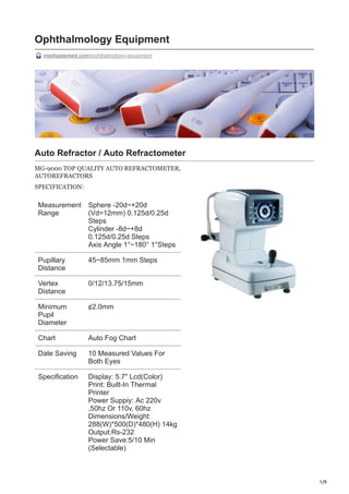
- 1. 1/9 Ophthalmology Equipment medigatemed.com/ophthalmology-equipment Auto Refractor / Auto Refractometer MG-9000 TOP QUALITY AUTO REFRACTOMETER, AUTOREFRACTORS SPECIFICATION: Measurement Range Sphere -20d~+20d (Vd=12mm) 0.125d/0.25d Steps Cylinder -8d~+8d 0.125d/0.25d Steps Axis Angle 1°~180° 1°Steps Pupillary Distance 45~85mm 1mm Steps Vertex Distance 0/12/13.75/15mm Minimum Pupil Diameter ¢2.0mm Chart Auto Fog Chart Date Saving 10 Measured Values For Both Eyes Specification Display: 5.7" Lcd(Color) Print: Built-In Thermal Printer Power Suppiy: Ac 220v ,50hz Or 110v, 60hz Dimensions/Weight: 288(W)*500(D)*480(H) 14kg Output:Rs-232 Power Save:5/10 Min (Selectable)
- 2. 2/9 SLIT LAMP OPTICARE Technical Data o There are iphone or Sumsung mobile adapter optional, can take images by mobile. o Type Parallel Galilean o Eyepiece 12.5X o Total Magnification 6X, 10X, 16X, 25X, 40X o Diameter of Visual Field Φ 37, Φ 23, Φ 14, Φ 8.7, Φ 5.7mm o Diopter Adjustment +5D–5D o Slit Width 0mm-14mm continuously adjustable o Slit Height 1mm-14mm continuously adjustable o Aperture Diameter Φ 14, Φ 10, Φ 5, Φ 3, Φ 2, Φ 1, Φ 0.2mm o Slit Angle 0° -180° o Illumination Tilting 0° , 5° , 10° , 15° , 20° o Filter Heat absorption, grey, redfree(green), cobalt blue o Lamp: German OSRAM Halogen Tungsten Lamp o Longitudinal movement: 90mm o Lateral movement: 100mm o Vertical movement: 30mm o Fine base movement: 15mm o Input Voltage 110V/220V 60/50Hz o Input Power 60VA o Illumination Bulb 12V/50W halogen bulb, led lamp optional o Fixation Red LED OPHTHALMIC OCT (OCT-500) OPTICARE Technical Data Built-in computer Simple, fast and easy to operate Posterior segment OCT. The OCT is an ophthalmic optical coherence tomography scanner tailored for rapid screening of fundus diseases in outpatient clinics. It is easy to use, clear in image, smooth and delicate in operation, and equipped with professional analysis software to meet the requirements of OCT in the clinical examination and analysis of ophthalmology. Features:
- 3. 3/9 1.Compact Design: Everything is inside this compact body. No external computer is needed. Plug in the power cable, then you are ready to go. 2.PC Inside: Data acquisition and processing are accomplished by the internal computer. The data can be transported via ethernet or external harddrive. Peripheral devices, such as keyboard or printer, can be connected to the computer ports. 3.Easy Installation: No complex connection or setup. Compact body can fit in even small space. Methodology Spectral domain OCT Axial resolution ≤ 6 µm (in tissue) Transverse resolution ≤ 20 µm (in tissue) Scan depth ≥ 6 mm Scan speed ≥ 24,000 A-scans/sec, up to 36,000 A-scans/sec Scan modes 30, Raster, Circle Fundus image OCT en face Focus adjustment -150 to +150 Pupil diameter ≥ 3 mm OCT light source 840 nm SLD Optical power 750 µW (at cornea) Operation 13.3''touch screen, optional external mouse or keyboard Power supply 100-240 V, 50/60 Hz Dimensions 497 mm × 395 mm × 490 mm (L× W × H) Weight LED dot matrix External Fixtion 34 kg (75 lbs) A/B SCAN Ophthalmic AB Scan.AB-1000-OPTICARE Feature: 1).12.1″ color and touch screen. 2). With the same software as Quantel, top one in China With advanced intelligent digital software and parameters of freezed and Stored images could be adjusted voluntarily. 3). Can have different results on the report according to different constants A Scan A measures data for every part of the eyeball as anterior chamber depth, lens thickness axial length and so on which are needed in ophthalmic surgeries, and calculates IOL by axial length.
- 4. 4/9 4). B scanner use electromagnetic, better save B probe life. Scan B displays profile images of the eyeball clearly and directly. Scanning anatomical forms and nidi inside the eyeball, doctors can diagnose accurately for examination of cataract, vitreous body disease, ocular trauma, detachment of retina or choroid, macula disease, and intraocular tumor, etc 5). B scanner, video playback of 100 images. 6). A scanner, one group with 10 data to get average, At the same time the accuracy is 0.05mm TECHNICAL DATA: A Scan biometry Probe frequency 10MHz Measuring mode contact Measuring range Over 5mm-40mm Measuring error ≤±0.05mm Measuring type Length of optic axis , automatic calculation , result analysis auto , Image freezing auto+preservable, manual Intraocular lens implantation (IOL)formula SRK-T, SRX II, HOLLADAY, BINKHORST-II, HOFFER- Q,HAIGIS Gain 20-110dB Time gain adjustment 0-30dB B Scan patterns Probe frequency 10MHz Probing depth ≥50mm Axial Resolution ≤0.2mm Lateral resolution ≤0.3mm Gain adjustment 20-110dB Time gain adjustment 0-30dB
- 5. 5/9 Operation mode Eyecup, gel, aqueous capsule Post-processing measuring tools Area, length, mark and annotation Review 100 images available Size 340mm*320mm*115mm weight 10kg Power supply 100-240V 50/60Hz AUTOLENSOMETER AUTO LENS METER OPTICARE Features: 1.Measurement of PD, PH, and PCL (progressive channel Length). D910 Auto Lens meter 2.New capacitive touch panel with high resolution 3.The wavelength of UV and green light can be measured simultaneously . D910 Auto Lens meter 4. Bluetooth, RS232 serial port, USB and WIFI ,4 forms of data communication 5. Better performance .Finer marking pens and lens holder. Magnetic and stable sliding nosepiece D910 Auto Lens meter TECHNICAL DATA: Sphere -25.00D-+25.OOD Axis 0°-180° (1°step) CYLINDER 0 - ±9.99D 0.01/0.12/0.25D steps Prism Degree 0 - 15D 0.01/0.12/0.25D steps ADD 0 - ±9.99D 0.01/0.12/0.25D steps Cylinder +, +/-, - Prism X-Y, P-B Contact lens Soft/hard Measuring Mode Single/progressive/automatic recognition
- 6. 6/9 Diameter of Lens φ12-φ112mm PD 40~90mm, 0.5mm steps Speed of measurement 0.1s Display TFT LCD (7.0") 640*480 Printer Thermal Printer Dimension 180(L) * 255(W) * 455 (H) mm Package 620(L) * 390(W) * 390 (H) mm Weight About 12kgs Power Supply 100~240V 50~60H OPTHALMIC VISUAL FIELD APS6000 Aps-6000cerTraditional Perimeter; LED Specifications: Aps-6000cerTraditional Perimeter; LED Specifications Radius of stimulator: 300mm±5mm 1.Stimulating source of LED 2.Two visual lights: yellow and red. 3.Stimulating strength: From 0nt (0asb) to318.310nt (1000asb), have 14 degree to adjust, the error is±10%. (1.)Error of background brightness: 4asb±10% (2.)Light spot: Diameter is 2mm +/- 0.25mm. (3.)The number of stimulating and the stimulating time: A.388 spots (Red light: 61spots, yellow: 327spots); B: stimulating retention time: 0.2s–2.0s, the program can adjust (±5%); C: stimulating spacing interval: 0.5s –2.0s, the program can adjust (±5%); 4.Window of eye-position tracking: White-black CCD, directly tracking the testing eye; 5.The length of chin rest: up-down: 80mm±10%; right-left 115mm±10%Eye position tracking: When blinking, the system will alarm automatically. TECHNICAL DATA: Sphere -25.00D-+25.OOD Axis 0°-180° (1°step) CYLINDER 0 - ±9.99D 0.01/0.12/0.25D steps
- 7. 7/9 Prism Degree 0 - 15D 0.01/0.12/0.25D steps ADD 0 - ±9.99D 0.01/0.12/0.25D steps Cylinder +, +/-, - Prism X-Y, P-B Contact lens Soft/hard Measuring Mode Single/progressive/automatic recognition Diameter of Lens φ12-φ112mm PD 40~90mm, 0.5mm steps Speed of measurement 0.1s Display TFT LCD (7.0") 640*480 Printer Thermal Printer Dimension 180(L) * 255(W) * 455 (H) mm Package 620(L) * 390(W) * 390 (H) mm Weight About 12kgs Power Supply 100~240V 50~60H AUTO KERATOMETER OPT-V018-N OPTICARE auto refractometer with keratometer Features: OPT-V018-N OPTICARE auto refractometer with keratometer • Sphere -20~+20D(VD=12) 0.125D • Step Cylinder -8~+8D 0.125D • Step Axis 0°~180° (1°step) PD 045- 88mm,1mm • Step VD 0mm,12mm,13.75mm • Min Pupil Size ø2.0mm • Keratometry Radius of curvature 5.0~10mm(increment:0.01mm) • Comal power 33.75~67.50D(when comer equivalent refractive index is 1.337) GREEN LASER ARGON -APPASSAMI Specifications:
- 8. 8/9 • GREEN LASER PHOTO COAGULATOR FOR RETINAL DISORDERS • PATENTED SOLID – STATE TECHNOLOGY • EXTENDED LIFE AND EFFICIENCY • TRUE CONTINUOUS WAVE • TOUCH SCREEN INTERFACE • Special Features • Light weight and portable. • LCD touch screen display with mounting option on table and also on the console. • Foot switch with LED. • Fiber connecting port is illuminated with LED. • Metal shielded laser delivery fiber optic cable TECHNICAL DATA: Model AMOGH PLUS Treatment Laser Diode pumped, frequency doubled, true cw & solid state. Laser Module Capacity 3 Watts Aiming Beam 635 nm, Semiconductor diode laser,0 to 1 mW (variable) Power Adjustment Variable from 10 to 1500 mW Electrical Requirements 110V/230 VAC 50/60Hz, 2A, single phase Cooling Thermo electric cooling (peltier) and air cooled Operation Mode Repeat pulse, single pulse, continuous mode Touch Screen Display LCD screen with feather touch Lifetime 10,000 Working hours Dimensions 290(H) x 175(W) x 365(D) mm Weight 8 Kg Power Consumption 100 Watts Pulse Duration 0.01 – 10 Sec. Pulse Interval 0.01 – 10 Sec. Treatment Laser Safety Class IV
- 9. 9/9