Anatomy & physiology of thyroid gland
•Télécharger en tant que PPTX, PDF•
135 j'aime•28,296 vues

The thyroid gland is located in the neck and produces thyroid hormones that regulate metabolism. It consists of two lobes connected by an isthmus. During development it arises from an endodermal diverticulum. The thyroid traps iodine from the blood and uses it along with the amino acid tyrosine to produce the hormones thyroxine (T4) and triiodothyronine (T3) via a series of coupling reactions within the thyroid follicles. T4 makes up 90% of secretion but T3 is the active hormone. Thyroid hormone production is regulated by TSH from the pituitary gland.

Recommandé
Contenu connexe
Tendances
Tendances (20)
Similaire à Anatomy & physiology of thyroid gland
Similaire à Anatomy & physiology of thyroid gland (20)
Plus de Jinu Iype
Dernier
Dernier (20)
Anatomy & physiology of thyroid gland
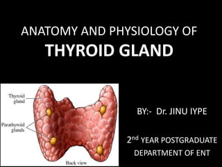
- 1. ANATOMY AND PHYSIOLOGY OF THYROID GLAND BY:- Dr. JINU IYPE 2nd YEAR POSTGRADUATE DEPARTMENT OF ENT
- 2. • Scott Brown 7th edition vol 1 • Textbook otorhinolaryngology-Zakir Hussain • Textbook of Sabiston 19 edition • Guyton textbook of physiology 12th edition
- 3. FROM GREEK thyreoeides = SHIELDSHAPE
- 4. • Butterfly/H shaped • Brownish-Red • Highly vascular • Ductless gland • Adult gland weighing 20 to 25gm. • Larger in female • Enlarges further during puberty, menstruation & pregnancy
- 5. • Consist of right and left cone shaped lobes 5*3*2cm • Connected by a narrow region of gland - Isthmus 1.2*1.2cm • Situated anteriorly in the visceral compartment of the neck at the level of C5-T1 vertebrae
- 6. Location: C5 T1 / 4th or 5th tracheal ring 2nd Trachealrings 4th
- 7. DEVELOPMENT OF THYROID GLAND •Median endodermal thyroid diverticulum(median anlage) Foramen caecum Thyroglossal duct(usually reabsorbed after 6 weeks of age) The very distal end of this remnant may be retained and mature as a pyramidal lobe in the adult thyroid
- 8. THE THYROID begins to function- end of 3month, at which time, the first follicles containing colloid can be seen • Ultimobranchial body gives parafollicular C cells to thyroid gland - Parafollicular cells(Ccells)from the neuralcrest reachthethyroidviatheultimobranchial body-arises from 4th & 5th Brachial pouch. • Superior parathyroid gland- arises from 4th Brachial pouch.
- 9. • Inferior parathyroid gland – arises from 3rd brachial pouch
- 11. • A lateral or posterior projection of the thyroid lobe, known as the tubercle of Zuckerkandl, identified in up to 60 % of surgical dissections • It is represent the point of embryological fusion of the ultimobranchial body and median anlage. Its surgical importance is : (a) RLN runs medial to it; (b) the superior parathyroid gland attached to its cranial aspect;
- 12. CAPSULES • Truecapsule (fibrous) –contains the parenchyma & sends fine septaebetween lobules of the thyroid gland. • Arteries and plexus of veins deep toit • Falsecapsule–pretracheal fascia
- 14. Suspensory ligament of berry The pretracheal layeris thin alongthe posterior border of the lobes, but thick on theinner surface of thegland where it forms a suspensory ligament of berry which connects the gland to the cricoidcartilage
- 15. Why thyroid moves withdeglutition? • During 1st stage ofdeglutition • Hyoid bone movesup • Pulls pre-tracheal fasciaup • This pulls ligament of berryupward • This pulls thyroidupward
- 16. • A fibromuscular band levatorglandulae thyroideae descendfrom thebodyofthe hyoid bone to isthmus or topyramidal lobe
- 17. RELATIONS • The lobes are conical in shape having – Apex – Base – Three surfaces : Lateral, Medial, Posterolateral – Two borders : Anterior & posterior
- 18. • APEX: Directed upward & slightly laterally. limited superiorly – sternothyroid on the oblique line of thyroid cartilage • BASE: At the level of 4th or 5th tracheal length
- 19. • Lateral surface –convex, • Coveredwith sternohyoid, SCM, superior belly of omohyoid, sternothyroid
- 20. • Medial surface- Trachea & Oesophagus 2muscles-cricothyroid, inferior constrictor 2 nerves-external laryngeal &recurrent laryngeal N • Posterolateral surface –carotidsheath
- 21. • Ant border – anterior branch of Superior thyroid artery
- 22. • Posteriorborder - • Thick and round 1. InferiorThyroidArtery 2. Anastomosisb/w STA &ITA 3. Parathyroid glands 4. Thoracic duct onleft
- 23. ISTHMUS • 2 surfacesAnt& Post • 2 borders Sup& Inf • Anteriorsurface –skin & fascia - anterior jugularveins - R& L sternohyoid & sternothyroid • Posteriorsurface- 2nd –3rd trachealrings • Supborder –anastomosis b/w R&L SupThyroidArtery • Inf border -ITVleave
- 24. Blood supply Superior thyroid artery: • 1st anterior branch of external carotid artery • Runs downwards & forward with close relation with external laryngeal nerve • Pierces pretracheal fascia- upper pole of lobe anterior Posterior branch
- 25. • Anterior branch descends on the anterior border of the lobe anastomosing branch which runs along the upper border of the isthmus to anastomosis with opp. side
- 26. • Posterior branch – Posterior border of the lobe Anastomosis with ascending branch of inferior thyroid artery Superior thyroid artery Supplies Upper 1/3rd of the lobe Upper ½ of the isthmus
- 27. Inferior thyroid artery: • Branch of thyrocervival trunk Subclavian artery • Runs forward then medially & finally downward to reach lower pole of the gland. • Pass behind carotid sheath, Middle CervicalGanglion and in front of vertebral vessels • Close related Recurrent laryngeal Nerve
- 28. • Artery divides into 4 or 5 Glandular branch pierces the fascia separately to reach the lower part of the gland • Ascending branch anastomoses with posterior branch of superior thyroid artery Supply Parathyroid gland Inferior thyroid artery Supplies Lower 2/3rd of the lobe Lower ½ of the isthmus & Parathyroid gland
- 29. THYROIDEA IMA ARTERY ( lowest thyroid artery) • 12 % present ascending in front of the trachea to end at the isthmus. • most commonly arises from the brachiocephalic artery • it can also originate from the aorta, the right common carotid, subclavian or internal thoracic arteries.
- 30. VENOUS DRAINAGE : The superior, middle and inferior thyroid vein Superior thyroid vein (STV) •Accompany SupThyroid Artery •Drain to IJV/ facial vein
- 31. Middle thyroid vein • Very short, may be double or absent. • They receive blood from the inferior and antero-lateral part of the gland as well as the larynx and trachea. • most commonly cross the common carotid artery • Drain to IJV
- 32. Inferior thyroid vein •Plexus anterior surface of the trachea on leaving the gland. • usually drain into right and left inferior veins, superior vena cava or left brachiocephalic
- 33. 4THVEINOFKOCHER’S • Seen b/w middle and inferior thyroidvein • drain intoIJV
- 34. LYMPHATIC DRAINAGE The thyroid gland contains a rich network of lymphatics The lateral aspects of the gland drain into levels III and IV and those of the posterior triangle (level V). The more medial aspects of the gland also drain into the nodes of the anterior compartment of the neck (level VI), drain into those of the superior mediastinum (level VII).
- 36. Nerve supply: • Parasympathetic fibers –fromVagus • Sympatheticfibers–from superior,middle,and inferiorganglia ofthe sympathetictrunk • Entertheglandalong withthebloodvessels.
- 37. Recurrent laryngeal nerves • The recurrent laryngeal nerve is variable in size 1.5-4 mm in diameter. • Identified by its whitish appearance, characteristic longitudinal vessel flattened, rounded surface. In up to 39 % of cases the nerve divides into 2 (and occasionally up to 6) terminal branches between 6 and 35 mm from the cricoid cartilage.
- 38. A non recurrent laryngeal nerve is found in 0.2- 0.4% of patients It tends to be thicker than a normally sited nerve Usually associated with a vascular anomaly of the subclavian artery on the right side Transposition of the great vessels on the left side.
- 39. Recurrent laryngeal nerves • Branches of the vagus nerve, which supply all the intrinsic muscles of the larynx except the cricothyroid muscle.(External laryngeal N – Superior Laryngeal N) • They also supply sensory fibres to the mucous membrane below the level of the vocal folds • Accidentaldamage tothis nerveduring surgery causes ipsilateralvocal cord paralysis& difficultyin phonation
- 40. Left Recurrent laryngeal nerves • The approximate length of the left RLN is 12 cms • The nerve leaves the vagus in the mediastinum anterior to the arch of the aorta passing behind the ligamentum arteriosum & then posteriorly under the concavity of the arch before passing superiorly to lie in the tracheo-oesophageal groove.
- 41. • It most usually passes behind the inferior thyroid artery • Then posterior to the ligament of Berry before passing under or between the fibres of the cricopharyngeal part of the inferior constrictor
- 42. Right Recurrent laryngeal nerves • The approximate length of right RLN 6 cms • Rtside itoriginates from vagus crosses firstpartof subclavian artery . • More oblique course to the tracheo- oesophageal groove.
- 43. SUPERIORLARYNGEALNERVE • Arises from inferior ganglion of vagus • Descends behind internal carotid artery • At the level of greater cornua of hyoid it divides:- Internal branch(sensory) Externalbranch(motor)
- 45. • The external branch of the superior laryngeal nerve, which supplies the cricothyroid muscle, runs parallel to the superior thyroid vessels • The Internal branch of the superior laryngeal nerve supply sensory fibres to the mucous membrane above the level of the vocal folds
- 47. External branch of superior laryngeal nerve and joll’s triangle • Joll's triangle is used to identify the location of external branch of superior laryngeal nerve during thyroid surgeries. • Damage to this nerve during the surgical procedure may reduce the voice range in those patients. • This triangle is also known as sternothyrolaryngeal triangle.
- 48. Boundaries of Joll's triangle : Lateral - Upper pole of thyroid gland and superior thyroid vessels Superior - Attachment of the strap muscles and deep investing layer of fascia to the hyoid Medial - Midline Floor - Cricothyroid muscle External branch of superior laryngeal nerve lies within this triangle.
- 49. Beahrs Triangle or Riddle’s triangle Boundaries Medial :The RL nerve in the lower part of tracheo - oesophageal groove Lateral :Common carotid Superior: Inferior thyroid artery
- 50. Microscopic anatomy • The thyroid gland consists mainly of follicular cells, one cell thick around a central pool of colloid to form follicles. • The follicles spherical in shape & 0.02- 0.9 mm in diameter • A thyroid lobule consists of 20 to 40 follicles and is supplied by a lobular artery.
- 51. When the gland is relatively inactive, the cells are flattened and the colloid is abundant, dense. On prolonged and excessive TSH stimulation, the follicular cells become hypertrophied and hyperplastic and they adopt a more columnar shape. This cellular enlargement is associated with development of microvilli which helps in reduction in the size of the follicular lumen.
- 53. PHYSIOLOGY OF THE THYROID The thyroid follicles secretestri-iodothyronine (T3) and thyroxin(T4) Synthesis involves combination of iodine with tyrosine group toform mono and di-iodotyrosine which are coupled to form T3 andT4. The hormones are stored in follicles bound to thyrogobulin
- 54. When hormones released in the blood they are bound to plasma proteins and small amount remain free in the plasma The metabolic effect of thyroid hormones are due to free (unbound)T3 andT4. 90%of secreted hormones is T4 butT3is the active hormone so, T4is converted to T3peripherally.
- 55. Regulation of thyroid gland metabolism CirculatingT3 andT4exert - - ve feedback mechanism on hypothalamus and anterior pituitary gland So, in hyperthyroidism where hormone level in blood is high ,TSH production is suppressed and vice versa.
- 56. STEPS OF THYROID HORMONE SYNTHESIS 1.Thyroglobulin Synthesis 2.Iodine trapping 3.Oxidation 4.Iodination 5.Coupling 6.Storage & Release
- 57. 1. Thyroglobulin Synthesis • Endoplasmic reticulum and Golgi apparatus in the follicular cells of thyroid gland synthesize and secrete thyroglobulin continuously. • Thyroglobulin molecule is a large glycoprotein containing 140 molecules of amino acid tyrosine. • After synthesis, thyroglobulin is stored in the follicle.
- 58. 2. Iodine trapping Iodide is actively transported from blood into follicular cell, against electrochemical gradient. This process is called iodide trapping. Iodide is transported into the follicular cell along with sodium by Sodium iodide (Na + / I-) symporter , which is also called iodide pump. Whereby two sodium ions are transported for each iodide ion.
- 59. The accumulated iodide in the follicular cells is then transferred to the apical plasma membrane down an electrochemical gradient. TSH stimulation increases adenosine triphosphate (ATP) and ATPase activity at the apex of the cell increasing the efflux of iodide into the colloid down a further electrical gradient. Iodine available through certain foods (eg, seafood, bread, dairy products), iodizedsalt, or dietarysupplementsetc
- 60. 3. Oxidation of Iodide • Iodide must be oxidized to elementary iodine, because only iodine is capable of combining with tyrosine to form thyroid hormones. • The oxidation of iodide into iodine occurs inside the follicular cells in the presence of thyroid peroxidase.
- 61. 4. Iodination of Tyrosine • Iodine is transported from follicular cells into the follicular cavity, where it binds with thyroglobulin. • Then, iodine (I) combines with tyrosine, which is already present in thyroglobulin • Tyrosine is iodized first into monoiodotyrosine (MIT) and later into di-iodotyrosine (DIT)- thyroglobulin(Tg) tyrosine residues
- 62. 5. Coupling Reactions i. One molecule of DIT and one molecule of MIT combine to form tri-iodothyronine (T3) DIT + MIT = Tri-iodothyronine (T3) ii. One molecule of MIT and one molecule of DIT combine to produce another form of T3 called reverse T3 or rT3. Reverse T3 is only 1% of thyroid output MIT + DIT = Reverse T3 iii. Two molecules of DIT combine to form tetraiodothyronine (T4) thyroxine. DIT + DIT = Tetraiodothyronine or Thyroxine (T4)
- 65. FUNCTIONS OF THYROID HORMONES 1. Action on basal metabolic rate (BMR) Thyroid hormones (specificallyT3)regulate rate of overall bodymetabolism –T3increases basal metabolicrate Calorigeniceffects –T3increases oxygen consumption bymost peripheraltissues –Increases body heatproduction
- 66. 2. Action on carbohydrate metabolism • Thyroxine stimulates almost all processes involved in the metabolism of carbohydrate. Thyroxine: i. Increases the absorption of glucose from GI tract ii. Enhances the glucose uptake by the cells, by accelerating the transport of glucose through the cell membrane iii. Increases the breakdown of glycogen into glucose iv. Accelerates gluconeogenesis.
- 67. 3. Action on fat metabolism Thyroxine decreases the fat storage by mobilizing it from adipose tissues and fat depots. Thus, thyroxine increases the free fatty acid level in blood.
- 68. 4.Action on Growth andDevelopment • Thyroid hormone is essential for normal braindevelopment • Essential for childhoodgrowth –Untreated congenital hypothyroidism or chronic hypothyroidism during childhood can result in incomplete development and mentalretardation
- 69. 5.Action on CNS • Thyroid hormones are essential for neural development and maturation and function ofthe CNS • Decreased thyroid hormone concentrations maylead to alterations in cognitivefunction –Patients with hypothyroidism may develop impairment of attention,slowed motorfunction, and poormemory –Thyroid-replacementtherapy may improve cognitive function when hypothyroidism is present
- 70. 6.Action on BoneGrowth – T3also may participate in osteoblast differentiation and proliferation, and chondrocyte maturation leading to boneossification
- 71. 7.Action on femaleReproductiveSystem • Normal thyroid hormone function is important for reproductivefunction – Hypothyroidism may be associated with menstrual disorders, infertility, risk of miscarriage, and other complications of pregnancy
- 72. 8.ACTION ON GASTROINTESTINAL TRACT • Generally, thyroxine increases the appetite and food intake. • It also increases the secretions and movements of GI tract. • So, hypersecretion of thyroxine causes diarrhoea and the lack of thyroxine causes constipation.
- 73. 9. Actions on:- • Heart In Increase cardiac output, heart rate • Adipose tissue Stimulate lipolysis • Muscle Increase protein breakdown
- 74. APPLIEDANATOMY • Presenceof thyroidae imaA-chanceof profusebleeding procedures in neckbelow isthmus • Thyroglossal cysts– Remnants of thyroglossal ducts at anypoint in the wayof descent,(midline nearhyoid) • Pyramidal lobe and presenceof levator glandulae thyroidae
- 75. • Ectopic thyroid glands–lingual/higher placed • Non neoplastic, noninflammatory enlargement – goiter • pressuresymptoms and nerve involvments are common in goiter and carcinoma
- 76. THANK YOU!