
Recommandé
Contenu connexe
Tendances
Tendances (20)
Similaire à THE SKELETON SYSTEM ANATOMY AND PHYSIOLOGY SLIDESHARE
Similaire à THE SKELETON SYSTEM ANATOMY AND PHYSIOLOGY SLIDESHARE (20)
Plus de Jitendra Bhargav
Plus de Jitendra Bhargav (20)
Dernier
Dernier (20)
THE SKELETON SYSTEM ANATOMY AND PHYSIOLOGY SLIDESHARE
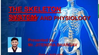
- 1. THE SKELETON SYSTEM ANATOMY AND PHYSIOLOGY Presented by... Mr. JITENDRA BHARGAV .
- 2. Presented by... Mr. JITENDRA BHARGAV . Ass.Lecturer
- 8. Classification of Bones • Long Bones :- • Ilustration mapping the different components of a long bone • The bones of the body come in a variety of sizes and shapes. • The four principal types of bones are long, short, flat and irregular. • Bones that are longer than they are wide are called long bones. • They consist of a long shaft with two bulky ends or extremities.
- 11. Short Bones :- Short bones are roughly cube shaped with vertical and horizontal dimensions approximately equal. They consist primarily of spongy bone, which is covered by a thin layer of compact bone. Short bones include the bones of the wrist and ankle.
- 13. • Flat Bones :- • Flat bones are thin, flattened, and usually curved. • Most of the bones of the cranium are flat bones.
- 15. • Irregular Bones:- • Bones that are not in any of the above three categories are classified as irregular bones. • They are primarily spongy • bone that is covered with a thin layer of compact bone. • The vertebrae and some of the bones in the skull are irregular bones.
- 20. Axial Skeleton (80 bones) Skull (28) Illustration mapping cranial bones • Cranial Bones • Parietal (2) • Temporal (2) • Frontal (1) • Occipital (1) • Ethmoid (1) • Sphenoid (1
- 22. • Frontal bone. This is the flat bone that makes up your forehead. It also forms the upper portion of your eye sockets. • Parietal bones. This a pair of flat bones located on either side of your head, behind the frontal bone. • Temporal bones. This is a pair of irregular bones located under each of the parietal bones. • Occipital bone. This is a flat bone located in the very back of your skull. It has an opening that allows your spinal cord to connect to your brain. • Sphenoid bone. This is an irregular bone that sits below the frontal bone. It spans the width of your skull and forms a large part of the base of your skull. • Ethmoid bone. This is an irregular bone located in front of the sphenoid bone. It makes up part of your nasal cavity.
- 23. • Facial Bones :- • • Maxilla (2) • Zygomatic (2) • Mandible (1) • Nasal (2) • Platine (2) • Inferior nasal concha (2) • Lacrimal (2) • Vomer (1)
- 24. The two palatine bones (L., palatum “palate”) form portions of the hard palate, lateral walls of the nasal cavity, and floors of the orbits. Palatine bone These small, L-shaped, facial bones are located between the palatine processes of the maxilla bones and the pterygoid processes of the
- 25. The two maxilla or maxillary bones (maxillae, plural) form the upper jaw (L., mala, jaw). Each maxilla has four processes (frontal, zygomatic, alveolar, and palatine) and helps form the orbit, roof of the mouth, and the lateral walls of the nasal cavity. Maxilla Bone Orbital surface (process) – posterior extension from body that forms much of orbit floor. [Anterior view]
- 26. Some of the smallest are the two nasal bones, two inferior turbinates (nasal conchae), and the single vomer bone. Nasal, Vomer Nasal bones – are two small bones that form the bridge of the nose. Vomer bone – a thin bone that runs vertically along the midline of the nasal cavity.
- 27. Hyoid bone The hyoid bone (Gr. hyoeides, U-shaped) is a small, U- shaped bone that is located between the mandible and larynx and anterior to the third cervical vertebra. It does not directly articulate with any other bones. Instead, the hyoid bone is loosely held in place by several ligaments and muscles that attach to the skull, mandible, tongue, larynx, and scapula.
- 28. The Mandible (L., mandere – to chew) is the facial bone that forms the lower jaw and contains the lower teeth. It consists of right and left halves that fuse together early in life. The anterior portion of the mandible, called the body, is horseshoe-shaped and runs horizontally.
- 29. The zygomatic bones (Gr., zygoma – yoke) are two facial bones that form the cheeks and the lateral walls of the orbits. They are also commonly referred to as the cheekbones or malar bones (L., mala – the cheek). Each zygomatic bone articulates with the temporal bone, frontal bone, maxilla, and sphenoid bones.
- 30. Lacrimal Bone The lacrimal bone (L., lacrima – tear) is a small facial bone that forms a portion of the anterior medial wall of the orbit Lacrimal fossa (or lacrimal sulcus; fossa for lacrimal sac) – depression along the junction of the lacrimal bone and maxilla bone that holds the lacrimal sac; tears formed by the sac drain through a duct into the nasal cavity. Orbital plate (or orbital surface) – thin plate of bone that forms a portion of the medial wall of the orbit posterior to lacrimal fossa.
- 31. • Auditory Ossicles :- • • Malleus (2) • Incus (2) • Stapes (2)
- 32. • Vertebral Column • • Cervical vertebrae (7) • Thoracic vertebrae (12) • Lumbar vertebrae (5) • Sacrum (1) • Coccyx (1)
- 34. • Thoracic Cage • • Sternum (1) • • Ribs (24)
- 36. • Appendicular Skeleton (126 bones) • • Illustration mapping the bones of the pectoral girdles • Pectoral girdles • • Clavicle (2) • • Scapula (2)
- 38. ( 2) U ln a ( 2) C a r p al s ( 1 6) M e t a c a r p al • Upper Extremity • • Humerus (2) • Radius (2) • Ulna (2) • Carpals (16) • Metacarpals (10) • Phalanges (28)
- 41. BUSINESS BUSINESS • Pelvic Girdle • • Coxal, innominate, or hip bones (2)
- 44. • Illustration mapping the bones of the lower extremety • Lower Extremity • • Femur (2) • Tibia (2) • Fibula (2) • Patella (2) • Tarsals (14) • Metatarsals (10) • Phalanges (28)
- 45. Review: Introduction to the Skeletal System • The human skeleton is well-adapted for the functions it must perform. • Functions of bones include support, protection, movement, mineral storage, and formation of blood • There are two types of bone tissue: compact and spongy. • Compact bone consists of closely packed osteons, or haversian system. • Spongy bone consists of plates of bone, called trabeculae, around irregular spaces that contain red bone marrow.
- 46. • Osteogenesis is the process of bone formation. • Three types of cells, osteoblasts, osteocytes, and osteoclasts, are involved in bone formation and remodeling • In intramembranous ossification, connective tissue membranes are replaced by bone. • This process occurs in the flat bones of the skull. • In endochondral ossification, bone tissue replaces hyaline cartilage models. • Most bones are formed in this manner.
- 47. • Bones grow in length at the epiphyseal plate between the diaphysis and the epiphysis. • When the epiphyseal plate completely ossifies, bones no longer increase in length. • Bones may be classified as long, short, flat, or irregular. • The diaphysis of a long bone is the central shaft. • There is an epiphysis at each end of the diaphysis.
- 48. • The adult human skeleton usually consists of 206 named bones and these bones can be grouped in two divisions: axial skeleton and appendicular skeleton. • The bones of the skeleton are grouped in two divisions: axial skeleton and appendicular skeleton.
- 49. • There are three types of joints in terms of the amount of movement they allow: synarthroses (immovable), amphiarthroses (slightly movable), and diarthroses (freely movable).
- 51. Muscle Types BUSINESS • Skeletal Muscle :- • Skeletal muscle, attached to bones, is responsible for skeletal movements. • The peripheral portion of the central nervous system (CNS) controls the skeletal muscles. • Thus, these muscles are under conscious, or voluntary, control. • The basic unit is the muscle fiber with many nuclei. These muscle fibers are striated (having transverse streaks) and each acts independently of neighboring
- 53. • Smooth Muscle • Smooth muscle, found in the walls of the hollow internal organs such as blood vessels, the gastrointestinal tract, bladder, and uterus, is under control of the autonomic nervous system. • Smooth muscle cannot be controlled consciously and thus acts involuntarily. • The non-striated (smooth) muscle cell is spindle- shaped and has one central nucleus. • Smooth muscle contracts slowly and rhythmically.
- 54. • Cardiac Muscle • Cardiac muscle, found in the walls of the heart, is also under control of the autonomic nervous system. • The cardiac muscle cell has one central nucleus, like smooth muscle, but it also is striated, like skeletal muscle. • The cardiac muscle cell is rectangular in shape. • The contraction of cardiac muscle is involuntary, strong, and rhythmical.
- 55. Function of the skeletal system :- • supports the body • facilitates movement • protects internal organs • produces blood cells • stores and releases minerals
- 58. Joints are classified by their range of movement: • Immovable, or fibrous, joints • Partially movable, or cartilaginous • Freely movable, or synovial
- 59. • Hinge joints :- allow movement in one direction, as seen in the knees and elbows. • Pivot joints:-allow a rotating or twisting motion, like that of the head moving from side to side • Ball-and-socket joints :-allow the greatest freedom of movement. • The hips and shoulders have this type of joint, in which the round end of a long bone fits into the hollow of another bone.
- 60. method of design. It is a blank space. It is the most common in minimalist design. Keeping white space sounds very simple and then use it properly simple and then use it properly White space is an advanced method of design. It is a blank space. It is the most common in minimalist design. Keeping white space sounds very simple and then use it properly simple and then use it properly Presented by... Mr. JITENDRA BHARGAV . THE SKELETON SYSTEM