1. SHORT COMMUNICATION
Protective effect of fallopian tubal fluid against activated
leucocyte-induced sperm DNA fragmentation: preliminary
results
P. Navarrete Gomez1,6, J. Espinoza Ruiz1,5, J. Parodi Rivera1, J. G. Alvarez2,3 & R. Sanchez Gutierrez4
´ ´ ´
1 ´ ´
Centro de Excelencia de Biotecnologıa de la Reproduccion (CEBIOR), Universidad de La Frontera, Temuco, Chile;
2 `
Instituto Marques, Barcelona, Barcelona, Spain;
3 ´ `
Fundacion Leonardo Marques, Barcelona, Spain;
4 ´
Departamento de Ciencias Preclınicas, Universidad de La Frontera, Temuco, Chile;
5 Tesista y;
6 ´ ´
Estudiante del Programa de Doctorado en Ciencias Mencion Biologıa Celular y Molecular Aplicada, Becarios CONICYT
Keywords: Summary
Activated leucocytes—DNA
fragmentation—human sperm—ROS— The integrity of the paternal genome is of paramount importance in the initia-
tubal fluid tion and maintenance of a viable pregnancy. Oxygen radicals (ROS) have been
identified as one of the main factors responsible for the induction of sperm
Correspondence DNA damage. Spermatozoa are mainly protected against ROS-induced damage
´ ´ ´
Dr Raul Sanchez Gutierrez, Facultad de
by seminal plasma. However, this protective effect disappears once spermatozoa
Medicina, Universidad de La Frontera, Manuel
Montt 112, Temuco, Chile.
enter the female genital tract. The fallopian tube mucosa may play a protective
Tel.: +56 45 744248; role against ROS-induced sperm damage. The main objective of this study was
Fax: +56 45 325600; to determine whether human tubal explants and tubal fluid exert a protective
E-mail: rsanchez@ufro.cl effect on ROS-induced sperm DNA damage. Spermatozoa were exposed to
tubal explants and/or tubal fluid in the presence of phorbol myristate acetate
Accepted: January 13, 2009 (PMA)-activated polymorphonuclear leucocytes or control medium and sperm
DNA fragmentation was measured using the TdT-mediated dUTP-biotin nick
end labelling (TUNEL) test. Exposure of human spermatozoa to PMA-activated
leucocytes resulted in a 2-fold increase in sperm DNA fragmentation. Co-incu-
bation of spermatozoa with tubal explants did not reduce this damage.
However, pre-incubation of spermatozoa with tubal fluid resulted in a statis-
tically significant reduction in sperm DNA fragmentation levels, comparable to
those observed in control. In conclusion, tubal fluid appears to protect against
activated leucocyte-induced sperm DNA fragmentation, thus preserving the
integrity of the paternal genome.
fluid exert a protective effect against ROS-induced sperm
Introduction
DNA damage.
The integrity of the paternal genome is vital to the onset
and maintenance of a viable pregnancy to term
Materials and methods
(Angelopoulou et al., 2007). The previous studies have
shown that exposure of human spermatozoa to tubal/ Tubal explants were obtained from the fallopian tubes of
oviductal epithelial cells protects them from ROS-induced four healthy women subjected to hysterectomy. Tubal
damage, thus increasing sperm survival (El Mouatassim fluid was obtained from the explants and incubated at
et al., 2000). Despite all these studies, however, it is still 37 °C for 24 h in supplemented DMEM medium
not known whether the oviduct exerts a protective effect (sDMEM). Semen samples were obtained from healthy
on sperm DNA fragmentation. The primary aim of this donors, processed for standard swim-up and incubated
study was to determine if human tubal explants or tubal with tubal fluid during 12 h.
196 ª 2009 Blackwell Verlag GmbH Æ Andrologia 41, 196–198
2. ´
P. Navarrete Gomez et al. Tubal fluid protects against sperm DNA damage
Polymorphonuclear (PMN) leucocytes were activated (a)
with phorbol-12-miristate-13-acetate (PMA). Sperm DNA
fragmentation was determined using the TUNEL test
according to the manufacturer’s instructions (Roche,
Grenzach-Wyhlen, Germany). Following TUNEL labelling,
propidium iodide was added and the samples analysed by
flow cytometry. TUNEL test data were analysed using the
graphpad prism 5.0 program (GraphPad Software, La
Jolla, CA, USA).
Differences between the groups were analysed using the
Kruskal–Wallis and Dunn’s post-tests. A P-value less than
0.05 (P < 0.05) was considered as statistically significant.
(b)
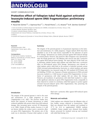
Results and discussion
Incubation of human spermatozoa in sDMEM did not
result in any significant increase in DNA fragmentation
compared with the control with HTF medium (P > 0.05)
(Fig. 1a). Treatment with DNAse I produced a fragmenta-
tion level of 95 ± 3.06% (P < 0.05). Incubation of human
spermatozoa with activated leucocytes resulted in an
increase in DNA fragmentation levels, although these dif-
ferences were not statistically significant (P > 0.05).
Moreover, incubation of the spermatozoa with PMN,
PMA and DMSO did not produce any significant effect
(c)
(P > 0.05) on sperm DNA fragmentation. Incubation of
human spermatozoa with tubal explants produced a sig-
nificant decrease in sperm DNA fragmentation levels dur-
ing incubation at 37 °C (P < 0.05) (Fig. 1b), whereas
incubation of human spermatozoa with tubal medium
did not result in any significant effect on sperm DNA
fragmentation (P > 0.05). Incubation with activated leu-
cocytes did not produce any significant decrease
(P > 0.05) in activated leucocyte-induced sperm DNA
fragmentation levels, whereas incubation with tubal fluid,
after incubation with activated leucocytes, produced a sig-
nificant decrease in sperm DNA fragmentation levels
(P < 0.05) comparable to those observed in the control
(Fig. 1c).
Fig. 1 Effect of incubation in sDMEM, tubal explants (TU) or tubal
The main findings of this study are that (i) tubal ex-
fluid (CM) on sperm DNA fragmentation. (a) Percentage of TUNEL-
plants decrease sperm DNA fragmentation during incuba- positive spermatozoa after incubation in sDMEM (*P < 0.05); (b) Per-
tion at 37 °C, whereas tubal fluid had no effect; and (ii) centage of TUNEL-positive spermatozoa after incubation with TU or
tubal fluid prevents sperm DNA fragmentation induced CM; (c) Percentage of TUNEL-positive spermatozoa after incubation
by PMA-activated leucocytes, whereas tubal explants had with tubal TU or CM prior to incubation with activated leucocytes and
no effect. The previous studies have shown that human their respective controls.
spermatozoa can establish direct contact with the tubal
epithelium and that this contact increases motility, pre- effect on activated leucocyte-induced sperm DNA frag-
vents the premature acrosome reaction and increases the mentation might be related to tubal cell damage induced
fertility capacity of spermatozoa (Morales et al., 1996; by the specific ROS produced by activated leucocytes,
Green et al., 2001). This could be related to a possible which could inactivate enzymatic antioxidant defences of
antioxidant effect of the tubal cells that would neutralise these cells. In the case of tubal fluid, the antioxidant
the ROS produced by spermatozoa in the extracellular defences would be mainly nonenzymatic soluble factors
medium. In contrast, the tubal explants’ lack of protective that are not susceptible to inactivation by the high levels
ª 2009 Blackwell Verlag GmbH Æ Andrologia 41, 196–198 197
3. Tubal fluid protects against sperm DNA damage ´
P. Navarrete Gomez et al.
of ROS produced by the activated leucocytes. Studies are ´ ´ ´
El Mouatassim S, Guerin P, Menezo Y (2000) Mammalian
underway to try to answer the question of why the tubal oviduct and protection against free oxygen radicals:
fluid protects against activated leucocyte-induced DNA expression of genes encoding antioxidant enzymes in
damage and not that produced during incubation with human and mouse. Eur J Obstet Gynecol Reprod Biol 89:
nonactivated leucocytes. 1–6.
Green CE, Bredl J, Holt WV, Watson PF, Fazeli A (2001)
Carbohydrate mediation of boar sperm binding to oviductal
References epithelial cells in vitro. Reproduction 122:305–315.
´
Morales P, Palma V, Salgado AM, Villalon M (1996) Sperm
Angelopoulou A, Plastira K, Msaouel P (2007) Spermatozoal
sensitive biomarkers to defective protaminosis and frag- interaction with human oviductal cells in vitro. Hum Reprod
mented DNA. Reprod Biol Endocrinol 5:1–15. 11:1504–1509.
198 ª 2009 Blackwell Verlag GmbH Æ Andrologia 41, 196–198