
Recommandé
Recommandé
Contenu connexe
Tendances
Tendances (20)
Similaire à URINE SPECIMEN.pptx
Similaire à URINE SPECIMEN.pptx (20)
Dernier
Dernier (20)
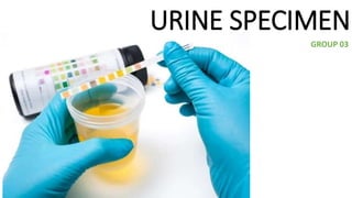
URINE SPECIMEN.pptx
- 2. Why urine collection. It's used to detect and manage a wide range of disorders, such as urinary tract infections, kidney disease and diabetes. In microbiology we deal with microorganisms causing infections in urine. Possible ones examined in urine are; 1. Mostly gram negative bacteria - Enterobacteriaceae Escherichia coli Proteus species Pseudomonas aeruginosa Klebsiella strains Salmonella Typhi Salmonella Para-typhi Neisseria gonorrhoeae
- 3. 2. Gram positive bacteria Staphylococcus saprophyticus Hemolytic streptococci 3. Protozoans T. vaginalis, Schistosoma haematobium, micofilaria. 4. Fungi species Candida species but may also be caused by Cryptococcus neoformans, Aspergillus species, and the endemic mycoses.
- 4. Urine collecting methods. sterile urine bag, urethral catheterization (CATH), suprapubic aspiration (SPA), or clean-catch (CC), dry sterile leak proof container.
- 5. Types of urine Specimen 1. First Morning Specimen - a specimen obtained during the first Best for: • Nitrite • Protein • Microscopic examination 2. Postprandial – is a specimen obtained 2 hours after meal. Good for glucose level examination. 3. 24- Hour specimen - a specimen obtained within 24 hours. • determination of protein levels quantitatively.
- 6. 4. Mid- stream Specimen - a specimen obtained from the middle part of the first urine. • It is commonly used for routine urinalysis. • It is also important for bacteriological urine culture. Midstream urine (MSU) for microbiological examination is collected as follows: 1. Give the patient a sterile, dry, wide-necked, leak-proof container and request a 10–20 ml specimen. Female patients: Wash the hands. Cleanse the area around the urethral opening with clean water, dry the area with a sterile gauze pad, and collect the urine with the labia held apart. Male patients: Wash the hands before collecting a specimen (middle of the urine flow). 2 Label the container with the date, the name and number of the patient, and the time of collection. As soon as possible, deliver the specimen with a request form to the laboratory. When immediate delivery to the laboratory is not possible, refrigerate the urine at 4–6 °C. When a delay in delivery of more than 2 hours is antici- pated, add boric acid preservative to the urine.
- 8. Mix the urine sample to re-suspend microorganism present. Dip a 1 μl calibrated loop in vertical position in the urine and remove the loop and use the collected fluid to inoculate CLED, Blood and MacConkey agars respectively. Culturing Procedure
- 10. . •Contains lactose as substrate and Neutral red as an indicator •Bacteria fermenting lactose produce acid and this changes the colour of the indicator and the colonies turn pink Used: To differentiate lactose fermenters (E. coli, Klebsiella) from non-lactose fermenters (Salmonella, Shigella) 5/30/2023 10 1. Mac Conkey’s agar
- 11. Results of Lactose Fermentation on MacConkey 5/30/2023 11
- 12. 2. Blood agar • Used to differentiate hemolytic and non hemolytic bacteria • Most gram negative bacteria show no hemolysis on blood agar which are mostly found in urine specimen i.e klebsiella, e-coli and proteus ssp. klebsiella E-coli proteus
- 14. Cystine lactose electrolyte-deficient (CLED) agar is widely used by laboratories to isolate urinary pathogens because: It gives consistent results and allows the growth of both Gram negative and Gram positive pathogens. The indicator in CLED agar is bromothymol blue and therefore lactose fermenting colonies appear yellow. The medium is electrolyte-deficient to prevent the swarming of Proteus species.
- 15. 1. E-coli are lactose fermenters on CLED yellowish colonies and reddish/pinkish colonies in MCA Biochemical test a. indole test – using Kovac reagent if red ring is formed its indole positive 2. Klebsiella Are lactose fermenters large mucoid colonies Biochemical test a. Citrate test – if it turns blue its citrate positive b. Urea test – if it turns pinkish its positive 3. Proteus mirabilis On blood agar it swarms but not in CLED (PREFERED MEDIA), its non lactose fermenter Biochemical test; a. Urea test – its positive pinkish 4. Pseudomonas aeruginosa Causative agent of UTI, its non lactose fermenter Biochemical test. a. Oxidase test = its positive
- 16. A plate count of 100,000 CFU/ml of pure culture should be considered positive and isolated organism should be identified and sensitivity test will be performed. A plate count between 10,000 – 100,000 CFU/ml is considered suspected . A plate count less than 10,000 CFU/ml is considered negative. ml) in plated, (volume (dilution) counted colonies # sample original in ml CFU Colony counting
- 18. Count the approximate number of colonies. Estimate the number of bacteria, i.e. colony-forming units (CFU) per ml of urine. Report the bacterial count as: ● Less than 10 000 organisms/ml (104/ml), not significant. ● 10 000–100 000/ml (104–105/ml), doubtful significance (suggest repeat specimen) ● More than 100 000/ml (105/ml), significant bacteriuria. Interpretation of quantitative urine culture result.
- 20. EXAMPLE If 25 E. coli colonies are counted and a 1/1000 ml loop was used, the approximate number of CFU per ml of urine: 1000 X 25 = 25 000, Such a count would be reported as: 10 000 – 100,000 E. coli/ml NOTE: It is generally agreed that if more than 10 per 5 colonies/mL are cultivated from a properly collected and properly cultured urine specimen, this constitutes strong evidence of active urinary tract infection.
- 21. Sensitivity tests. We use MHA in performing sensitivity tests using four to five antibiotic drugs. Drugs for gram negative rods are; 1. Tetracycline 2. Contrimazole 3. Ciproflaxone 4. Gentamycin 5. Erythromycin 6. Penicilin Incubate over night and look for zone of inhibition. We report in mm in diameter.