Keratometry is used to measure the curvature of the cornea. It works by reflecting light off the cornea and measuring the size of the reflected image. Dynamic retinoscopy objectively determines the refractive state of the eye when it is accommodating to view a near target. It provides information about the eye's accommodative response and ability to focus at near. Dynamic retinoscopy techniques include MEM, Nott retinoscopy, and Bell retinoscopy which use different targets and methods to evaluate accommodation.
3. Zones of the Cornea
Central Zone (apical zone/corneal
cap/central spherical zone) – 4 mm,
radius of curvature does not vary by
more than 1 D or 0.05 mm
• Area where refraction differs by
<0.25 D
• Paracentral zone – 4 - 8 mm.
• Peripheral zone – 8 - 11 mm.
• Limbal zone – rim of cornea, 0.55
mm wide.
4. What is Keratometry?
Measurement of the anterior surface of the
cornea, across the fixed chord length, usually
2-3mm, which lies within the optical spherical
zone of the cornea.
5. Principle of Keratometry
Anterior surface of the cornea- CONVEX MIRROR
higher the Curvature = smaller Image size
Image (ie.1st Purkinje image) formed in cornea
With this image the radius of the curvature of the
cornea can be calculated.
7. Principle…
Due to presence of involuntary miniature eye
movements during fixation of a eye
The image formed by anterior surface of the cornea
also moves –
Use of Doubling principle
CHALLENGE
8. DOUBLING PRINCIPLE
Measurement of image height
Doubling device - Plano prism
Lateral disp. of doubled image = IMAGE HEIGHT
Prism is moved along the optical axis until two images
are just touching
At this point, the prismatic displacement is exactly equal
to the size of the image
10. DOUBLING SYSTEMS
FIXED DOUBLING –
VARIABLE
Image size and mire separation
FIXED
Object height and doubling device distance
Ex. B & L , Topcon & Magnon
► VARIABLE DOUBLING –
Fixed
image size & mire separation
Variable
object size & doubling device
distance
Ex. Haag streit & Javal Schiotz
11. Conversion to corneal Power
Radius of curvature can be converted into corneal
power using equation:
K = (n – 1) / r
K = corneal power (D)
n = refractive index of cornea = 1.3375
r = radius of curvature of anterior corneal surface (m)
Refractive index of the cornea is actually 1.376 but we use n = 1.3375 to
compensate for the -ve power of the posterior corneal
surface
∴K = 0.3375/r
Or for r in mm K= 337.5/r
23. Preparation of Keratometry
Focus the eyepiece of the keratometer
for the examiner’s eye
Set the adjustable eyepiece as far
counter-clockwise as possible
Place a white sheet of paper in front of
the instrument’s objective lens to
retroilluminate the reticle (i.e., cross hairs)
Turn the eyepiece clockwise until the
reticle is first seen in sharp focus
24. Adjust height of patient’s chair & instrument to a
comfortable position for both patient & examiner.
Instruct patient to place chin on chin rest & forehead
against forehead rest & adjust for the patient.
Raise or lower chin rest until patient’s outer canthus is
aligned with hash mark on upright support of instrument.
From outside instrument, roughly align barrel with
patient’s eye by raising or lowering instrument and by
moving it to left or right until a reflection of mire is seen
on patient’s cornea.
Preparation--- Adjust instrument for patient
25. Procedure:- Instruct patient
• Keep eyes open wide and blink
normally.
• Try not to move the head nor
speak.
• Look at the reflection of own eye in the
keratometer barrel.
26. Procedure cont..
Look into the keratometer
and refine the alignment of
the image of the mires
(three circles) on the
patient’s cornea.
27. Procedure cont..
Focus the mires and adjust
the instrument so that the
reticle is centered in the
lower right hand circle.
28. Procedure cont..
Adjust the horizontal
power wheels until the
horizontal mires are in
close apposition.
Adjust the vertical power
wheels until the vertical
mires are in close
apposition.
29. Oblique Astigmatism
2 + signs will not be aligned
Entire optical instrument is rotated till
the two plus signs are aligned
Procedure (for oblique astigmatism)
30. Extended Keratometry
Range from 36 Ds to 52 Ds
If K reading is very high
For very high
Place +1.25 Ds trial lens over eye piece –
increase range by 9 D or
Multiply k reading by 1.185
Ex: if with +1.25 D, dial reading = 49 D, Actual
K = +58 D
Precise 49 D x 1.185 = + 58.07 Ds
31. To expand the range of measurement
For very low
Place – 1.00 Ds trail lens over the eye piece –
shift 6 D
Multiply k reading by 0.840
Ex: With –1.0Ds, dial reading = +38 Ds , Actual
reading = +32 Ds
Accurate, +38 x 0.840 = + 31.92 Ds
32. Types of Keratometers
One-position keratometers:
that don’t require rotation through 90 ̊ in
order to measure the second principal
meridian
the principal meridians are assumed to be at right
angles to each other.
Doubling device variable & object height
constant.
Ex: B & L or Magnon
33. Types of Keratometers
Two-position keratometers:
that require rotation through 90 ̊ in order to
measure the second principal meridian
Fixed amount of image doubling & object
height adjusted.
Ex: Javal-Schiotz Keratometer manufactured by
Haag -Streit
35. Caliberation
Should be done regularly to ensure the
accuracy of “K” readings
Mount a 5/8 inch steel ball bearing at the
position close to that normally of the patient’s
eye.
The steel ball has a known radius of
curvature, which upon proper calibration of
the keratometer, can be correctly read.
36.
37. Keratometry
Calibration Index:
The keratometer uses a specific
refractive index to account for both the
front and back surface corneal
curvatures.
The calibration index adopted is normally
1.332 or 1.3375.
38. Uses of keratometer
Measurement of corneal astigmatism. ie. Diff in
power btn two Principle meridians= the amount
of corneal astigmatism
In contact lens fitting
Assess integrity of tear film
Monitors the shape of cornea- Keratoconus,
Keratoglobus.
Assess refractive error in cases of hazy media.
IOL power calculation.
To monitor pre-& post –surgical astigmatism.
Used for differential diagnosis of axial versus
curvatural anismetropia.
39. Limitations of Keratometry
Measures refractive status of a very small central area of
cornea (3 mm), ignoring the peripheral corneal zones.
Accuracy lost when measuring very flat or very steep cornea.
Small corneal irregularities would preclude the use of
keratometer due to irregular
High astigmatism.
One position instruments assume regular astigmatism.
Distance to focal point is approximated by distance to the
image.
Autokeratometers do not evaluate the quality of cornea
40. Some of the troubles shooting tips
PROBLEMS SOLUTIONS
Keratometric mires not visible Align the instrument with the patients eye follow
cantal marking
Clarity of the mires are not stable. Allow the patient to blink and quickly take the
movement .
Not getting the Knob after full rotation. Adjust headrest by rotating its knob.
Patients gaze is changing. Occlude the other eye.
+ + & - - signs are not overlapping Patient is having irregular astigmatism
Only one – sign is visible Patients eye is drooping; widen the eye
Only one + sign is visible. Occluder is coming on the way; take it away
42. What is retinoscope ?
Is an instrument used to determine the refractive error
Is an objective method
What is retinoscopy ?
The purpose of retinoscopy is to obtain an objective
measurement of patient’s refractive state
it is based on the fact that when the light is reflected from a
mirror into the eye, the direction in which the light will
travel across the pupil will depend upon the refractive state
of the eye
43. Types of retinoscopy
Static retinoscopy: the patient is looking at a
distance object, with accommodation relaxed
Dynamic retinoscopy: the patient is looking at a
near object ,with accommodation active
Near retinoscopy: the patients is looking at a near
object, with accommodation relaxed
44. Dynamic retinoscopy
Objectively determines the point that is conjugate
to the retina when the pt. is viewing a particular
target
NO WORKING DISTANCE POWER IS ADDED OR
SUBSTRACTED FROM THE FINDING
45. Movements
same as that of static retinoscopy
With movement : eye conjugate to a point either
behind the eye or behind the retinoscope.
Against movement : eye conjugate to a point
between the eye (patient’s) and retinoscope.
Neutrality : eye conjugate with retinoscope
46. History
Early 1900s, various investigators began utilizing the
retinoscope to determine the amplitude or status of
accommodation in non-verbal patients - term
dynamic retinoscope emerged
A.J. Cross is credited with introducing the basic theory
and method for dynamic retinoscopy
Sheard, Nott, and Skeffington - elaborated on the
theory and procedure
47. Goals
to determine accommodative Response
also helped to determine the most appropriate near
prescription with testing conditions
Reveals the degree to which accommodation is
fluctuating when attending to a near target & if the
eyes are balanced equally at near
provide the information and insights regarding the
patient’s abilities and level of visual processing at the
chosen distance
48. Accomodation
Accomodative stimulus is defined by the near target
stimulus
Because of depth of focus and depth of field the
accommodative response is generally less than the
stimulus
Near point is usually located around 10-17cm
beyond near target at 40cm
49. Accommodation
Accomodative demand is provided by the target
distance as well as the refractive error
Over minus or under plussed: has extra
accommodative demand required to see target clearly
Under minused :does not have to accommodate as
much
50. Accommodation
Accommodative response is a measure of the actual
accommodation that is present
If your accommodative system likes to “hang
out”
Right on the target accommodative
response = stimulus
In front of the target accommodative
response >stimulus (i.e. accommodative lead)
Behind the target accommodative
response< stimulus ( i.e.accommodative lag)
51. Lag of accommodation
Time lapse between the presentation of an
accommodative stimulus and occurrence of the
accommodative response
Average time
- Far to near accommodation is 0.64 seconds
- Near to far accommodation is 0.56 seconds
52. Lag of accommodation
Accommodative lag = accommodative demand (
+2.50D at 40 cm) – accommodative response
Lags are greater when closer test distances are used
Lag of accommodation exhibits a slow but
progressive increase to adult levels
Binocular accommodative system normally respond
with only +1.75D to +2.00D of increased plus power
53. Normal Lag: +0.50 or +0.75 diopters
High Lag: +1.00 diopters or higher
Lead : +0.25 diopters or less
54. Lag > +0.75D/ High Lag
Inadequate accommodative response:-
as a result of :- near esophoria
poor negative vergences
accommodative insufficiency
uncorrected hyperopia
Patient is Overminused
55. Low Lag /lead of accommodation <
+0.50
Overaccommodating
As a result of :- near exophoria
spasm of accommodation
Over Plus Correction
inadequate positive vergences
57. MEM (monocular estimated
method)
Founder Dr. Harold Haynes
Clinician neutralize the reflex of the eye while
patient accommodates to fixate a target placed at
the patient’s customary reading distance (usually
at 40cm)
58. Materials
series of cards with a central aperture mounted on
a retinoscope
cards can have printed letters, or words, or pictures
that range in size from 20/160 (6/120) to 20/30 (6/9)
Arranged around the aperture
59.
60. Procedures
instructed to keep the targets clear
sweeps the retinoscope beam
observes the motion of the retinoscopic reflex
quickly interposes a trial lens at the spectacle plane
61. Interpretation
“lag of accommodation” is the amount of plus
lens that neutralizes the reflex
has been found to accurately measure the lag
of accommodation in an objective manner
Example
If the retinoscopic reflex is neutralized by
+1.75D then lag is
ADD = +1.75 – (+0.75)
= +1.00
62. Limitation
Plus lenses – relaxation of
accommodation – accommodative
response measured by this value found to
be 10% less
No longer than one fifth of a second
63. Bell retinoscopy
Developed by Drs. W.R. Henry and R.J. Appel
Evaluate the performance of the
accommodative system under moving & real life
conditions in free space
cognitive demand is low
term “Bell” is used because the procedure was
done originally using a cat-bell suspended on a
string.
64. Materials
Three dimensional viewing target
a small, highly reflective bell dangling from
String – replaced with a Wolff Wand(½ inch
diameter, metal ball mounted on the end of a
rod)
65. Procedures
wand is held by the examiner
moved closer to and farther from the patient -
slower than 2 inches/sec
retinoscope is positioned at a fixed distance of 50
cm (20 inches)
patient fixates the target and the examiner notes
the direction of the reflex
66. Contd…
target is moved closer to the patient there will
be a point where the motion changes from
“with” to“against’’
Target is again moved away from patient until
with motion is observed
67. Interpretation
The two measurements are recorded as a fraction
e.g. 30/40 (meaning that the inward change from
“with” to “against” occurred at 30cm and the
outward change from “against” to “with” occurred
at 40cm.
The expected values for Bell retinoscopy are:
Inward shift at 42.5 to 35cm and outward shift at
37.5 to 45cm.
If the lag of accommodation does not fall within
these ranges, the procedure is repeated with plus
lenses. Lenses which normalize these ranges are
considered an acceptable nearpoint prescription.
68. Contd..
eye movement control can be assessed by
judging the extent to which the ball can be
fixated
eye-hand coordination can be evaluated by
asking the patient to touch the Wolff Ball during
the procedure
NPC can be determined by the normal means
Limitation
patient converges - scoping more off axis
69. Nott’s retinoscopy
developed by I. S. Nott in the 1920s
main purpose is identical to the MEM method
cognitive demand is moderate
71. Procedures
Patient wearing their best correction is
instructed to view a detailed and high contrast
target placed on the retinoscope
Retinoscopic reflex is examined from the plane
of target and retinoscope is moved closer or
farther away from the target until neutrality is
achieved
72. Interpretation
Dioptric difference between these two distances
equals the lag of accommodation
Example
Distance from the target to spectacle plane = 40cm
Distance from retinoscope to spectacle plane = 50cm
Lag of accommodation = +2.50D – 2.00D
= +0.50D
73. Book retinoscopy
Also known as Getman
retinoscopy.
Developed at Gesell institute of
child development at Yale
university.
Develop to obtain information
about the visual processing of
nonverbal infants .
Cognitive demand is high.
74. Getman and Kephart described the following response levels
with this technique.
A. free reading level : Desirable , reflex varies from neutral to
with
B. Instructional level : more demanding than the free reading
level , reflex is a varying fast against motion. •
C. Frustration level : Even though the subject is “focused” on
the page he is not interpreting the information properly slow
against motion
Reflex color is bright and white when the words are
understood.
75. Contd..
Reflex color is more pink and dims slightly if
the patient is struggling to comprehend a
word or passage.
Reflex color is dull and brick colored when
the patient has given up on comprehending
a word or reading passage.
76. Cross retinoscopy
Andrew J. Cross (1911) •
Start with static retinoscopy finding .
Patient made to view target at 40cm .
Examiner performs retinoscopy adding plus lens
till neutrality.
A alternative to cycloplegic refraction
Method of adding plus lens power to obtain a
reversal
77. Determining the correction in cases of
Astigmatism
Presbyopia
Subnormal accommodation in young
patients
78. Limitation
A measurement of negative relative
accommodation
Plus power recommended – patient
would not persist
79. Sheard’s method
Charles Sheard (1920)
Introduced the concept of “ Lag of accommodation”
add plus lens power until neutrality occurred
80. Tait’s method
Tait(1953)
Working distance = 33cm
Fogging with a considerable amount of plus
lens power and then approaches neutral by
reducing the plus lens power
Found an average of approximately +1.50 D
more than sheard system , thus total lag of
accommodation = +2.25 D
Close to +2.50D i.e Negative relative
accommodation.
81. Low neutral and high neutral
methods
Sheard ( low neutral method)
The end point is the least plus power required for
a neutral reflex to be observed.
Cross ( high neutral method)
Addition of plus power beyond neutrality until a
reversal occurs.
82. Stress point retinoscopy
developed by Harmon and Kraskin
evaluate the response of the entire
organism to stress
in stress-point retnoscopy - looking at
the change in reflex quality
Cognitive demand is moderate to high
83. reasoning behind stress-point retinoscopy is that
vision is intimately related to the whole body and that
a physiological change in stress occurring in the body
can be perceived through a change in the retinal
reflex
Three things occur when near-point stress is
experienced
Firstly - there is a change in the individual's pulse
Secondly - there is an inner canthal twitch and
lastly - change in the colour of the retinal reflex is
observed
84. Procedures
Wolff ball is moved closer to the patient - looks
at which distance the reflex "pops"
initially brightened and then became dull and
finally brightened again - termed "popping" of
the reflex - about 4 inches in front of the patient
distance is noted and then different lenses are
placed binocularly and the procedure is repeated
85. ideal lens is the one which makes the stress point
as close to the subject as possible
more desirable to have the stress-point closer to
the patient - they are not working under
physiological stress
For example; if the stress-point of a subject is
40cm and they habitually read at 30cm they
would be under constant near-point stress
86. plus lenses move the stress-
point closer to the subject and
minus lenses move it away
in children the stress-point
should be 10cm closer to the
subject than the Harmon
distance.
In adults, the stress point is 20
to 22.5cms from face.
87. Near retinoscopy by
Mohindra
Near retinoscopy by Mohindra in 1977.
For use in determining the refractive state of
infants and children
The stimulus or fixation is the dimmed light
source of the retinoscope in a darkened
room.
The retinoscope is held at a distance of 50
cm with hand-held trial lenses.
88. Near retinoscopy differs from other forms of
dynamic retinoscopy in the following ways:
1. it is performed in complete darkness , the only
illumination in the room is supplied by retinoscope
with child fixating at retinoscope light .
2. It is monocular procedure that is eye not being
examined is occluded.
3. The adjustment factor of -1.25 D is algebrically
combined with the spherical component of the
gross sphero - cylindrical lens powers.
91. Source of error
Same as those with static: scissors, small
pupils, dim media (cataracts, etc.), angle
More sensitive to physical arrangement for
the measurement (distance, lens adaptation),
instructions given and patient’s cooperation
Changes in patient’s fixation or accommodative
level (often related to failure to understand task
or to cooperate)
92. Patient looking at a target at a different
distance than requested
A +0.50 to +0.75 lag is not normal if not
testing at 40cm
Lag increases as fixation distance is
reduced
Adaptation to lenses with MEM: relaxes with
plus lenses, stimulates with minus lenses
93. Refrences..
o Clinical Procedures in Optometry by J.D. Bartlett, J.B.
Eskridge, J.F. Amos
o Theory and Practice of Squint and Orthoptics by
A.K.Khurana
o Borish’s Clinical Refraction by W.J. Benjamin
o Internet
Notes de l'éditeur
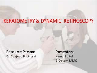
Good morning everyone
The topic of our presentation is ...
I would like to thank Dr. sanjib bhattarai for his kind guidence.
Also called ophthalmometer
Optical zones of cornea. Central zone is 4 mm in diameter
This is area whre refrection differs by less than 0.25D
paracentral zone the dizmeter ranges from 4-8mm
Here a preson is doing keratometry in our clinics
Image formed by the anterior surface of the cornea
= 1st Purkinje image- with this image the radius of the curvature of the cornea can be calculated
This is the picture of Optical principle of keratometry here BP =....... If AB is at infinity then A’B’ will be very small and situated at the focus F. Therefore B’P will be the focal distance or we can simply say ½ of the curvature of the mirror. U ie. object distance is known and also O object size is known. Only we need to find the imaze size inorder to find the curvature of the Mirror or we can say the curvature of Cornea…
It is very difficult to measure the image size against a reticule( ie, a reading line inside the keratometer), which make the entire process more difficult In order to overcome this challenge the idea of doubling principle has given by Helmholtz in 1854
Based on the measurement of image height.
In essence, a keratometer measures reflecting power and infers refracting power.
If we place 2 prisms base to base and position them such that the baseline splits the pupil, the observer will see 2 images separated by a fixed amount (depending on the power of the prisms).
Thus, any oscillation of the cornea during measurement will affect both doubled images equally-that is, motion of the eye will not cause the separation between the doubled images to change. This allows the observer to adjust knobs on the keratometer to arrive at the "contact" position despite small eye movements. This technique is called the doubling principle
Types of doubling systems here we are discuss only first 2 types
B = Bausch & L= Lomb keratometer , In Fixed doubling system the variable are Imaze size and mire separation and fixed are object height and doubling device distance
We use 1.3375 instead beacuse
Here we can see patient cornea , objective mire ,image in cornea is 1st Purkinji image ,reflecting mirror, objective lens , Aperture diaphragms , Doublings Prisms( base up & base out) , Doubled images,
Eye piece and examiner eye.
when the light is on.
reflected by reflecting mirror
falls on objective mire
Image in the eye of the mires or the 1st purkinji images
Image get reflected through the hole into the objetive lens.
The rays of Image passes through the left aperture falls on Base up prism.
Which double the central mire.
Similarly rays of image passes through the right aperture and falls on Base out prism and hence double the image.
the image are magnified by the telescopic system of keratometer.
which is seen by examiner.
Alingment of outer canthus and alignment marker
Reflection of mire on patient cornea
Here the central mire is not sharply focused on cornea so doubled images are seen
Normal range of Keratometry is 36-52D
the actual k reading will be. +58
for very low K reading .shift K reading by 6D
Keratometers that do not require rotation through 90° in order to measure the second principal meridian are known as one position keratometers
Keratometers that require rotation through 90 ̊ in order to measure the second principal meridian are known as two position keratometers
Picture of Calibreating keratometer
If keratometric mires not visible
] The Harmon distance is measured from the elbow to the knuckle of the middle finger (Figure 1). Consider it as the distance from fist at chin to the elbow on the desk