SlideShare une entreprise Scribd logo
1  sur  9
Télécharger pour lire hors ligne
High performance liquid chromatography coupled to mass spectrometry
for profiling and quantitative analysis of folate monoglutamates
in tomato
Kamal Tyagi, Pallawi Upadhyaya, Supriya Sarma, Vajir Tamboli, Yellamaraju Sreelakshmi,
Rameshwar Sharma ⇑
Repository of Tomato Genomics Resources, Department of Plant Sciences, School of Life Sciences, University of Hyderabad, Hyderabad 500046, India
a r t i c l e i n f o
Article history:
Received 21 October 2014
Received in revised form 20 January 2015
Accepted 23 January 2015
Available online 31 January 2015
Keywords:
Tomato
Folates
HPLC–MS/MS
Folate extraction
a b s t r a c t
Folates are essential micronutrients for animals as they play a major role in one carbon metabolism.
Animals are unable to synthesize folates and obtain them from plant derived food. In the present study,
a high performance liquid chromatography coupled to mass spectrometric (HPLC–MS/MS) method was
developed for the high throughput screening and quantitative analysis of folate monoglutamates in
tomato fruits. For folate extraction, several parameters were optimized including extraction conditions,
pH range, amount of tri-enzyme and boiling time. After processing the extract was purified using
ultra-filtration with 10 kDa membrane filter. The ultra-filtered extract was chromatographed on a RP
Luna C18 column using gradient elution program. The method was validated by determining linearity,
sensitivity and recovery. This method was successfully applied to folate estimation in spinach, capsicum,
and garden pea and demonstrated that this method offers a versatile approach for accurate and fast
determination of different folate monoglutamates in vegetables.
Ó 2015 Elsevier Ltd. All rights reserved.
1. Introduction
Folate (folic acid) is one of the B groups of vitamins that are
essential for the human health. The dietary insufficiency of folate
can lead to several health related disorders such as megaloblastic
anemia (Gough, Read, McCarthy, & Waters, 1963), exacerbation
of cardiovascular disease (Kolb & Petrie, 2013) and some types of
cancer (Blount et al., 1997). It is also required for the development
of a healthy fetus and its deficiency affects formation of fetus’
spinal cord and brain (Blom, Shaw, den Heijer, & Finnell, 2006).
Folate cannot be synthesized by animals and therefore it is solely
obtained from diet.
Plants are the major source of dietary folates. Green leafy vege-
tables, legumes and some fruits are among the richest sources of
folate. In several countries the cereal based food products are
mandatorily fortified with folic acid to prevent folate-related disor-
ders. In addition, transgenic approaches have been used to increase
the biosynthesis of folates in tomato fruits, potato tubers and rice
grains to remove dietary constraints of low folate levels in diet
(Blancquaert, De Steur, Gellynck, & Van Der Straeten, 2014).
Folate is comprised of a pterin moiety attached by a methylene
bridge to para-amino benzoic acid, which is coupled to one or more
glutamyl residues. In vivo folates exist as tetrahydrofolate (THF)
and its derivatives (5-methyl, methylene, methenyl, or 10-formyl)
which vary in oxidation states, single carbon substituents, and
with a variable number of glutamyl residues (Rébeillé et al.,
2006), which are collectively called – folate or vitamin B9. THF
plays a key role in one-carbon transfer reactions in all living organ-
isms participating in diverse metabolic reactions such as amino
acid metabolism, pantothenate synthesis, purines and thymidylate
synthesis etc.
For quantitative determination of total folates, microbiological
assay is the most commonly used method which is also recom-
mended by Association of Official Analytical Communities Interna-
tional (AOAC, 2000). The main weakness of this method is that it
cannot distinguish diverse forms of folate present in food samples.
Above limitation has been overcome by using chromatography-
based methods allowing separation of different vitamers. The
folate determination has been carried out using high-performance
liquid chromatography (HPLC) coupled with UV (Pfeiffer, Rogers, &
Gregory, 1997), electrochemical (Bagley & Selhub, 2000), and
fluorescence detection (Ndaw, Bergaentzlé, Aoudé-Werner,
http://dx.doi.org/10.1016/j.foodchem.2015.01.110
0308-8146/Ó 2015 Elsevier Ltd. All rights reserved.
⇑ Corresponding author. Tel.: +91 40 23010514; fax: +91 40 23010120.
E-mail addresses: tyagi.kamal6672@gmail.com (K. Tyagi), pravas43@gmail.com
(P. Upadhyaya), supu.megha@gmail.com (S. Sarma), vajirchem@gmail.com (V.
Tamboli), syellamaraju@gmail.com (Y. Sreelakshmi), rameshwar.sharma@gmail.
com (R. Sharma).
Food Chemistry 179 (2015) 76–84
Contents lists available at ScienceDirect
Food Chemistry
journal homepage: www.elsevier.com/locate/foodchem
Lahély, & Hasselmann, 2001). The folate level in tomatoes was esti-
mated using HPLC coupled with fluorescence detector (Zhang et al.,
2003). However, HPLC is unable to quantify folate forms occurring
in very low amounts.
In recent years, HPLC/UPLC hyphenated with mass spectrometry
has allowed both qualitative profiling and quantification of folate
derivatives in several foods, because of its high sensitivity, selectiv-
ity, specificity, and accuracy (De Brouwer, Zhang, Storozhenko, Van
Der Straeten, & Lambert, 2007; Garratt et al., 2005; Patring &
Jastrebova, 2007; Rychlik & Freisleben, 2002; Zhang, Storozhenko,
Van Der Straeten, & Lambert, 2005). The gas chromatography mass
spectrometry (GC–MS) has also been used for quantitative analysis
of total folates using acidic extraction (Dueker et al., 2000). How-
ever, GC–MS requires derivatization of all folate to para-amino ben-
zoyl glutamate prior to analysis hindering distinction of different
folate forms, thereby limiting its usage.
The precise analysis of folate derivatives present in plants/food
samples is a complicated task stemming from multiplicity of folate
derivatives, bound/unbound forms, low levels of folate and differ-
ences in plant matrix such as seeds, leaf, fruits or tuber. The deter-
mination of folate in food is also strongly influenced by the sample
preparation and extraction methods. Given that each plant matrix
is distinct, there is no standard method for extraction of folate from
plant material owing to difficulties in sample preparation, extrac-
tion, deconjugation and purification. This entails that for each
plant/tissue the extraction protocol has to be independently opti-
mized to enable precise qualitative and quantitative determination
of folate levels (Arcot & Shrestha, 2005). Currently limited methods
are available to extract and specifically quantify different folate
derivatives present in plants, such as in spinach (Zhang et al.,
2005), rice (De Brouwer et al., 2008), and potato (Van Daele
et al., 2014).
The aim of this study was to develop an accurate, reproducible,
and quantitative method for high throughput screening of large
populations of different cultivars, mutants and natural accessions
of tomato. We report here optimization of extraction, validation,
and application of a high performance liquid chromatographic
method with mass spectrometric (HPLC–MS/MS) detection for
quantification of folate monoglutamates in tomato. The all ion
fragmentation approach of the Orbitrap mass spectrometer
allowed confirmation of all targeted folate monoglutamates at
the same time without pre-selection. The above method was also
used for quantification of folates from plants such as capsicum
fruits and spinach leaf.
2. Materials and methods
2.1. Chemicals and reagents
5-Methyltetrahydrofolate (5-CH3-THF), tetrahydrofolate (THF),
5,10-methenyltetrahydrofolate (5,10-CH+
THF), 5-formyltetrahy-
drofolate (5-CHO-THF) and 5,10-methylenetetrahydrofolate
(5,10-CH2-THF) were purchased from Schirck’s Laboratory (Jona,
Switzerland). Folic acid (FA) and methotrexate (MTX) were pur-
chased from Sigma–Aldrich Co. (St. Louis, USA). Supplementary
Fig. 1 shows the structure of various folate forms and MTX.
LC–MS grade acetonitrile was purchased from Sigma–Aldrich
Co. (St. Louis, USA). Ultrapure water (18.2 mX at 25 °C) was
obtained from Milli-Q water purification system (Millipore, Brad-
ford, USA). Ascorbic acid and 2-mercaptoethanol (ME) were pur-
chased from Sigma–Aldrich Co. (St. Louis, USA). Formic acid
(HCOOH) and acetic acid (CH3COOH) of LC–MS grade were
purchased from Fisher Scientific (Loughborough, UK). Potassium
dihydrogen phosphate, dipotassium hydrogen phosphate and acti-
vated charcoal were purchased from HiMedia (Mumbai, India).
2.2. Preparation of standard and enzyme solutions
Stock solutions of folate standards (1 mg/mL) were prepared in
50 mM potassium phosphate solution, pH 4.5 containing 1% (w/v)
of ascorbic acid and 0.5% (w/v) of 2-mercaptoethanol except FA
and MTX, which were dissolved in neutral or basic pH buffer.
The standard stock solutions were diluted appropriately in the
extraction buffer to prepare working solutions. The remaining
stock solutions were flushed with nitrogen gas, and small aliquots
were stored at À80 °C.
Protease (from Streptomyces griseus, RM6186) and a-amylase
(from Bacillus sp., A6814) were purchased from HiMedia (Mumbai,
India) and Sigma–Aldrich Co. (St. Louis, USA) respectively. Protease
(2 mg/mL) and a-amylase (20 mg/mL) were dissolved in ultrapure
water, and aliquots were stored at À20 °C. Rat plasma was pur-
chased from National Institute of Nutrition (NIN), Hyderabad,
India. To remove endogenous folate from rat plasma, 100 mL of
rat plasma and a-amylase were mixed with 5 g of activated char-
coal separately, stirred on ice for 1 h followed by centrifugation
at 5000g for 10 min at room temperature. The supernatant was fil-
tered through a 0.22 lm filter, divided into 1 mL aliquots, and
stored at À20 °C. Protease was used without pre-treatment and
was stored at À20 °C. For ultra-filtration, 10 kDa membrane filters
(Pall Corporation, USA) were used.
2.3. Plant material: growth conditions and tissue harvesting
Tomato plants (Solanum lycopersicum L. cv. Arka Vikas) were
grown in a greenhouse of University of Hyderabad at 25 ± 2 °C
day and ambient temperature in night. For extraction procedure,
method development, and validation experiments, different tissues
were selected including tomato fruit and leaf tissue, garden pea
seeds, capsicum fruit and spinach leaf. Fresh garden pea, spinach
and capsicum were purchased from the local market. The juvenile
leaves from six-weeks-old plants and fruit tissue at mature green
and red ripe stage were harvested from tomato plants. Tomato
fruits, pea seeds, and capsicum fruits were homogenized to powder
after freezing in liquid nitrogen using a homogenizer (IKA A11,
Germany). The tomato and spinach leaves were manually homog-
enized in liquid nitrogen using pre-chilled mortar and pestle.
Homogenized powder was used for extraction of folates.
2.4. Sample extraction: homogenization, enzyme treatments and
purification
The basic extraction procedure for folates was adopted from De
Brouwer et al. (2008) with modifications. Several parameters were
evaluated to obtain the best recovery of folates from tomato fruit
and leaf. These modifications included selection of appropriate
pH and concentration of extraction buffer, amount of enzyme
and different boiling treatment to deactivate enzyme activity.
The extraction buffer (pH 4.5) consisting of 50 mM potassium
phosphate, 1% (w/v) ascorbic acid, 0.5% (v/v) b-mercaptoethanol
and 1 mM calcium chloride was freshly prepared for the sample
extraction and flushed with nitrogen for 20 s. Initially, 650 lL of
extraction buffer was mixed with 100 mg of plant homogenate in
2 mL Eppendorf tube. To the extract 2.7 ng/mL of individual folates
were added at the beginning of extraction process for recovery
calculation.
The tubes were capped tightly and after mixing by vortexing
were placed in a boiling water bath for 10 min. After cooling on
ice, the extract was incubated with 10 lL of a-amylase at
room temperature for 10 min to digest complex carbohydrates.
Thereafter, 2.5 lL of protease was added and tubes were incubated
at 37 °C for 1 h, then transferred to boiling water bath for 5 min.
After cooling on ice, the deconjugation of folate polyglutamates
K. Tyagi et al. / Food Chemistry 179 (2015) 76–84 77
to monoglutamates was carried out by adding 100 lL of rat plasma
to tubes and incubating at 37 °C for 2 h. Thereafter the sample was
boiled for 5 min followed by cooling on ice. The tubes were centri-
fuged for 30 min (14,000g, 4 °C) and the supernatant was filtered
through the 0.22 lm filter. Finally, the filtrate was clarified by
ultra-filtration at 12,000g for 12 min using 10 kDa cut-off mem-
brane filter. Immediately, the resulting extract was transferred to
autosampler vials in HPLC and a 7.5 lL aliquot was directly
injected on the column.
2.5. Liquid chromatography settings
The chromatographic separation of folate derivatives was car-
ried out using Waters Acquity™ UPLC system (Milford, USA) run-
ning in HPLC mode, coupled to a binary pump, an autosampler,
and controlled by Xcalibur 3.0 software (Thermo Fisher Scientific,
San Jose, USA). The sample manager was configured with a 10 lL
sample injection loop and a 100 lL sample syringe with the partial
loop with needle overfill function. Prior to sample injections, the
syringe was rinsed with 500 lL of weak (water/acetonitrile, 95:5)
and strong (water/acetonitrile, 5:95) solvents, respectively. The
auto-sampler (set at 4 °C) was covered to minimize any degrada-
tion of light sensitive folates during sample analysis.
Folate derivatives were separated on a reversed phase Luna C18
column (5 lm particle size, 250 mm  4.60 mm ID) (Phenomenex,
USA) using gradient elution program. The gradient comprised of a
binary solvent system consisting 0.1% (v/v) of formic acid in water
(solvent A) and acetonitrile (solvent B) at a flow rate of 500 lL
minÀ1
. The injection volume was 7.5 lL, run time 25 min and col-
umn temperature was set at 24 °C. The starting condition of gradi-
ent was 95% of solvent A and 5% of solvent B. Subsequently, solvent
B was linearly increased to 10% in 4 min, then to 30% in 6 min and
to 95% in 7 min. Thereafter, the mobile phase was reverted to the
initial condition in 3 min and held for 5 min for re-equilibration
of column before the next injection. After sample injection for first
8 min, the solvent flow from LC pump was diverted to waste posi-
tion to minimize the suppression of MS signals by salts present in
the extract. Thereafter, the solvent flow was switched to mass
detector position for next 12 min and again diverted to waste posi-
tion for 5 min till completion of the total run.
2.6. Mass spectrometry settings
For all experiments, Exactive™ Plus Orbitrap mass spectrometer
(Thermo Fisher Scientific, USA) was operated in alternating full
scan and all ion fragmentation (AIF) mode equipped with positive
heated electrospray ionization (ESI). The source parameters were
optimized as follows: ion spray voltage, 4 kV; capillary tempera-
ture, 380 °C and heater temperature, 300 °C. Sheath and auxiliary
gas flows were 60 and 20 arbitrary units (au) respectively. Capil-
lary, tube lens, and skimmer voltages were 65, 60, and 24 V respec-
tively. For full scan experiment, the mass scan in the range of m/z
120–800 was monitored, while for AIF experiment, it was in the
range of m/z 50–450. The resolution for full scan experiment was
set to 70,000 full width at half maximum (FWHM) and for the
AIF experiment it was set to 35,000 FWHM. The automatic gain
control (AGC) target for full scan and AIF experiments were set
to high dynamic range of 1e6
and 5e5
respectively. Similarly, the
maximum injection time was set to 100 and 50 ms respectively.
All ions entering into the mass spectrometer (MS) were frag-
mented using nitrogen gas in the High-energy collision-induced
dissociation (HCD) cell at normalized collision energy (CE) 20
(arbitrary unit) with 20% stepped CE, and finally detected in the
Orbitrap mass analyzer. The fore vacuum, high vacuum and
ultra-high vacuum were maintained around 1.5 mbar, from
2.5eÀ5
to 3.5eÀ5
mbar and below 4eÀ10
mbar, respectively.
Instrument control and data processing were carried out by Xcali-
bur 3.0 software (Thermo Fisher Scientific, San Jose, USA).
Prior to sample analysis, the external calibration of instrument
was performed to ensure the mass accuracy by directly infusing
calibration solutions in both positive and negative ion modes.
The positive mode calibration solution consisted of caffeine, Met-
Arg-Phe-Ala acetate salt (MRFA), n-butyl amine and Ultramark™
1621, while negative mode calibration solution consisted of
sodium dodecyl sulfate, sodium taurocholate and Ultramark™
1621. These solutions were freshly prepared using acetonitrile,
methanol, and water containing 1% (v/v) acetic acid. All these cal-
ibration solutions were mixed at room temperature and directly
infused in the source using a syringe pump (Chemyx Fusion 100,
Thermo Fisher Scientific, USA) with a flow rate of 5 lL/min. In posi-
tive ion mode, the external lock mass (polydimethyl cyclosiloxane,
exact mass = 455.12003) was also tested for the mass analyzer
recalibration.
2.7. Recovery studies
In the absence of specific guidelines for analysis of folate deriv-
atives in tomato plant tissue, the International Conference on
Harmonization (ICH) guideline was used for the validation of ana-
lytical method which includes accuracy, sensitivity and linearity.
To determine the accuracy of method, recovery test was performed
by spiking of tomato extract with known amount of individual
folate derivatives and calculating their final content in extract.
The recovery (R) was calculated as B À C/A Â 100, where A = peak
area of neat folates standard, B = peak area of spiked standard,
C = peak area of extract.
2.8. Limit of detection and limit of quantification
Sensitivity was confirmed by evaluating the limit of detection
(LOD; calculated as: 3.3r/S, where r is the standard deviation
and S is the slope of calibration curve) and limit of quantification
(LOQ; calculated as: 10r/S). The linearity of each folate derivative
was evaluated by plotting the peak area at different concentra-
tions. The sample concentrations were calculated from the equa-
tion y = mx + c, as determined by the linear regression analysis.
2.9. Statistical analysis
All results are expressed as mean value ± SE of three or more
replicates based on fresh weight (FW). A t-test was used for deter-
mining significant difference between the mean values. The differ-
ences were considered to be significant for P < 0.05. Statistical
analysis of data was performed using SigmaPlot 11.0.
3. Result and discussion
3.1. Optimization of HPLC and MS/MS conditions
It has been reported that reverse phase C18 column is the most
appropriate column (based on resolution and retention time) for
the separation and detection of folate derivatives. Initially, both
organic solvents (acetonitrile and methanol) with 0.1% (v/v) formic
acid and 0.1% (v/v) acetic acid were evaluated. The results show
that usage of acetonitrile with 0.1% (v/v) formic acid increased
the resolution with higher signal to noise ratio and proved to be
the best eluent for ionization of folate derivatives. The optimized
chromatographic conditions are described in Section 2.5. Under
these conditions, separation of THF, 5-CH3-THF, 5,10-CH+
THF,
MTX, 5-CHO-THF, and FA were achieved at 10.28, 10.84, 11.35,
12.87, 13.02, 13.16 min respectively (Fig. 1). Interfering
78 K. Tyagi et al. / Food Chemistry 179 (2015) 76–84
compounds present in the plant extract (sugars, salt, and other
matrices) may suppress ionization. To eliminate them, a divert
valve was used during first 8 min and last 5 min of chromato-
graphic run to redirect the major impurities to the waste instead
to the mass detector.
Prior to determining the optimal conditions, individual folate
standard derivatives and internal standards were directly infused
to ESI in both positive and negative ion modes. Comparison of both
modes revealed that the positive ion mode provided better sensi-
tivity than negative ion mode in conformity with earlier reports
(De Brouwer et al., 2007; Patring & Jastrebova, 2007; Rychlik &
Freisleben, 2002). Therefore, positive ion mode was selected for
subsequent experiments. The optimized MS parameters are
described in Section 2.6. For identification of each compound, accu-
rate mass of the corresponding ion, elemental composition, and
specific retention times were considered. In full scan mode, the
ions detected at m/z 446.1779, 460.1942, 456.1632, 455.1786,
474.1729 and 442.1467 corresponded to the THF, 5-CH3-THF,
5,10-CH+
THF, MTX, 5-CHO-THF and FA respectively. Fig. 1 shows
the extracted ion chromatogram. The accurate masses of corre-
sponding ions were also compared with the theoretical masses cal-
culated by Xcalibur 3.0 software. In addition, all ions were
fragmented without pre-selection in HCD cell at different collision
energy ($20–40 au). Interestingly, several fragment ions were
detected at m/z 299.1249 (THF), 313.1404 (5-CH3-THF), 308.1256
(MTX), 327.1198 (5-CHO-THF), and 295.0936 (FA) indicating the
loss of glutamate moiety (Supplementary Table 1) except
412.1530 (5,10-CH+
THF). Using HCD, the sample analysis time
was substantially reduced by confirming the observed m/z values
at the same time without pre-selection.
3.2. Optimization of sample extraction in tomato
For optimization of folate extraction/estimation from tomato,
several factors were considered like type and composition of sam-
ple matrix, nature, amount, and stability of the compounds for
extraction and fine tuning of the LC–MS parameters for their detec-
tion. Considering that folates are very labile compounds, their
extractions differ for each kind of matrix. Tomato fruit is a very
complex matrix having many compounds to which folates bind.
We optimized the folate extraction from tomato by modifying
the method of De Brouwer et al. (2008) originally developed for
rice. Largely for the folate extraction from a variety of food
matrices, a trienzyme treatment (amylase, protease and conjugase)
developed by Martin, Landen, Soliman, and Eitenmiller (1989) is
used to release the bound folate from matrix. The trienzyme treat-
ment accomplishes more complete extraction of folates that are
bound to matrices of proteins or polysaccharides by including pro-
tease and a-amylase in addition to conjugase.
Considering matrix complexity, several parameters were evalu-
ated for optimal folate extraction from tomato fruits such as pH
and molarity of the extraction buffer, amount of a-amylase, prote-
ase, rat plasma conjugase, and duration of boiling time after
enzyme treatments. Fig. 2 shows optimization of different param-
eters for folate extraction. The pH of extraction buffer greatly
affects the stability of folates in extracts. Therefore, folate extrac-
tion was compared using two different pH, 4.5 and 7.0. No signifi-
cant difference in recovery of total folate content was observed
using these two pH (P value 0.0519) (Fig. 2A).
Stability of the folate derivatives greatly varies with respect to
pH, thus requiring selection of a pH that is optimal for efficient
extraction of most folate derivatives (De Brouwer et al., 2007).
The optimal pH for protease and amylase is also different from
optimal pH for rat plasma conjugase (Arcot & Shrestha, 2005).
Since multiple pH adjustments prolong the extraction process
and increase the chances of oxidation of folate, the usage of differ-
ent pH to achieve optimal trienzyme treatment is not feasible
while screening a large number of samples. The amount of total
folate extracted at pH 4.5 and 7.0 was nearly similar, however, at
pH 4.5, we detected THF in tomato leaf but not in tomato fruit tis-
sue. Given the variability in stability of different folate derivatives,
it is difficult to have an ideal pH for extraction of all folate
derivatives in their native state from plant matrix. Considering that
5-CH3-THF and 5-CHO-THF, the most predominant folate forms
present in the plants are more stable at low pH, therefore we used
pH 4.5 for subsequent extraction of folates. The recovery of folate is
also influenced by the buffer composition; however, phosphate
buffer is most widely used for folate extraction. We compared both
50 and 100 mM phosphate buffer and found that both were equally
efficient. Taking cognizance of fact that high salt concentrations in
buffer interfere with the ionization process in the mass spectrom-
eter, we used 50 mM phosphate buffer for all extraction
procedures.
Considering that the fruit and vegetable matrices are complex,
possessing compounds that may possibly bind with the metabolite
of interest and improve/inhibit the extraction efficiency, the trien-
zyme method of folate extraction was also optimized. The optimi-
zation involved alteration of enzyme concentrations and duration
of treatments. We compared the extraction of folate with and
without respective enzymes for all three enzymes used for folate
Fig. 1. Representative chromatogram of five folates standards and an internal standard. (A) THF, (B) 5-CH3-THF; (C) 5,10-CH+
THF; (D) MTX; (E) 5-CHO-THF and (F) FA.
K. Tyagi et al. / Food Chemistry 179 (2015) 76–84 79
extraction. It was observed that without a-amylase treatment,
$85% of the total folate content was extracted (P value 0.0281)
compared to the a-amylase treated control. Similarly, addition of
protease and incubation at 37 °C for 1 h did not significantly
improve the extraction of total folate content (P value 0.050)
(Fig. 2B). Among the three enzymes used in this study, the rat
plasma conjugase was found to be the most important for folate
deglutamylation. Different amounts of rat plasma conjugase was
added to the extract (per 100 mg of fresh tissue), that is, 0, 25,
50 lL which resulted in 17%, 52% and 88% recovery of total folate
pool respectively compared with addition of 100 lL rat plasma
serving as control. Taking into account that increase from 50 to
100 lL of rat plasma lead to 12% improvement in the folate
recovery, 100 lL of rat plasma was used for all subsequent exper-
iments (Fig. 2C).
It is reported that boiling the extract prior to enzyme treatment
reduces the overall yield of folate from food matrices. Moreover,
the addition of antioxidants such as ME and ascorbic acid in the
extraction buffer protects folate from degradation during boiling
treatment that is needed to denature the enzymes besides improv-
ing the stability of folate in an autosampler. The increase in the
boiling time from 5 to 10 min post-enzyme treatment did not
enhance the recovery of folate, therefore, boiling for 5 min was
used for all enzymatic deactivation steps to minimize any possible
degradation of folates (Fig. 2D).
3.3. Optimization of purification
Different folate vitamers differ in their functional groups thus
vary in their chemical properties and therefore can be better sepa-
rated by HPLC. It is reported that sample purification prior to sep-
aration in HPLC leads to better recovery of folate and also
distinction between different forms. The presence of different
endogenous constituents in food extracts interfere with folates
and hamper the determination of some folate forms. Considering
that folate is also present at low level in most food matrices,
removal of the interfering compounds during purification
improves the detection limit and selectivity of the folate in HPLC.
Solid phase extraction (SPE) is the most commonly used method
for folate purification using either a strong anion exchange (SAX)
sorbent or C18 SPE cartridges for food extract prior to HPLC–MS
analysis (Chandra-Hioe, Bucknall, & Arcot, 2013; Stokes & Webb,
1999). Use of SAX sorbent for purification of food extracts provides
high recovery of different folate forms, however, pre-concentration
of sample is not possible as large buffer volumes are required to
elute all the folates from SAX column. In addition, high concentra-
tion of salt used in elution step interferes with subsequent MS
analysis.
Recently, affinity chromatography using immobilized folate-
binding protein has been used as a highly selective purification
method for folates, especially for complex food matrices such as
cereals (Pfeiffer et al., 1997). Though affinity chromatography
enables quantification at 10-fold lower concentration than SAX,
the lack of commercial availability of FBP columns precludes their
use for folate analysis. Moreover, folate-binding protein exhibits
low affinity to 5-HCO-H4 folate, which may result in higher losses
of this folate form during the purification step.
Among the different methods of folate analysis, usage of LC–MS
has emerged as most preferred method owing to its specificity and
sensitivity to distinguish different forms of folate. Several studies
have used LC–MS for folate analysis, particularly from fortified
food samples (Alaburda, de Almeida, Shundo, Ruvieri, & Sabino,
2008; Patring, Wandel, Jägerstad, & Frølich, 2009). However, the
detection of folate derivatives by LC–MS needs robust purification
of folate vitamers for efficient and accurate analysis. In this study,
we optimized the purification of folates for subsequent analysis by
LC–MS. In earlier studies, SPE using SAX cartridge (Vishnumohan,
Arcot, & Pickford, 2011) or affinity column using folate binding
protein (Díaz de La Garza et al., 2004) have been used so far. How-
ever, these approaches are time consuming and cannot be applied
when handling large number of samples. To overcome these draw-
backs which hinder folate estimation for high throughput analysis,
the purification step was modified to a simple, efficient and also
cost effective method for quantification of folate in tomato by
using a molecular weight cut-off membrane filter according to
Zhang et al. (2005). For efficient sample cleanup, extract was fil-
tered through a 0.22 lm filter to remove the particulate matter
in the extract followed by ultra-filtration through a 10 kDa
Fig. 2. Optimization of folate extraction in tomato fruit. (A) Effect of pH, (B) extraction in presence (control) and absence of a-amylase and protease, (C) extraction with
increasing amount of rat plasma conjugase and (D) effect of duration of boiling (n P 3).
80 K. Tyagi et al. / Food Chemistry 179 (2015) 76–84
centrifugal device removing various polymeric compounds present
in the tomato extract. The usage of step wise increase in gradient
consisting of 0.1% (v/v) of formic acid in water (solvent A) and ace-
tonitrile (solvent B) on a C18 reverse phase column yielded good
resolution of different folate vitamers. To avoid contamination of
electrospray of MS machine by salt and matrix, which reduces
the sensitivity of the mass spectrometer, out of total runtime of
25 min, first 8 min and last 5 min eluent were discarded to the
waste and sample was allowed to pass through MS during inter-
vening 12 min.
Folates were analyzed in positive ion mode through mass spec-
trometry as the analysis indicated it to be superior to the negative
ion mode. All ion fragmentation (AIF) parameters including precur-
sor ions, product ions, and collision energy were optimized for frag-
mentation by injecting the folate standards. Electrospray ionization
(ESI) source temperature was 300 °C and capillary temperature was
maintained at 380 °C. For fragmentation of the folate standards in
HCD cell, collision energy of 20 or 40 (arbitrary unit, Supplementary
Table 1) was considered optimal depending on the folate com-
pound. The most abundant fragment resulting from all folates
was the one that was generated from the neutral loss of the gluta-
mate moiety (m/z 147), e.g. for FA m/z 442/295, for THF m/z 446/
299, for 5-CH3-THF m/z 460/313, for 5-CHO-THF m/z 474/327 and
for MTX m/z 455/308. Collision energy used for 5,10-CH+
THF was
40 arbitrary unit resulting in m/z 456/412. Supplementary Table 1
shows the fragmentation result of the folate standards. A represen-
tative chromatogram of folates standards is shown in Fig. 1.
3.4. Standard calibration curve and linearity
The linearity of the developed HPLC–MS method was evaluated
by preparing seven point calibration curves without matrix for
each folate derivative (THF; 5-CH3-THF; 5,10-CH+
THF; MTX;
5-CHO-THF; and FA) in triplicates. Correlation coefficients (R2
)
determined for all these folates were approximately 0.99, which
confirmed their good linearity within the considered concentration
ranges. For the folates derivatives, which were detected in the
tomato fruits, the limit of detection (LOD, S/N P 3.3) and quantita-
tion (LOQ, S/N P 10) were theoretically determined based on cali-
bration curve, relating concentrations with signal to noise ratios.
Table 1 shows the correlation coefficient, linear range, and slope
of calibration curve.
3.5. Recovery and stability
Accuracy of the developed method was determined by recovery
test. The mean recoveries (n = 3) for all individual folate derivatives
were in the range of 48–131% (Table 2). The observed value indi-
cates that the developed method is accurate, except for folic acid,
which is not a naturally occurring folate. Taking cognizance that
multiple samples were analyzed for folate levels, the stability of
the extracted samples was examined in the autosampler at 4 °C.
The results indicated that most of the folate derivatives are stable
up to 72 h of storage in the autosampler at 4 °C barring 5,10-CH+-
THF (Supplementary Fig. 2). It is reported that the presence of
ME and ascorbic acid in the extraction buffer enhances the stability
of the folate for a prolonged period (Vahteristo & Finglas, 2000).
3.6. Use of external and internal standard
In order to check the possible variations in MS response for dif-
ferent folate derivatives while screening of large number of sam-
ples, we used internal standards for accurate and reproducible
quantitation. Though the usage of stable isotope labeled folate
standards is the best option, these were not used owing to their
limited availability and high cost. In its place a known amount of
individual folate derivatives were used as external standards. The
MS response of folate derivatives were checked by analyzing sev-
eral tomato extracts and comparing the calculated concentrations
with initial values. Furthermore, we used two structurally similar
compounds, FA and MTX (Zhang et al., 2005) as internal standards.
Both these standards were mixed to the tomato homogenate prior
to initiation of extraction protocol and recoveries were calculated.
Considering that the recovery of MTX was higher than FA (Table 2),
it was selected as internal standard for our further screening.
3.7. Folate content in tomato fruit
The total folate level in mature green and red ripe fruits of
tomato cultivar Arka Vikas was 21.94 and 18.06 lg/100 g of FW
respectively (Fig. 3A). 5-CH3-THF was the most predominant folate
form comprising of 68% and 58% of total folate in mature green and
red ripe fruits respectively. These values were similar to those
reported by Díaz de La Garza, Gregory, and Hanson (2007) that
5-CH3-THF constitutes 70% of total folate present in tomato fruit.
In addition to 5-CH3-THF, other two folate derivatives 5-CHO-
THF and 5,10-CH+
THF were also detected in tomato fruit. Among
these, 5-CHO-THF was the second predominant folate form present
in both mature green and red ripe stage fruits. The level of 5-CH3-
THF significantly decreased during tomato fruit ripening from
mature green to red ripe stage (P value 0.035). In contrast, the con-
tent of 5,10-CH+
THF and 5-CHO-THF in the mature green and red
ripe stage of tomato fruit did not show any significant changes
(Fig. 3B). Considering that 10-CHO-THF cannot be identified in
the LC–MS due to its conversion to 5,10-CH+
THF in mobile phase
containing 0.1% formic acid (Fazili & Pfeiffer, 2004; Goyer et al.,
2005), it can be assumed that 5,10-CH+
THF peak likely included
both 10-CHO-THF and any preexisting 5,10-CH+
THF. Consistent
Table 1
Calibration and sensitivity data of folate standards. All experiments were done in triplicates.
Compounds LOD (ng/mL) LOQ (ng/mL) Slope (n = 6 or 7) mean ± SE R2
Linear range (ng/mL)
FA 0.15 0.47 38095.5 ± 450.5 0.999 1–30
THF 0.25 0.77 30541.5 ± 577.5 0.997 2–30
5,10-CH+
THF 0.34 0.92 10757.3 ± 70.7 0.984 0.5–30
5-CH3-THF 0.17 0.53 36024.3 ± 682.2 0.998 1–30
5-CHO-THF 0.37 1.12 19204.0 ± 122.3 0.994 1–30
Table 2
Percentage recovery of spiked folate standards in tomato fruit
extract. 2.7 ng/mL of folate standards were spiked into extract.
Compound % recovery mean ± S.E.
FA 48.97 ± 8.48
THF 70.89 ± 7.10
5,10-CH+
THF 94.72 ± 8.29
5-CH3-THF 131.31 ± 8.78
5-CHO-THF 115.90 ± 1.59
MTX 95.27 ± 11.52
K. Tyagi et al. / Food Chemistry 179 (2015) 76–84 81
with earlier report (Iniesta, Perez-Conesa, Garcia-Alonso, Ros, &
Periago, 2009), THF was not detected in tomato fruit extract.
Tomato is consumed in both cooked and raw form by the people.
Taking the folate content of fully red ripe tomato fruit (18.06 lg/
100 g of FW) in account, its percentage contribution to the RDA
(400 lg/day) is 4.51. However, the folate content in a given food
matrix such as tomato fruit is not constant as several factor affects
its level such as nature of cultivar, developmental stage, and envi-
ronmental conditions during ripening (Iniesta et al., 2009;
Jägerstad et al., 2005).
The extent of basal polyglutamylation status of folate helps in
its retention in the cellular compartments as well as increases its
affinity toward the enzymes with which they interact (Akhtar
et al., 2010). The polyglutamylation of individual folate derivatives
was evaluated by comparing the monoglutamate amounts of each
folate derivative detected in tomato red ripe fruits with and with-
out the addition of rat plasma during folate extraction. 83% of 5-
CH3-THF was present in polyglutamylated form. Similarly, $90%
of total 5-CHO-THF was present in the polyglutamylated form
(Fig. 3C), whereas $50% 5,10-CH+
THF was present in polyglutamy-
lated form.
3.8. Application of the method
Fruits and vegetables are good sources of naturally occurring
folate, primarily 5-CH3-THF. To check the efficiency and applicabil-
ity of the above method for other plant materials, the folate levels
were determined in tomato juvenile leaf, spinach leaf, garden pea
seeds and capsicum fruit (Table 3). The major folate detected in
these varieties was 5-CH3-THF. Other forms observed were
5-CHO-THF and 5,10-CH+
THF with traces of THF and FA. 5-CHO-
THF was the second most predominant form detected in all of
them. Contrary to the report that recovery of THF was low at acidic
pH but increased as the pH of extraction buffer increased to 7.0
(Zhang et al., 2005), no THF was detected in tomato fruit tissue
using pH 7.0 extraction buffer. Compared to fruits, the juvenile
leaves of tomato showed very high level of folates, which may be
related to active cell division and expansion in these leaves.
Interestingly, folic acid or other oxidation products of natural folate
forms were not detected indicating that extraction procedure used
in this study led to minimal degradation or loss of folate.
The total folate content mean (±SE) in tomato juvenile leaf,
spinach leaf, garden pea seeds and capsicum fruit was 429.54
(61.49), 287.16 (55.14), 188.33 (14.06) and 39.44 (2.97) lg/100 g
FW respectively (Table 3). Considering that these values are higher
than the reported values in literature supports that our extraction
method leads to maximum recovery of folates from matrix. Total
folate content of spinach reported in earlier studies varied from
194–364 lg/100 g of FW assessed through microbiological assay
(Lin & Lin, 1999) to 48–177 lg/100 g of FW through HPLC
(Johansson, Jägerstad, & Frølich, 2007; Vahteristo, Lehikoinen,
Ollilainen, & Varo, 1997). Our results showed nearly 110 lg higher
value of folate than the earlier reported value for spinach. While
these differences in the folate levels could be attributed to cultivar,
Table 3
Folate content of tomato fruit, leaf and other vegetables (lg/100 g FW).
Sample THF (mean ± SE) 5-CH3-THF (mean ± SE) 5,10-CH+
THF (mean ± SE) 5-CHO-THF (mean ± SE) Total folate (mean ± SE)
Tomato fruit (mature green) – 14.97 ± 0.74 1.15 ± 0.08 5.81 ± 0.56 21.93 ± 1.17
Tomato fruit (red ripe) – 10.61 ± 1.42 1.55 ± 0.19 5.89 ± 0.59 18.05 ± 2.01
Tomato leaf 4.12 ± 0.10 226.36 ± 29.95 13.13 ± 2.92 185.93 ± 28.14 429.54 ± 61.49
Spinach 0.24 ± 0.09 159.17 ± 30.79 19.21 ± 4.05 108.54 ± 20.71 287.16 ± 55.14
Garden pea – 156.19 ± 7.62 4.28 ± 0.78 27.86 ± 2.67 188.33 ± 14.06
Green capsicum – 32.36 ± 2.76 2.06 ± 0.14 5.02 ± 0.55 39.44 ± 2.97
Fig. 3. Folate content in tomato fruits. (A) Total folate content in mature green (MG) and red ripe (RR) fruits (n P 3). (B) Relative distribution of different folate forms in
mature green and red ripe fruit (n P 3). (C) Polyglutamylation status of different folate derivatives in red ripe fruit.
82 K. Tyagi et al. / Food Chemistry 179 (2015) 76–84
variety, and geographical area, climatic and seasonal differences
among study groups (Shohag et al., 2011), it is also likely that sam-
ple preparation and extraction protocol reported in this study may
have led to more efficient extraction and minimal losses of folate
from spinach.
4. Conclusion
An accurate HPLC–MS/MS method has been successfully devel-
oped and validated for folate analysis in tomato fruits, which is
simple, fast and cost effective and can be applied to a large number
of samples for folate analysis. Our method has wide applicability as
the folate levels determined in different types of tissue used in the
study were either comparable or more than those reported in pre-
vious studies. We found that 5-CH3-THF is the main folate deriva-
tive in tomato. This method is fairly simple to execute as the
sample cleanup involves ultra-filtration instead of SPE or affinity
chromatography, without compromising on the amount or forms
of folates detected. The validation experiments showed linearity
of the method and total folate concentration in mature green and
red ripe tomato fruit was 21.93 and 18.06 lg/100 g of fresh weight
with acceptable precision and accuracy. We have used MTX as an
internal standard in screening of multiple samples for reproducible
quantitation. Most of the folates were stable for more than 24 h
when kept at 4 °C in autosampler. Total folate profile in tomato leaf
differed considerably from that of tomato fruit tissue indicating
that this extraction procedure can be used to analyze other food
matrices too.
Acknowledgments
This work was supported by the Department of Biotechnology
(Grant No. BT/PR11671/PBD/16/828/2008 to R.S. and Y.S.), the
Council of Scientific and Industrial Research (research fellowship
to KT), University Grants Commission (research fellowship to PU
and SS).
Appendix A. Supplementary data
Supplementary data associated with this article can be found, in
the online version, at http://dx.doi.org/10.1016/j.foodchem.2015.
01.110.
References
Akhtar, T. A., Orsomando, G., Mehrshahi, P., Lara-Núñez, A., Bennett, M. J., Gregory, J.
F. III,, et al. (2010). A central role for gamma-glutamyl hydrolases in plant folate
homeostasis. The Plant Journal, 64, 256–266.
Alaburda, J., de Almeida, A. P., Shundo, L., Ruvieri, V., & Sabino, M. (2008).
Determination of folic acid in fortified wheat flours. Journal of Food Composition
and Analysis, 21, 336–342.
AOAC (2000). In AOAC (Ed.). Official methods for analysis of AOAC International (17th
ed). Gaithersburg, MD: Association of Official Analytical Chemists International.
Arcot, J., & Shrestha, A. (2005). Folate: Methods of analysis. Trends in Food Science
and Technology, 16, 253–266.
Bagley, P. J., & Selhub, J. (2000). Analysis of folate form distribution by affinity
followed by reversed-phase chromatography with electrochemical detection.
Clinical Chemistry, 46, 404–411.
Blancquaert, D., De Steur, H., Gellynck, X., & Van Der Straeten, D. (2014). Present and
future of folate biofortification of crop plants. Journal of Experimental Botany, 65,
895–906.
Blom, H. J., Shaw, G. M., den Heijer, M., & Finnell, R. H. (2006). Neural tube defects
and folate: Case far from closed. Nature Reviews Neuroscience, 7, 724–731.
Blount, B. C., Mack, M. M., Wehr, C. M., MacGregor, J. T., Hiatt, R. A., Wang, G., et al.
(1997). Folate deficiency causes uracil misincorporation into human DNA and
chromosome breakage: Implications for cancer and neuronal damage.
Proceedings of the National Academy of Sciences United States of America, 94,
3290–3295.
Chandra-Hioe, M. V., Bucknall, M. P., & Arcot, J. (2013). Folic acid-fortified flour:
Optimised and fast sample preparation coupled with a validated high-speed
mass spectrometry analysis suitable for a fortification monitoring program.
Food Analytical Methods, 6, 1416–1423.
De Brouwer, V., Storozhenko, S., Van De Steene, J. C., Wille, S. M., Stove, C. P., Van Der
Straeten, D., et al. (2008). Optimisation and validation of a liquid
chromatography–tandem mass spectrometry method for folates in rice.
Journal of Chromatography A, 1215, 125–132.
De Brouwer, V., Zhang, G. F., Storozhenko, S., Van Der Straeten, D., & Lambert, W. E.
(2007). PH stability of individual folates during critical sample preparation steps
in prevision of the analysis of plant folates. Phytochemical Analysis, 18, 496–508.
Díaz de La Garza, R., Gregory, J. F., & Hanson, A. D. (2007). Folate biofortification of
tomato fruit. Proceedings of the National Academy of Sciences United States of
America, 104, 4218–4222.
Díaz de La Garza, R. I., Quinlivan, E. P., Klaus, S. M., Basset, G. J., Gregory, J. F., &
Hanson, A. D. (2004). Folate biofortification in tomatoes by engineering the
pteridine branch of folate synthesis. Proceedings of the National Academy of
Sciences United States of America, 101, 13720–13725.
Dueker, S. R., Lin, Y., Jones, A. D., Mercer, R., Fabbro, E., Miller, J. W., et al. (2000).
Determination of blood folate using acid extraction and internally standardized
gas chromatography–mass spectrometry detection. Analytical Biochemistry, 283,
266–275.
Fazili, Z., & Pfeiffer, C. M. (2004). Measurement of folates in serum and
conventionally prepared whole blood lysates: Application of an automated
96-well plate isotope-dilution tandem mass spectrometry method. Clinical
Chemistry, 50, 2378–2381.
Garratt, L. C., Ortori, C. A., Tucker, G. A., Sablitzky, F., Bennett, M. J., & Barrett, D. A.
(2005). Comprehensive metabolic profiling of mono-and polyglutamated
folates and their precursors in plant and animal tissue using liquid
chromatography/negative ion electrospray ionisation tandem mass
spectrometry. Rapid Communications in Mass Spectrometry, 19, 2390–2398.
Gough, K. R., Read, A., McCarthy, C., & Waters, A. (1963). Megaloblastic anaemia due
to nutritional deficiency of folic acid. Quarterly Journal of Medicine, 32, 243–256.
Goyer, A., Collakova, E., de la Garza, R. D., Quinlivan, E. P., Williamson, J., Gregory, J.
F., et al. (2005). 5-Formyltetrahydrofolate is an inhibitory but well tolerated
metabolite in Arabidopsis leaves. Journal of Biological Chemistry, 280,
26137–26142.
Iniesta, M. D., Perez-Conesa, D., Garcia-Alonso, J., Ros, G., & Periago, M. J. (2009).
Folate content in tomato (Lycopersicon esculentum). Influence of cultivar,
ripeness, year of harvest, and pasteurization and storage temperatures.
Journal of Agricultural and Food Chemistry, 57, 4739–4745.
International Conference on Harmonization (ICH) (1996). Guideline on validation of
analytical procedures: Methodology Q2B, Geneva, 1.
Jägerstad, M., Piironen, V., Walker, C., Ros, G., Carnovale, E., Holasova, M., et al.
(2005). Increasing natural food folates through bioprocessing and
biotechnology. Trends in Food Science and Technology, 16, 298–306.
Johansson, M., Jägerstad, M., & Frølich, W. (2007). Folates in lettuce: A pilot study.
Scandinavian Journal of Food and Nutrition, 51, 22–30.
Kolb, A. F., & Petrie, L. (2013). Folate deficiency enhances the inflammatory response
of macrophages. Molecular Immunology, 54, 164–172.
Lin, B., & Lin, R. (1999). Effect of Chinese stir-fry cooking on folate contents of
vegetables. Journal-Chinese Agricultural Chemical Society, 37, 443–469.
Martin, J., Landen, W., Jr., Soliman, A., & Eitenmiller, R. (1989). Application of a tri-
enzyme extraction for total folate determination in foods. Journal-Association of
Official Analytical Chemists, 73, 805–808.
Ndaw, S., Bergaentzlé, M., Aoudé-Werner, D., Lahély, S., & Hasselmann, C. (2001).
Determination of folates in foods by high-performance liquid chromatography
with fluorescence detection after precolumn conversion to 5-
methyltetrahydrofolates. Journal of Chromatography A, 928, 77–90.
Patring, J. D., & Jastrebova, J. A. (2007). Application of liquid chromatography–
electrospray ionisation mass spectrometry for determination of dietary folates:
Effects of buffer nature and mobile phase composition on sensitivity and
selectivity. Journal of Chromatography A, 1143, 72–82.
Patring, J., Wandel, M., Jägerstad, M., & Frølich, W. (2009). Folate content of
Norwegian and Swedish flours and bread analysed by use of liquid
chromatography–mass spectrometry. Journal of Food Composition and Analysis,
22, 649–656.
Pfeiffer, C. M., Rogers, L. M., & Gregory, J. F. (1997). Determination of folate in cereal-
grain food products using trienzyme extraction and combined affinity and
reversed-phase liquid chromatography. Journal of Agricultural and Food
Chemistry, 45, 407–413.
Rébeillé, F., Ravanel, S., Jabrin, S., Douce, R., Storozhenko, S., & Van Der Straeten, D.
(2006). Folates in plants: Biosynthesis, distribution, and enhancement.
Physiologia Plantarum, 126, 330–342.
Rychlik, M., & Freisleben, A. (2002). Quantification of pantothenic acid and folates
by stable isotope dilution assays. Journal of Food Composition and Analysis, 15,
399–409.
Shohag, M., Wei, Y.-Y., Yu, N., Zhang, J., Wang, K., Patring, J., et al. (2011). Natural
variation of folate content and composition in spinach (Spinacia oleracea)
germplasm. Journal of Agricultural and Food Chemistry, 59, 12520–12526.
Stokes, P., & Webb, K. (1999). Analysis of some folate monoglutamates by high-
performance liquid chromatography–mass spectrometry. I. Journal of
Chromatography A, 864, 59–67.
Vahteristo, L., & Finglas, P. (2000). Chromatographic determination of folates.
Chromatographic Science Series, 84, 301–324.
Vahteristo, L., Lehikoinen, K., Ollilainen, V., & Varo, P. (1997). Application of an HPLC
assay for the determination of folate derivatives in some vegetables, fruits and
berries consumed in Finland. Food Chemistry, 59, 589–597.
Van Daele, J., Blancquaert, D., Kiekens, F., Van Der Straeten, D., Lambert, W. E., &
Stove, C. P. (2014). Folate profiling in potato (Solanum tuberosum) tubers by
K. Tyagi et al. / Food Chemistry 179 (2015) 76–84 83
ultrahigh-performance liquid chromatography–tandem mass spectrometry.
Journal of Agricultural and Food Chemistry, 62, 3092–3100.
Vishnumohan, S., Arcot, J., & Pickford, R. (2011). Naturally-occurring folates in
foods: Method development and analysis using liquid chromatography–tandem
mass spectrometry (LC–MS/MS). Food Chemistry, 125, 736–742.
Zhang, G.-F., Maudens, K. E., Storozhenko, S., Mortier, K. A., Van Der Straeten, D., &
Lambert, W. E. (2003). Determination of total folate in plant material by
chemical conversion into para-aminobenzoic acid followed by high
performance liquid chromatography combined with on-line postcolumn
derivatization and fluorescence detection. Journal of Agricultural and Food
Chemistry, 51, 7872–7878.
Zhang, G.-F., Storozhenko, S., Van Der Straeten, D., & Lambert, W. E. (2005).
Investigation of the extraction behavior of the main monoglutamate folates
from spinach by liquid chromatography–electrospray ionization tandem mass
spectrometry. Journal of Chromatography A, 1078, 59–66.
84 K. Tyagi et al. / Food Chemistry 179 (2015) 76–84

Contenu connexe

Tendances

Effects of Metformin, Pioglitazone and Aqueous Extract of Delonix Regia on Bl...
Effects of Metformin, Pioglitazone and Aqueous Extract of Delonix Regia on Bl...Effects of Metformin, Pioglitazone and Aqueous Extract of Delonix Regia on Bl...
Effects of Metformin, Pioglitazone and Aqueous Extract of Delonix Regia on Bl...iosrjce
 
Food and nutrition
Food and nutritionFood and nutrition
Food and nutritionPalani Appan
 
Biochemical effect of Aqueous Carica papaya Seed and Leaf Extracts on Serum B...
Biochemical effect of Aqueous Carica papaya Seed and Leaf Extracts on Serum B...Biochemical effect of Aqueous Carica papaya Seed and Leaf Extracts on Serum B...
Biochemical effect of Aqueous Carica papaya Seed and Leaf Extracts on Serum B...iosrjce
 
Hepatoprotective acitivity
Hepatoprotective acitivityHepatoprotective acitivity
Hepatoprotective acitivityMsAmiza
 
Extraction of Colorant From Dragon Fruit Peel and It's Application in a baker...
Extraction of Colorant From Dragon Fruit Peel and It's Application in a baker...Extraction of Colorant From Dragon Fruit Peel and It's Application in a baker...
Extraction of Colorant From Dragon Fruit Peel and It's Application in a baker...Salemir Hossain
 
30, diabetic 2, 4
30, diabetic 2, 430, diabetic 2, 4
30, diabetic 2, 4safaa saad
 
Antidiarrhoeal activity-of-moringa
Antidiarrhoeal activity-of-moringaAntidiarrhoeal activity-of-moringa
Antidiarrhoeal activity-of-moringaDrumstick Moringa
 
Fruit and Vegetable Waste Hydrolysates as Growth Medium for Higher Biomass an...
Fruit and Vegetable Waste Hydrolysates as Growth Medium for Higher Biomass an...Fruit and Vegetable Waste Hydrolysates as Growth Medium for Higher Biomass an...
Fruit and Vegetable Waste Hydrolysates as Growth Medium for Higher Biomass an...Premier Publishers
 
Proximate and Toxicological Analyses of Detoxified Jatropha Curcas Seeds
Proximate and Toxicological Analyses of Detoxified Jatropha Curcas SeedsProximate and Toxicological Analyses of Detoxified Jatropha Curcas Seeds
Proximate and Toxicological Analyses of Detoxified Jatropha Curcas Seedsiosrjce
 
The International Journal of Engineering and Science (The IJES)
The International Journal of Engineering and Science (The IJES)The International Journal of Engineering and Science (The IJES)
The International Journal of Engineering and Science (The IJES)theijes
 
Phytochemical, Proximate and Nutrient Analysis of Cassia Tora Seeds
Phytochemical, Proximate and Nutrient Analysis of Cassia Tora SeedsPhytochemical, Proximate and Nutrient Analysis of Cassia Tora Seeds
Phytochemical, Proximate and Nutrient Analysis of Cassia Tora Seedsinventionjournals
 
Protective Effect of Leaves of Ficus carica Against Carbon Tetrachloride-Indu...
Protective Effect of Leaves of Ficus carica Against Carbon Tetrachloride-Indu...Protective Effect of Leaves of Ficus carica Against Carbon Tetrachloride-Indu...
Protective Effect of Leaves of Ficus carica Against Carbon Tetrachloride-Indu...UKJPB Journal
 
IOSR Journal of Pharmacy (IOSRPHR)
IOSR Journal of Pharmacy (IOSRPHR)IOSR Journal of Pharmacy (IOSRPHR)
IOSR Journal of Pharmacy (IOSRPHR)iosrphr_editor
 
Thyroid hormone (T3 and T4) deficiency on the metabolism of vitamin A
Thyroid hormone (T3 and T4) deficiency on the metabolism of vitamin AThyroid hormone (T3 and T4) deficiency on the metabolism of vitamin A
Thyroid hormone (T3 and T4) deficiency on the metabolism of vitamin Aiosrphr_editor
 
2 2010-comparison of the functional properties of pea, chickpea and lentil pr...
2 2010-comparison of the functional properties of pea, chickpea and lentil pr...2 2010-comparison of the functional properties of pea, chickpea and lentil pr...
2 2010-comparison of the functional properties of pea, chickpea and lentil pr...Bảo Dung Phan
 

Tendances (20)

Effects of Metformin, Pioglitazone and Aqueous Extract of Delonix Regia on Bl...
Effects of Metformin, Pioglitazone and Aqueous Extract of Delonix Regia on Bl...Effects of Metformin, Pioglitazone and Aqueous Extract of Delonix Regia on Bl...
Effects of Metformin, Pioglitazone and Aqueous Extract of Delonix Regia on Bl...
 
STUDY OF PATHOLOGICAL, EFFECTS OF CRUDE EXTRACT OF PORTULACA OLERACEA L. IN T...
STUDY OF PATHOLOGICAL, EFFECTS OF CRUDE EXTRACT OF PORTULACA OLERACEA L. IN T...STUDY OF PATHOLOGICAL, EFFECTS OF CRUDE EXTRACT OF PORTULACA OLERACEA L. IN T...
STUDY OF PATHOLOGICAL, EFFECTS OF CRUDE EXTRACT OF PORTULACA OLERACEA L. IN T...
 
The Food metabolome
The Food metabolomeThe Food metabolome
The Food metabolome
 
Food and nutrition
Food and nutritionFood and nutrition
Food and nutrition
 
Biochemical effect of Aqueous Carica papaya Seed and Leaf Extracts on Serum B...
Biochemical effect of Aqueous Carica papaya Seed and Leaf Extracts on Serum B...Biochemical effect of Aqueous Carica papaya Seed and Leaf Extracts on Serum B...
Biochemical effect of Aqueous Carica papaya Seed and Leaf Extracts on Serum B...
 
Hepatoprotective acitivity
Hepatoprotective acitivityHepatoprotective acitivity
Hepatoprotective acitivity
 
Extraction of Colorant From Dragon Fruit Peel and It's Application in a baker...
Extraction of Colorant From Dragon Fruit Peel and It's Application in a baker...Extraction of Colorant From Dragon Fruit Peel and It's Application in a baker...
Extraction of Colorant From Dragon Fruit Peel and It's Application in a baker...
 
30, diabetic 2, 4
30, diabetic 2, 430, diabetic 2, 4
30, diabetic 2, 4
 
Antidiarrhoeal activity-of-moringa
Antidiarrhoeal activity-of-moringaAntidiarrhoeal activity-of-moringa
Antidiarrhoeal activity-of-moringa
 
Fruit and Vegetable Waste Hydrolysates as Growth Medium for Higher Biomass an...
Fruit and Vegetable Waste Hydrolysates as Growth Medium for Higher Biomass an...Fruit and Vegetable Waste Hydrolysates as Growth Medium for Higher Biomass an...
Fruit and Vegetable Waste Hydrolysates as Growth Medium for Higher Biomass an...
 
Proximate and Toxicological Analyses of Detoxified Jatropha Curcas Seeds
Proximate and Toxicological Analyses of Detoxified Jatropha Curcas SeedsProximate and Toxicological Analyses of Detoxified Jatropha Curcas Seeds
Proximate and Toxicological Analyses of Detoxified Jatropha Curcas Seeds
 
Moringa aphrodisiac-viagra
Moringa aphrodisiac-viagraMoringa aphrodisiac-viagra
Moringa aphrodisiac-viagra
 
The International Journal of Engineering and Science (The IJES)
The International Journal of Engineering and Science (The IJES)The International Journal of Engineering and Science (The IJES)
The International Journal of Engineering and Science (The IJES)
 
Phytochemical, Proximate and Nutrient Analysis of Cassia Tora Seeds
Phytochemical, Proximate and Nutrient Analysis of Cassia Tora SeedsPhytochemical, Proximate and Nutrient Analysis of Cassia Tora Seeds
Phytochemical, Proximate and Nutrient Analysis of Cassia Tora Seeds
 
Pooja
PoojaPooja
Pooja
 
Protective Effect of Leaves of Ficus carica Against Carbon Tetrachloride-Indu...
Protective Effect of Leaves of Ficus carica Against Carbon Tetrachloride-Indu...Protective Effect of Leaves of Ficus carica Against Carbon Tetrachloride-Indu...
Protective Effect of Leaves of Ficus carica Against Carbon Tetrachloride-Indu...
 
IOSR Journal of Pharmacy (IOSRPHR)
IOSR Journal of Pharmacy (IOSRPHR)IOSR Journal of Pharmacy (IOSRPHR)
IOSR Journal of Pharmacy (IOSRPHR)
 
Thyroid hormone (T3 and T4) deficiency on the metabolism of vitamin A
Thyroid hormone (T3 and T4) deficiency on the metabolism of vitamin AThyroid hormone (T3 and T4) deficiency on the metabolism of vitamin A
Thyroid hormone (T3 and T4) deficiency on the metabolism of vitamin A
 
Methyl donors
Methyl donorsMethyl donors
Methyl donors
 
2 2010-comparison of the functional properties of pea, chickpea and lentil pr...
2 2010-comparison of the functional properties of pea, chickpea and lentil pr...2 2010-comparison of the functional properties of pea, chickpea and lentil pr...
2 2010-comparison of the functional properties of pea, chickpea and lentil pr...
 

En vedette

КЭ анализ пространственного напряженного состояния прошитой стекловолокнистой...
КЭ анализ пространственного напряженного состояния прошитой стекловолокнистой...КЭ анализ пространственного напряженного состояния прошитой стекловолокнистой...
КЭ анализ пространственного напряженного состояния прошитой стекловолокнистой...Yury Novozhilov
 
Презентация 2.5 - "Сведения об инженерном оборудовании, о сетях инженерно-тех...
Презентация 2.5 - "Сведения об инженерном оборудовании, о сетях инженерно-тех...Презентация 2.5 - "Сведения об инженерном оборудовании, о сетях инженерно-тех...
Презентация 2.5 - "Сведения об инженерном оборудовании, о сетях инженерно-тех...Павел Ефимов
 
C:\fakepath\scad1
C:\fakepath\scad1C:\fakepath\scad1
C:\fakepath\scad1guest0cc536
 
Введение в midas GTS
Введение в midas GTSВведение в midas GTS
Введение в midas GTSndmidasit
 
tomato fruit show wide phenotypic diversity but fruit developmental gene show...
tomato fruit show wide phenotypic diversity but fruit developmental gene show...tomato fruit show wide phenotypic diversity but fruit developmental gene show...
tomato fruit show wide phenotypic diversity but fruit developmental gene show...Kamal Tyagi
 
Mismatch_Cleavage_by_CEL-1_1__final
Mismatch_Cleavage_by_CEL-1_1__finalMismatch_Cleavage_by_CEL-1_1__final
Mismatch_Cleavage_by_CEL-1_1__finalKamal Tyagi
 

En vedette (6)

КЭ анализ пространственного напряженного состояния прошитой стекловолокнистой...
КЭ анализ пространственного напряженного состояния прошитой стекловолокнистой...КЭ анализ пространственного напряженного состояния прошитой стекловолокнистой...
КЭ анализ пространственного напряженного состояния прошитой стекловолокнистой...
 
Презентация 2.5 - "Сведения об инженерном оборудовании, о сетях инженерно-тех...
Презентация 2.5 - "Сведения об инженерном оборудовании, о сетях инженерно-тех...Презентация 2.5 - "Сведения об инженерном оборудовании, о сетях инженерно-тех...
Презентация 2.5 - "Сведения об инженерном оборудовании, о сетях инженерно-тех...
 
C:\fakepath\scad1
C:\fakepath\scad1C:\fakepath\scad1
C:\fakepath\scad1
 
Введение в midas GTS
Введение в midas GTSВведение в midas GTS
Введение в midas GTS
 
tomato fruit show wide phenotypic diversity but fruit developmental gene show...
tomato fruit show wide phenotypic diversity but fruit developmental gene show...tomato fruit show wide phenotypic diversity but fruit developmental gene show...
tomato fruit show wide phenotypic diversity but fruit developmental gene show...
 
Mismatch_Cleavage_by_CEL-1_1__final
Mismatch_Cleavage_by_CEL-1_1__finalMismatch_Cleavage_by_CEL-1_1__final
Mismatch_Cleavage_by_CEL-1_1__final
 

Similaire à High performance liquid chromatography coupled to mass spectrometry for profiling and quantitative analysis of folate monoglutamate in tomato.PDF

Natural variation in folate levels among tomato (Solanum lycopersicum) Access...
Natural variation in folate levels among tomato (Solanum lycopersicum) Access...Natural variation in folate levels among tomato (Solanum lycopersicum) Access...
Natural variation in folate levels among tomato (Solanum lycopersicum) Access...Kamal Tyagi
 
Growth Pattern, Molecular Identification and Bio molecules Analysis of FOMITO...
Growth Pattern, Molecular Identification and Bio molecules Analysis of FOMITO...Growth Pattern, Molecular Identification and Bio molecules Analysis of FOMITO...
Growth Pattern, Molecular Identification and Bio molecules Analysis of FOMITO...journal ijrtem
 
1-s2.0-S0889157515001982-main
1-s2.0-S0889157515001982-main1-s2.0-S0889157515001982-main
1-s2.0-S0889157515001982-mainRyan McGinty
 
WHO guidelines for herbal drugs.pdf
WHO guidelines for herbal drugs.pdfWHO guidelines for herbal drugs.pdf
WHO guidelines for herbal drugs.pdfMayankpandey626786
 
Crude extract-from-taro-colocasia-esculenta-as-a-natural-source-of-bioactive-...
Crude extract-from-taro-colocasia-esculenta-as-a-natural-source-of-bioactive-...Crude extract-from-taro-colocasia-esculenta-as-a-natural-source-of-bioactive-...
Crude extract-from-taro-colocasia-esculenta-as-a-natural-source-of-bioactive-...racheltrans
 
Catarina Duarte / Design of nutraceuticals targeting cancer
Catarina Duarte / Design of nutraceuticals targeting cancerCatarina Duarte / Design of nutraceuticals targeting cancer
Catarina Duarte / Design of nutraceuticals targeting cancerBiocat, BioRegion of Catalonia
 
CYP2A6_HPLC_PK_2015 New Simple Method for Coumarin in Liver Cytochrome of Rats
CYP2A6_HPLC_PK_2015 New Simple Method for Coumarin in Liver Cytochrome of RatsCYP2A6_HPLC_PK_2015 New Simple Method for Coumarin in Liver Cytochrome of Rats
CYP2A6_HPLC_PK_2015 New Simple Method for Coumarin in Liver Cytochrome of RatsWael Ebied
 
detection and detoxification of aflatoxin
detection and detoxification of aflatoxindetection and detoxification of aflatoxin
detection and detoxification of aflatoxinMimranjaved1
 
Antiobesity activity-of-moringa-oleifera
Antiobesity activity-of-moringa-oleiferaAntiobesity activity-of-moringa-oleifera
Antiobesity activity-of-moringa-oleiferaDrumstick Moringa
 
Antiobesity activity-of-moringa-oleifera
Antiobesity activity-of-moringa-oleiferaAntiobesity activity-of-moringa-oleifera
Antiobesity activity-of-moringa-oleiferaDrumstick Moringa
 
Characterization of Dragon Fruit
Characterization of Dragon FruitCharacterization of Dragon Fruit
Characterization of Dragon FruitJanice Laforteza
 
Nutritional value composition_of_moringa_leaves
Nutritional value composition_of_moringa_leavesNutritional value composition_of_moringa_leaves
Nutritional value composition_of_moringa_leavesDrumstick Moringa
 
12.2 Goldsmith
12.2 Goldsmith12.2 Goldsmith
12.2 GoldsmithKate Jones
 
Advabced Breeding methods for quality improvement in solanceous vegetables.docx
Advabced Breeding methods for quality improvement in solanceous vegetables.docxAdvabced Breeding methods for quality improvement in solanceous vegetables.docx
Advabced Breeding methods for quality improvement in solanceous vegetables.docxPallavi Wani
 
Use of date syrup as alternative carbon source for microbial cultivation
Use of date syrup as alternative carbon source for microbial cultivationUse of date syrup as alternative carbon source for microbial cultivation
Use of date syrup as alternative carbon source for microbial cultivationPremier Publishers
 
Application of MALDI-TOF Mass Spectrometry for the Assessment of Prevalence o...
Application of MALDI-TOF Mass Spectrometry for the Assessment of Prevalence o...Application of MALDI-TOF Mass Spectrometry for the Assessment of Prevalence o...
Application of MALDI-TOF Mass Spectrometry for the Assessment of Prevalence o...Dr. Rahul Suryawanshi
 
Uchuva: una de las frutas exóticas con grandes propiedades nutricionales
Uchuva: una de las frutas exóticas con grandes propiedades nutricionalesUchuva: una de las frutas exóticas con grandes propiedades nutricionales
Uchuva: una de las frutas exóticas con grandes propiedades nutricionalesProColombia
 

Similaire à High performance liquid chromatography coupled to mass spectrometry for profiling and quantitative analysis of folate monoglutamate in tomato.PDF (20)

Natural variation in folate levels among tomato (Solanum lycopersicum) Access...
Natural variation in folate levels among tomato (Solanum lycopersicum) Access...Natural variation in folate levels among tomato (Solanum lycopersicum) Access...
Natural variation in folate levels among tomato (Solanum lycopersicum) Access...
 
Growth Pattern, Molecular Identification and Bio molecules Analysis of FOMITO...
Growth Pattern, Molecular Identification and Bio molecules Analysis of FOMITO...Growth Pattern, Molecular Identification and Bio molecules Analysis of FOMITO...
Growth Pattern, Molecular Identification and Bio molecules Analysis of FOMITO...
 
1-s2.0-S0889157515001982-main
1-s2.0-S0889157515001982-main1-s2.0-S0889157515001982-main
1-s2.0-S0889157515001982-main
 
WHO guidelines for herbal drugs.pdf
WHO guidelines for herbal drugs.pdfWHO guidelines for herbal drugs.pdf
WHO guidelines for herbal drugs.pdf
 
Crude extract-from-taro-colocasia-esculenta-as-a-natural-source-of-bioactive-...
Crude extract-from-taro-colocasia-esculenta-as-a-natural-source-of-bioactive-...Crude extract-from-taro-colocasia-esculenta-as-a-natural-source-of-bioactive-...
Crude extract-from-taro-colocasia-esculenta-as-a-natural-source-of-bioactive-...
 
(37)
(37)(37)
(37)
 
Catarina Duarte / Design of nutraceuticals targeting cancer
Catarina Duarte / Design of nutraceuticals targeting cancerCatarina Duarte / Design of nutraceuticals targeting cancer
Catarina Duarte / Design of nutraceuticals targeting cancer
 
CYP2A6_HPLC_PK_2015 New Simple Method for Coumarin in Liver Cytochrome of Rats
CYP2A6_HPLC_PK_2015 New Simple Method for Coumarin in Liver Cytochrome of RatsCYP2A6_HPLC_PK_2015 New Simple Method for Coumarin in Liver Cytochrome of Rats
CYP2A6_HPLC_PK_2015 New Simple Method for Coumarin in Liver Cytochrome of Rats
 
detection and detoxification of aflatoxin
detection and detoxification of aflatoxindetection and detoxification of aflatoxin
detection and detoxification of aflatoxin
 
Antiobesity activity-of-moringa-oleifera
Antiobesity activity-of-moringa-oleiferaAntiobesity activity-of-moringa-oleifera
Antiobesity activity-of-moringa-oleifera
 
Antiobesity activity-of-moringa-oleifera
Antiobesity activity-of-moringa-oleiferaAntiobesity activity-of-moringa-oleifera
Antiobesity activity-of-moringa-oleifera
 
Characterization of Dragon Fruit
Characterization of Dragon FruitCharacterization of Dragon Fruit
Characterization of Dragon Fruit
 
Nutritional value composition_of_moringa_leaves
Nutritional value composition_of_moringa_leavesNutritional value composition_of_moringa_leaves
Nutritional value composition_of_moringa_leaves
 
12.2 Goldsmith
12.2 Goldsmith12.2 Goldsmith
12.2 Goldsmith
 
Advabced Breeding methods for quality improvement in solanceous vegetables.docx
Advabced Breeding methods for quality improvement in solanceous vegetables.docxAdvabced Breeding methods for quality improvement in solanceous vegetables.docx
Advabced Breeding methods for quality improvement in solanceous vegetables.docx
 
SRA. Citric Acid
SRA. Citric AcidSRA. Citric Acid
SRA. Citric Acid
 
Use of date syrup as alternative carbon source for microbial cultivation
Use of date syrup as alternative carbon source for microbial cultivationUse of date syrup as alternative carbon source for microbial cultivation
Use of date syrup as alternative carbon source for microbial cultivation
 
Application of MALDI-TOF Mass Spectrometry for the Assessment of Prevalence o...
Application of MALDI-TOF Mass Spectrometry for the Assessment of Prevalence o...Application of MALDI-TOF Mass Spectrometry for the Assessment of Prevalence o...
Application of MALDI-TOF Mass Spectrometry for the Assessment of Prevalence o...
 
Foods 02-00043
Foods 02-00043Foods 02-00043
Foods 02-00043
 
Uchuva: una de las frutas exóticas con grandes propiedades nutricionales
Uchuva: una de las frutas exóticas con grandes propiedades nutricionalesUchuva: una de las frutas exóticas con grandes propiedades nutricionales
Uchuva: una de las frutas exóticas con grandes propiedades nutricionales
 

High performance liquid chromatography coupled to mass spectrometry for profiling and quantitative analysis of folate monoglutamate in tomato.PDF

  • 1. High performance liquid chromatography coupled to mass spectrometry for profiling and quantitative analysis of folate monoglutamates in tomato Kamal Tyagi, Pallawi Upadhyaya, Supriya Sarma, Vajir Tamboli, Yellamaraju Sreelakshmi, Rameshwar Sharma ⇑ Repository of Tomato Genomics Resources, Department of Plant Sciences, School of Life Sciences, University of Hyderabad, Hyderabad 500046, India a r t i c l e i n f o Article history: Received 21 October 2014 Received in revised form 20 January 2015 Accepted 23 January 2015 Available online 31 January 2015 Keywords: Tomato Folates HPLC–MS/MS Folate extraction a b s t r a c t Folates are essential micronutrients for animals as they play a major role in one carbon metabolism. Animals are unable to synthesize folates and obtain them from plant derived food. In the present study, a high performance liquid chromatography coupled to mass spectrometric (HPLC–MS/MS) method was developed for the high throughput screening and quantitative analysis of folate monoglutamates in tomato fruits. For folate extraction, several parameters were optimized including extraction conditions, pH range, amount of tri-enzyme and boiling time. After processing the extract was purified using ultra-filtration with 10 kDa membrane filter. The ultra-filtered extract was chromatographed on a RP Luna C18 column using gradient elution program. The method was validated by determining linearity, sensitivity and recovery. This method was successfully applied to folate estimation in spinach, capsicum, and garden pea and demonstrated that this method offers a versatile approach for accurate and fast determination of different folate monoglutamates in vegetables. Ó 2015 Elsevier Ltd. All rights reserved. 1. Introduction Folate (folic acid) is one of the B groups of vitamins that are essential for the human health. The dietary insufficiency of folate can lead to several health related disorders such as megaloblastic anemia (Gough, Read, McCarthy, & Waters, 1963), exacerbation of cardiovascular disease (Kolb & Petrie, 2013) and some types of cancer (Blount et al., 1997). It is also required for the development of a healthy fetus and its deficiency affects formation of fetus’ spinal cord and brain (Blom, Shaw, den Heijer, & Finnell, 2006). Folate cannot be synthesized by animals and therefore it is solely obtained from diet. Plants are the major source of dietary folates. Green leafy vege- tables, legumes and some fruits are among the richest sources of folate. In several countries the cereal based food products are mandatorily fortified with folic acid to prevent folate-related disor- ders. In addition, transgenic approaches have been used to increase the biosynthesis of folates in tomato fruits, potato tubers and rice grains to remove dietary constraints of low folate levels in diet (Blancquaert, De Steur, Gellynck, & Van Der Straeten, 2014). Folate is comprised of a pterin moiety attached by a methylene bridge to para-amino benzoic acid, which is coupled to one or more glutamyl residues. In vivo folates exist as tetrahydrofolate (THF) and its derivatives (5-methyl, methylene, methenyl, or 10-formyl) which vary in oxidation states, single carbon substituents, and with a variable number of glutamyl residues (Rébeillé et al., 2006), which are collectively called – folate or vitamin B9. THF plays a key role in one-carbon transfer reactions in all living organ- isms participating in diverse metabolic reactions such as amino acid metabolism, pantothenate synthesis, purines and thymidylate synthesis etc. For quantitative determination of total folates, microbiological assay is the most commonly used method which is also recom- mended by Association of Official Analytical Communities Interna- tional (AOAC, 2000). The main weakness of this method is that it cannot distinguish diverse forms of folate present in food samples. Above limitation has been overcome by using chromatography- based methods allowing separation of different vitamers. The folate determination has been carried out using high-performance liquid chromatography (HPLC) coupled with UV (Pfeiffer, Rogers, & Gregory, 1997), electrochemical (Bagley & Selhub, 2000), and fluorescence detection (Ndaw, Bergaentzlé, Aoudé-Werner, http://dx.doi.org/10.1016/j.foodchem.2015.01.110 0308-8146/Ó 2015 Elsevier Ltd. All rights reserved. ⇑ Corresponding author. Tel.: +91 40 23010514; fax: +91 40 23010120. E-mail addresses: tyagi.kamal6672@gmail.com (K. Tyagi), pravas43@gmail.com (P. Upadhyaya), supu.megha@gmail.com (S. Sarma), vajirchem@gmail.com (V. Tamboli), syellamaraju@gmail.com (Y. Sreelakshmi), rameshwar.sharma@gmail. com (R. Sharma). Food Chemistry 179 (2015) 76–84 Contents lists available at ScienceDirect Food Chemistry journal homepage: www.elsevier.com/locate/foodchem
  • 2. Lahély, & Hasselmann, 2001). The folate level in tomatoes was esti- mated using HPLC coupled with fluorescence detector (Zhang et al., 2003). However, HPLC is unable to quantify folate forms occurring in very low amounts. In recent years, HPLC/UPLC hyphenated with mass spectrometry has allowed both qualitative profiling and quantification of folate derivatives in several foods, because of its high sensitivity, selectiv- ity, specificity, and accuracy (De Brouwer, Zhang, Storozhenko, Van Der Straeten, & Lambert, 2007; Garratt et al., 2005; Patring & Jastrebova, 2007; Rychlik & Freisleben, 2002; Zhang, Storozhenko, Van Der Straeten, & Lambert, 2005). The gas chromatography mass spectrometry (GC–MS) has also been used for quantitative analysis of total folates using acidic extraction (Dueker et al., 2000). How- ever, GC–MS requires derivatization of all folate to para-amino ben- zoyl glutamate prior to analysis hindering distinction of different folate forms, thereby limiting its usage. The precise analysis of folate derivatives present in plants/food samples is a complicated task stemming from multiplicity of folate derivatives, bound/unbound forms, low levels of folate and differ- ences in plant matrix such as seeds, leaf, fruits or tuber. The deter- mination of folate in food is also strongly influenced by the sample preparation and extraction methods. Given that each plant matrix is distinct, there is no standard method for extraction of folate from plant material owing to difficulties in sample preparation, extrac- tion, deconjugation and purification. This entails that for each plant/tissue the extraction protocol has to be independently opti- mized to enable precise qualitative and quantitative determination of folate levels (Arcot & Shrestha, 2005). Currently limited methods are available to extract and specifically quantify different folate derivatives present in plants, such as in spinach (Zhang et al., 2005), rice (De Brouwer et al., 2008), and potato (Van Daele et al., 2014). The aim of this study was to develop an accurate, reproducible, and quantitative method for high throughput screening of large populations of different cultivars, mutants and natural accessions of tomato. We report here optimization of extraction, validation, and application of a high performance liquid chromatographic method with mass spectrometric (HPLC–MS/MS) detection for quantification of folate monoglutamates in tomato. The all ion fragmentation approach of the Orbitrap mass spectrometer allowed confirmation of all targeted folate monoglutamates at the same time without pre-selection. The above method was also used for quantification of folates from plants such as capsicum fruits and spinach leaf. 2. Materials and methods 2.1. Chemicals and reagents 5-Methyltetrahydrofolate (5-CH3-THF), tetrahydrofolate (THF), 5,10-methenyltetrahydrofolate (5,10-CH+ THF), 5-formyltetrahy- drofolate (5-CHO-THF) and 5,10-methylenetetrahydrofolate (5,10-CH2-THF) were purchased from Schirck’s Laboratory (Jona, Switzerland). Folic acid (FA) and methotrexate (MTX) were pur- chased from Sigma–Aldrich Co. (St. Louis, USA). Supplementary Fig. 1 shows the structure of various folate forms and MTX. LC–MS grade acetonitrile was purchased from Sigma–Aldrich Co. (St. Louis, USA). Ultrapure water (18.2 mX at 25 °C) was obtained from Milli-Q water purification system (Millipore, Brad- ford, USA). Ascorbic acid and 2-mercaptoethanol (ME) were pur- chased from Sigma–Aldrich Co. (St. Louis, USA). Formic acid (HCOOH) and acetic acid (CH3COOH) of LC–MS grade were purchased from Fisher Scientific (Loughborough, UK). Potassium dihydrogen phosphate, dipotassium hydrogen phosphate and acti- vated charcoal were purchased from HiMedia (Mumbai, India). 2.2. Preparation of standard and enzyme solutions Stock solutions of folate standards (1 mg/mL) were prepared in 50 mM potassium phosphate solution, pH 4.5 containing 1% (w/v) of ascorbic acid and 0.5% (w/v) of 2-mercaptoethanol except FA and MTX, which were dissolved in neutral or basic pH buffer. The standard stock solutions were diluted appropriately in the extraction buffer to prepare working solutions. The remaining stock solutions were flushed with nitrogen gas, and small aliquots were stored at À80 °C. Protease (from Streptomyces griseus, RM6186) and a-amylase (from Bacillus sp., A6814) were purchased from HiMedia (Mumbai, India) and Sigma–Aldrich Co. (St. Louis, USA) respectively. Protease (2 mg/mL) and a-amylase (20 mg/mL) were dissolved in ultrapure water, and aliquots were stored at À20 °C. Rat plasma was pur- chased from National Institute of Nutrition (NIN), Hyderabad, India. To remove endogenous folate from rat plasma, 100 mL of rat plasma and a-amylase were mixed with 5 g of activated char- coal separately, stirred on ice for 1 h followed by centrifugation at 5000g for 10 min at room temperature. The supernatant was fil- tered through a 0.22 lm filter, divided into 1 mL aliquots, and stored at À20 °C. Protease was used without pre-treatment and was stored at À20 °C. For ultra-filtration, 10 kDa membrane filters (Pall Corporation, USA) were used. 2.3. Plant material: growth conditions and tissue harvesting Tomato plants (Solanum lycopersicum L. cv. Arka Vikas) were grown in a greenhouse of University of Hyderabad at 25 ± 2 °C day and ambient temperature in night. For extraction procedure, method development, and validation experiments, different tissues were selected including tomato fruit and leaf tissue, garden pea seeds, capsicum fruit and spinach leaf. Fresh garden pea, spinach and capsicum were purchased from the local market. The juvenile leaves from six-weeks-old plants and fruit tissue at mature green and red ripe stage were harvested from tomato plants. Tomato fruits, pea seeds, and capsicum fruits were homogenized to powder after freezing in liquid nitrogen using a homogenizer (IKA A11, Germany). The tomato and spinach leaves were manually homog- enized in liquid nitrogen using pre-chilled mortar and pestle. Homogenized powder was used for extraction of folates. 2.4. Sample extraction: homogenization, enzyme treatments and purification The basic extraction procedure for folates was adopted from De Brouwer et al. (2008) with modifications. Several parameters were evaluated to obtain the best recovery of folates from tomato fruit and leaf. These modifications included selection of appropriate pH and concentration of extraction buffer, amount of enzyme and different boiling treatment to deactivate enzyme activity. The extraction buffer (pH 4.5) consisting of 50 mM potassium phosphate, 1% (w/v) ascorbic acid, 0.5% (v/v) b-mercaptoethanol and 1 mM calcium chloride was freshly prepared for the sample extraction and flushed with nitrogen for 20 s. Initially, 650 lL of extraction buffer was mixed with 100 mg of plant homogenate in 2 mL Eppendorf tube. To the extract 2.7 ng/mL of individual folates were added at the beginning of extraction process for recovery calculation. The tubes were capped tightly and after mixing by vortexing were placed in a boiling water bath for 10 min. After cooling on ice, the extract was incubated with 10 lL of a-amylase at room temperature for 10 min to digest complex carbohydrates. Thereafter, 2.5 lL of protease was added and tubes were incubated at 37 °C for 1 h, then transferred to boiling water bath for 5 min. After cooling on ice, the deconjugation of folate polyglutamates K. Tyagi et al. / Food Chemistry 179 (2015) 76–84 77
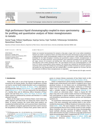
  • 3. to monoglutamates was carried out by adding 100 lL of rat plasma to tubes and incubating at 37 °C for 2 h. Thereafter the sample was boiled for 5 min followed by cooling on ice. The tubes were centri- fuged for 30 min (14,000g, 4 °C) and the supernatant was filtered through the 0.22 lm filter. Finally, the filtrate was clarified by ultra-filtration at 12,000g for 12 min using 10 kDa cut-off mem- brane filter. Immediately, the resulting extract was transferred to autosampler vials in HPLC and a 7.5 lL aliquot was directly injected on the column. 2.5. Liquid chromatography settings The chromatographic separation of folate derivatives was car- ried out using Waters Acquity™ UPLC system (Milford, USA) run- ning in HPLC mode, coupled to a binary pump, an autosampler, and controlled by Xcalibur 3.0 software (Thermo Fisher Scientific, San Jose, USA). The sample manager was configured with a 10 lL sample injection loop and a 100 lL sample syringe with the partial loop with needle overfill function. Prior to sample injections, the syringe was rinsed with 500 lL of weak (water/acetonitrile, 95:5) and strong (water/acetonitrile, 5:95) solvents, respectively. The auto-sampler (set at 4 °C) was covered to minimize any degrada- tion of light sensitive folates during sample analysis. Folate derivatives were separated on a reversed phase Luna C18 column (5 lm particle size, 250 mm  4.60 mm ID) (Phenomenex, USA) using gradient elution program. The gradient comprised of a binary solvent system consisting 0.1% (v/v) of formic acid in water (solvent A) and acetonitrile (solvent B) at a flow rate of 500 lL minÀ1 . The injection volume was 7.5 lL, run time 25 min and col- umn temperature was set at 24 °C. The starting condition of gradi- ent was 95% of solvent A and 5% of solvent B. Subsequently, solvent B was linearly increased to 10% in 4 min, then to 30% in 6 min and to 95% in 7 min. Thereafter, the mobile phase was reverted to the initial condition in 3 min and held for 5 min for re-equilibration of column before the next injection. After sample injection for first 8 min, the solvent flow from LC pump was diverted to waste posi- tion to minimize the suppression of MS signals by salts present in the extract. Thereafter, the solvent flow was switched to mass detector position for next 12 min and again diverted to waste posi- tion for 5 min till completion of the total run. 2.6. Mass spectrometry settings For all experiments, Exactive™ Plus Orbitrap mass spectrometer (Thermo Fisher Scientific, USA) was operated in alternating full scan and all ion fragmentation (AIF) mode equipped with positive heated electrospray ionization (ESI). The source parameters were optimized as follows: ion spray voltage, 4 kV; capillary tempera- ture, 380 °C and heater temperature, 300 °C. Sheath and auxiliary gas flows were 60 and 20 arbitrary units (au) respectively. Capil- lary, tube lens, and skimmer voltages were 65, 60, and 24 V respec- tively. For full scan experiment, the mass scan in the range of m/z 120–800 was monitored, while for AIF experiment, it was in the range of m/z 50–450. The resolution for full scan experiment was set to 70,000 full width at half maximum (FWHM) and for the AIF experiment it was set to 35,000 FWHM. The automatic gain control (AGC) target for full scan and AIF experiments were set to high dynamic range of 1e6 and 5e5 respectively. Similarly, the maximum injection time was set to 100 and 50 ms respectively. All ions entering into the mass spectrometer (MS) were frag- mented using nitrogen gas in the High-energy collision-induced dissociation (HCD) cell at normalized collision energy (CE) 20 (arbitrary unit) with 20% stepped CE, and finally detected in the Orbitrap mass analyzer. The fore vacuum, high vacuum and ultra-high vacuum were maintained around 1.5 mbar, from 2.5eÀ5 to 3.5eÀ5 mbar and below 4eÀ10 mbar, respectively. Instrument control and data processing were carried out by Xcali- bur 3.0 software (Thermo Fisher Scientific, San Jose, USA). Prior to sample analysis, the external calibration of instrument was performed to ensure the mass accuracy by directly infusing calibration solutions in both positive and negative ion modes. The positive mode calibration solution consisted of caffeine, Met- Arg-Phe-Ala acetate salt (MRFA), n-butyl amine and Ultramark™ 1621, while negative mode calibration solution consisted of sodium dodecyl sulfate, sodium taurocholate and Ultramark™ 1621. These solutions were freshly prepared using acetonitrile, methanol, and water containing 1% (v/v) acetic acid. All these cal- ibration solutions were mixed at room temperature and directly infused in the source using a syringe pump (Chemyx Fusion 100, Thermo Fisher Scientific, USA) with a flow rate of 5 lL/min. In posi- tive ion mode, the external lock mass (polydimethyl cyclosiloxane, exact mass = 455.12003) was also tested for the mass analyzer recalibration. 2.7. Recovery studies In the absence of specific guidelines for analysis of folate deriv- atives in tomato plant tissue, the International Conference on Harmonization (ICH) guideline was used for the validation of ana- lytical method which includes accuracy, sensitivity and linearity. To determine the accuracy of method, recovery test was performed by spiking of tomato extract with known amount of individual folate derivatives and calculating their final content in extract. The recovery (R) was calculated as B À C/A  100, where A = peak area of neat folates standard, B = peak area of spiked standard, C = peak area of extract. 2.8. Limit of detection and limit of quantification Sensitivity was confirmed by evaluating the limit of detection (LOD; calculated as: 3.3r/S, where r is the standard deviation and S is the slope of calibration curve) and limit of quantification (LOQ; calculated as: 10r/S). The linearity of each folate derivative was evaluated by plotting the peak area at different concentra- tions. The sample concentrations were calculated from the equa- tion y = mx + c, as determined by the linear regression analysis. 2.9. Statistical analysis All results are expressed as mean value ± SE of three or more replicates based on fresh weight (FW). A t-test was used for deter- mining significant difference between the mean values. The differ- ences were considered to be significant for P < 0.05. Statistical analysis of data was performed using SigmaPlot 11.0. 3. Result and discussion 3.1. Optimization of HPLC and MS/MS conditions It has been reported that reverse phase C18 column is the most appropriate column (based on resolution and retention time) for the separation and detection of folate derivatives. Initially, both organic solvents (acetonitrile and methanol) with 0.1% (v/v) formic acid and 0.1% (v/v) acetic acid were evaluated. The results show that usage of acetonitrile with 0.1% (v/v) formic acid increased the resolution with higher signal to noise ratio and proved to be the best eluent for ionization of folate derivatives. The optimized chromatographic conditions are described in Section 2.5. Under these conditions, separation of THF, 5-CH3-THF, 5,10-CH+ THF, MTX, 5-CHO-THF, and FA were achieved at 10.28, 10.84, 11.35, 12.87, 13.02, 13.16 min respectively (Fig. 1). Interfering 78 K. Tyagi et al. / Food Chemistry 179 (2015) 76–84
  • 4. compounds present in the plant extract (sugars, salt, and other matrices) may suppress ionization. To eliminate them, a divert valve was used during first 8 min and last 5 min of chromato- graphic run to redirect the major impurities to the waste instead to the mass detector. Prior to determining the optimal conditions, individual folate standard derivatives and internal standards were directly infused to ESI in both positive and negative ion modes. Comparison of both modes revealed that the positive ion mode provided better sensi- tivity than negative ion mode in conformity with earlier reports (De Brouwer et al., 2007; Patring & Jastrebova, 2007; Rychlik & Freisleben, 2002). Therefore, positive ion mode was selected for subsequent experiments. The optimized MS parameters are described in Section 2.6. For identification of each compound, accu- rate mass of the corresponding ion, elemental composition, and specific retention times were considered. In full scan mode, the ions detected at m/z 446.1779, 460.1942, 456.1632, 455.1786, 474.1729 and 442.1467 corresponded to the THF, 5-CH3-THF, 5,10-CH+ THF, MTX, 5-CHO-THF and FA respectively. Fig. 1 shows the extracted ion chromatogram. The accurate masses of corre- sponding ions were also compared with the theoretical masses cal- culated by Xcalibur 3.0 software. In addition, all ions were fragmented without pre-selection in HCD cell at different collision energy ($20–40 au). Interestingly, several fragment ions were detected at m/z 299.1249 (THF), 313.1404 (5-CH3-THF), 308.1256 (MTX), 327.1198 (5-CHO-THF), and 295.0936 (FA) indicating the loss of glutamate moiety (Supplementary Table 1) except 412.1530 (5,10-CH+ THF). Using HCD, the sample analysis time was substantially reduced by confirming the observed m/z values at the same time without pre-selection. 3.2. Optimization of sample extraction in tomato For optimization of folate extraction/estimation from tomato, several factors were considered like type and composition of sam- ple matrix, nature, amount, and stability of the compounds for extraction and fine tuning of the LC–MS parameters for their detec- tion. Considering that folates are very labile compounds, their extractions differ for each kind of matrix. Tomato fruit is a very complex matrix having many compounds to which folates bind. We optimized the folate extraction from tomato by modifying the method of De Brouwer et al. (2008) originally developed for rice. Largely for the folate extraction from a variety of food matrices, a trienzyme treatment (amylase, protease and conjugase) developed by Martin, Landen, Soliman, and Eitenmiller (1989) is used to release the bound folate from matrix. The trienzyme treat- ment accomplishes more complete extraction of folates that are bound to matrices of proteins or polysaccharides by including pro- tease and a-amylase in addition to conjugase. Considering matrix complexity, several parameters were evalu- ated for optimal folate extraction from tomato fruits such as pH and molarity of the extraction buffer, amount of a-amylase, prote- ase, rat plasma conjugase, and duration of boiling time after enzyme treatments. Fig. 2 shows optimization of different param- eters for folate extraction. The pH of extraction buffer greatly affects the stability of folates in extracts. Therefore, folate extrac- tion was compared using two different pH, 4.5 and 7.0. No signifi- cant difference in recovery of total folate content was observed using these two pH (P value 0.0519) (Fig. 2A). Stability of the folate derivatives greatly varies with respect to pH, thus requiring selection of a pH that is optimal for efficient extraction of most folate derivatives (De Brouwer et al., 2007). The optimal pH for protease and amylase is also different from optimal pH for rat plasma conjugase (Arcot & Shrestha, 2005). Since multiple pH adjustments prolong the extraction process and increase the chances of oxidation of folate, the usage of differ- ent pH to achieve optimal trienzyme treatment is not feasible while screening a large number of samples. The amount of total folate extracted at pH 4.5 and 7.0 was nearly similar, however, at pH 4.5, we detected THF in tomato leaf but not in tomato fruit tis- sue. Given the variability in stability of different folate derivatives, it is difficult to have an ideal pH for extraction of all folate derivatives in their native state from plant matrix. Considering that 5-CH3-THF and 5-CHO-THF, the most predominant folate forms present in the plants are more stable at low pH, therefore we used pH 4.5 for subsequent extraction of folates. The recovery of folate is also influenced by the buffer composition; however, phosphate buffer is most widely used for folate extraction. We compared both 50 and 100 mM phosphate buffer and found that both were equally efficient. Taking cognizance of fact that high salt concentrations in buffer interfere with the ionization process in the mass spectrom- eter, we used 50 mM phosphate buffer for all extraction procedures. Considering that the fruit and vegetable matrices are complex, possessing compounds that may possibly bind with the metabolite of interest and improve/inhibit the extraction efficiency, the trien- zyme method of folate extraction was also optimized. The optimi- zation involved alteration of enzyme concentrations and duration of treatments. We compared the extraction of folate with and without respective enzymes for all three enzymes used for folate Fig. 1. Representative chromatogram of five folates standards and an internal standard. (A) THF, (B) 5-CH3-THF; (C) 5,10-CH+ THF; (D) MTX; (E) 5-CHO-THF and (F) FA. K. Tyagi et al. / Food Chemistry 179 (2015) 76–84 79
  • 5. extraction. It was observed that without a-amylase treatment, $85% of the total folate content was extracted (P value 0.0281) compared to the a-amylase treated control. Similarly, addition of protease and incubation at 37 °C for 1 h did not significantly improve the extraction of total folate content (P value 0.050) (Fig. 2B). Among the three enzymes used in this study, the rat plasma conjugase was found to be the most important for folate deglutamylation. Different amounts of rat plasma conjugase was added to the extract (per 100 mg of fresh tissue), that is, 0, 25, 50 lL which resulted in 17%, 52% and 88% recovery of total folate pool respectively compared with addition of 100 lL rat plasma serving as control. Taking into account that increase from 50 to 100 lL of rat plasma lead to 12% improvement in the folate recovery, 100 lL of rat plasma was used for all subsequent exper- iments (Fig. 2C). It is reported that boiling the extract prior to enzyme treatment reduces the overall yield of folate from food matrices. Moreover, the addition of antioxidants such as ME and ascorbic acid in the extraction buffer protects folate from degradation during boiling treatment that is needed to denature the enzymes besides improv- ing the stability of folate in an autosampler. The increase in the boiling time from 5 to 10 min post-enzyme treatment did not enhance the recovery of folate, therefore, boiling for 5 min was used for all enzymatic deactivation steps to minimize any possible degradation of folates (Fig. 2D). 3.3. Optimization of purification Different folate vitamers differ in their functional groups thus vary in their chemical properties and therefore can be better sepa- rated by HPLC. It is reported that sample purification prior to sep- aration in HPLC leads to better recovery of folate and also distinction between different forms. The presence of different endogenous constituents in food extracts interfere with folates and hamper the determination of some folate forms. Considering that folate is also present at low level in most food matrices, removal of the interfering compounds during purification improves the detection limit and selectivity of the folate in HPLC. Solid phase extraction (SPE) is the most commonly used method for folate purification using either a strong anion exchange (SAX) sorbent or C18 SPE cartridges for food extract prior to HPLC–MS analysis (Chandra-Hioe, Bucknall, & Arcot, 2013; Stokes & Webb, 1999). Use of SAX sorbent for purification of food extracts provides high recovery of different folate forms, however, pre-concentration of sample is not possible as large buffer volumes are required to elute all the folates from SAX column. In addition, high concentra- tion of salt used in elution step interferes with subsequent MS analysis. Recently, affinity chromatography using immobilized folate- binding protein has been used as a highly selective purification method for folates, especially for complex food matrices such as cereals (Pfeiffer et al., 1997). Though affinity chromatography enables quantification at 10-fold lower concentration than SAX, the lack of commercial availability of FBP columns precludes their use for folate analysis. Moreover, folate-binding protein exhibits low affinity to 5-HCO-H4 folate, which may result in higher losses of this folate form during the purification step. Among the different methods of folate analysis, usage of LC–MS has emerged as most preferred method owing to its specificity and sensitivity to distinguish different forms of folate. Several studies have used LC–MS for folate analysis, particularly from fortified food samples (Alaburda, de Almeida, Shundo, Ruvieri, & Sabino, 2008; Patring, Wandel, Jägerstad, & Frølich, 2009). However, the detection of folate derivatives by LC–MS needs robust purification of folate vitamers for efficient and accurate analysis. In this study, we optimized the purification of folates for subsequent analysis by LC–MS. In earlier studies, SPE using SAX cartridge (Vishnumohan, Arcot, & Pickford, 2011) or affinity column using folate binding protein (Díaz de La Garza et al., 2004) have been used so far. How- ever, these approaches are time consuming and cannot be applied when handling large number of samples. To overcome these draw- backs which hinder folate estimation for high throughput analysis, the purification step was modified to a simple, efficient and also cost effective method for quantification of folate in tomato by using a molecular weight cut-off membrane filter according to Zhang et al. (2005). For efficient sample cleanup, extract was fil- tered through a 0.22 lm filter to remove the particulate matter in the extract followed by ultra-filtration through a 10 kDa Fig. 2. Optimization of folate extraction in tomato fruit. (A) Effect of pH, (B) extraction in presence (control) and absence of a-amylase and protease, (C) extraction with increasing amount of rat plasma conjugase and (D) effect of duration of boiling (n P 3). 80 K. Tyagi et al. / Food Chemistry 179 (2015) 76–84
  • 6. centrifugal device removing various polymeric compounds present in the tomato extract. The usage of step wise increase in gradient consisting of 0.1% (v/v) of formic acid in water (solvent A) and ace- tonitrile (solvent B) on a C18 reverse phase column yielded good resolution of different folate vitamers. To avoid contamination of electrospray of MS machine by salt and matrix, which reduces the sensitivity of the mass spectrometer, out of total runtime of 25 min, first 8 min and last 5 min eluent were discarded to the waste and sample was allowed to pass through MS during inter- vening 12 min. Folates were analyzed in positive ion mode through mass spec- trometry as the analysis indicated it to be superior to the negative ion mode. All ion fragmentation (AIF) parameters including precur- sor ions, product ions, and collision energy were optimized for frag- mentation by injecting the folate standards. Electrospray ionization (ESI) source temperature was 300 °C and capillary temperature was maintained at 380 °C. For fragmentation of the folate standards in HCD cell, collision energy of 20 or 40 (arbitrary unit, Supplementary Table 1) was considered optimal depending on the folate com- pound. The most abundant fragment resulting from all folates was the one that was generated from the neutral loss of the gluta- mate moiety (m/z 147), e.g. for FA m/z 442/295, for THF m/z 446/ 299, for 5-CH3-THF m/z 460/313, for 5-CHO-THF m/z 474/327 and for MTX m/z 455/308. Collision energy used for 5,10-CH+ THF was 40 arbitrary unit resulting in m/z 456/412. Supplementary Table 1 shows the fragmentation result of the folate standards. A represen- tative chromatogram of folates standards is shown in Fig. 1. 3.4. Standard calibration curve and linearity The linearity of the developed HPLC–MS method was evaluated by preparing seven point calibration curves without matrix for each folate derivative (THF; 5-CH3-THF; 5,10-CH+ THF; MTX; 5-CHO-THF; and FA) in triplicates. Correlation coefficients (R2 ) determined for all these folates were approximately 0.99, which confirmed their good linearity within the considered concentration ranges. For the folates derivatives, which were detected in the tomato fruits, the limit of detection (LOD, S/N P 3.3) and quantita- tion (LOQ, S/N P 10) were theoretically determined based on cali- bration curve, relating concentrations with signal to noise ratios. Table 1 shows the correlation coefficient, linear range, and slope of calibration curve. 3.5. Recovery and stability Accuracy of the developed method was determined by recovery test. The mean recoveries (n = 3) for all individual folate derivatives were in the range of 48–131% (Table 2). The observed value indi- cates that the developed method is accurate, except for folic acid, which is not a naturally occurring folate. Taking cognizance that multiple samples were analyzed for folate levels, the stability of the extracted samples was examined in the autosampler at 4 °C. The results indicated that most of the folate derivatives are stable up to 72 h of storage in the autosampler at 4 °C barring 5,10-CH+- THF (Supplementary Fig. 2). It is reported that the presence of ME and ascorbic acid in the extraction buffer enhances the stability of the folate for a prolonged period (Vahteristo & Finglas, 2000). 3.6. Use of external and internal standard In order to check the possible variations in MS response for dif- ferent folate derivatives while screening of large number of sam- ples, we used internal standards for accurate and reproducible quantitation. Though the usage of stable isotope labeled folate standards is the best option, these were not used owing to their limited availability and high cost. In its place a known amount of individual folate derivatives were used as external standards. The MS response of folate derivatives were checked by analyzing sev- eral tomato extracts and comparing the calculated concentrations with initial values. Furthermore, we used two structurally similar compounds, FA and MTX (Zhang et al., 2005) as internal standards. Both these standards were mixed to the tomato homogenate prior to initiation of extraction protocol and recoveries were calculated. Considering that the recovery of MTX was higher than FA (Table 2), it was selected as internal standard for our further screening. 3.7. Folate content in tomato fruit The total folate level in mature green and red ripe fruits of tomato cultivar Arka Vikas was 21.94 and 18.06 lg/100 g of FW respectively (Fig. 3A). 5-CH3-THF was the most predominant folate form comprising of 68% and 58% of total folate in mature green and red ripe fruits respectively. These values were similar to those reported by Díaz de La Garza, Gregory, and Hanson (2007) that 5-CH3-THF constitutes 70% of total folate present in tomato fruit. In addition to 5-CH3-THF, other two folate derivatives 5-CHO- THF and 5,10-CH+ THF were also detected in tomato fruit. Among these, 5-CHO-THF was the second predominant folate form present in both mature green and red ripe stage fruits. The level of 5-CH3- THF significantly decreased during tomato fruit ripening from mature green to red ripe stage (P value 0.035). In contrast, the con- tent of 5,10-CH+ THF and 5-CHO-THF in the mature green and red ripe stage of tomato fruit did not show any significant changes (Fig. 3B). Considering that 10-CHO-THF cannot be identified in the LC–MS due to its conversion to 5,10-CH+ THF in mobile phase containing 0.1% formic acid (Fazili & Pfeiffer, 2004; Goyer et al., 2005), it can be assumed that 5,10-CH+ THF peak likely included both 10-CHO-THF and any preexisting 5,10-CH+ THF. Consistent Table 1 Calibration and sensitivity data of folate standards. All experiments were done in triplicates. Compounds LOD (ng/mL) LOQ (ng/mL) Slope (n = 6 or 7) mean ± SE R2 Linear range (ng/mL) FA 0.15 0.47 38095.5 ± 450.5 0.999 1–30 THF 0.25 0.77 30541.5 ± 577.5 0.997 2–30 5,10-CH+ THF 0.34 0.92 10757.3 ± 70.7 0.984 0.5–30 5-CH3-THF 0.17 0.53 36024.3 ± 682.2 0.998 1–30 5-CHO-THF 0.37 1.12 19204.0 ± 122.3 0.994 1–30 Table 2 Percentage recovery of spiked folate standards in tomato fruit extract. 2.7 ng/mL of folate standards were spiked into extract. Compound % recovery mean ± S.E. FA 48.97 ± 8.48 THF 70.89 ± 7.10 5,10-CH+ THF 94.72 ± 8.29 5-CH3-THF 131.31 ± 8.78 5-CHO-THF 115.90 ± 1.59 MTX 95.27 ± 11.52 K. Tyagi et al. / Food Chemistry 179 (2015) 76–84 81
  • 7. with earlier report (Iniesta, Perez-Conesa, Garcia-Alonso, Ros, & Periago, 2009), THF was not detected in tomato fruit extract. Tomato is consumed in both cooked and raw form by the people. Taking the folate content of fully red ripe tomato fruit (18.06 lg/ 100 g of FW) in account, its percentage contribution to the RDA (400 lg/day) is 4.51. However, the folate content in a given food matrix such as tomato fruit is not constant as several factor affects its level such as nature of cultivar, developmental stage, and envi- ronmental conditions during ripening (Iniesta et al., 2009; Jägerstad et al., 2005). The extent of basal polyglutamylation status of folate helps in its retention in the cellular compartments as well as increases its affinity toward the enzymes with which they interact (Akhtar et al., 2010). The polyglutamylation of individual folate derivatives was evaluated by comparing the monoglutamate amounts of each folate derivative detected in tomato red ripe fruits with and with- out the addition of rat plasma during folate extraction. 83% of 5- CH3-THF was present in polyglutamylated form. Similarly, $90% of total 5-CHO-THF was present in the polyglutamylated form (Fig. 3C), whereas $50% 5,10-CH+ THF was present in polyglutamy- lated form. 3.8. Application of the method Fruits and vegetables are good sources of naturally occurring folate, primarily 5-CH3-THF. To check the efficiency and applicabil- ity of the above method for other plant materials, the folate levels were determined in tomato juvenile leaf, spinach leaf, garden pea seeds and capsicum fruit (Table 3). The major folate detected in these varieties was 5-CH3-THF. Other forms observed were 5-CHO-THF and 5,10-CH+ THF with traces of THF and FA. 5-CHO- THF was the second most predominant form detected in all of them. Contrary to the report that recovery of THF was low at acidic pH but increased as the pH of extraction buffer increased to 7.0 (Zhang et al., 2005), no THF was detected in tomato fruit tissue using pH 7.0 extraction buffer. Compared to fruits, the juvenile leaves of tomato showed very high level of folates, which may be related to active cell division and expansion in these leaves. Interestingly, folic acid or other oxidation products of natural folate forms were not detected indicating that extraction procedure used in this study led to minimal degradation or loss of folate. The total folate content mean (±SE) in tomato juvenile leaf, spinach leaf, garden pea seeds and capsicum fruit was 429.54 (61.49), 287.16 (55.14), 188.33 (14.06) and 39.44 (2.97) lg/100 g FW respectively (Table 3). Considering that these values are higher than the reported values in literature supports that our extraction method leads to maximum recovery of folates from matrix. Total folate content of spinach reported in earlier studies varied from 194–364 lg/100 g of FW assessed through microbiological assay (Lin & Lin, 1999) to 48–177 lg/100 g of FW through HPLC (Johansson, Jägerstad, & Frølich, 2007; Vahteristo, Lehikoinen, Ollilainen, & Varo, 1997). Our results showed nearly 110 lg higher value of folate than the earlier reported value for spinach. While these differences in the folate levels could be attributed to cultivar, Table 3 Folate content of tomato fruit, leaf and other vegetables (lg/100 g FW). Sample THF (mean ± SE) 5-CH3-THF (mean ± SE) 5,10-CH+ THF (mean ± SE) 5-CHO-THF (mean ± SE) Total folate (mean ± SE) Tomato fruit (mature green) – 14.97 ± 0.74 1.15 ± 0.08 5.81 ± 0.56 21.93 ± 1.17 Tomato fruit (red ripe) – 10.61 ± 1.42 1.55 ± 0.19 5.89 ± 0.59 18.05 ± 2.01 Tomato leaf 4.12 ± 0.10 226.36 ± 29.95 13.13 ± 2.92 185.93 ± 28.14 429.54 ± 61.49 Spinach 0.24 ± 0.09 159.17 ± 30.79 19.21 ± 4.05 108.54 ± 20.71 287.16 ± 55.14 Garden pea – 156.19 ± 7.62 4.28 ± 0.78 27.86 ± 2.67 188.33 ± 14.06 Green capsicum – 32.36 ± 2.76 2.06 ± 0.14 5.02 ± 0.55 39.44 ± 2.97 Fig. 3. Folate content in tomato fruits. (A) Total folate content in mature green (MG) and red ripe (RR) fruits (n P 3). (B) Relative distribution of different folate forms in mature green and red ripe fruit (n P 3). (C) Polyglutamylation status of different folate derivatives in red ripe fruit. 82 K. Tyagi et al. / Food Chemistry 179 (2015) 76–84
  • 8. variety, and geographical area, climatic and seasonal differences among study groups (Shohag et al., 2011), it is also likely that sam- ple preparation and extraction protocol reported in this study may have led to more efficient extraction and minimal losses of folate from spinach. 4. Conclusion An accurate HPLC–MS/MS method has been successfully devel- oped and validated for folate analysis in tomato fruits, which is simple, fast and cost effective and can be applied to a large number of samples for folate analysis. Our method has wide applicability as the folate levels determined in different types of tissue used in the study were either comparable or more than those reported in pre- vious studies. We found that 5-CH3-THF is the main folate deriva- tive in tomato. This method is fairly simple to execute as the sample cleanup involves ultra-filtration instead of SPE or affinity chromatography, without compromising on the amount or forms of folates detected. The validation experiments showed linearity of the method and total folate concentration in mature green and red ripe tomato fruit was 21.93 and 18.06 lg/100 g of fresh weight with acceptable precision and accuracy. We have used MTX as an internal standard in screening of multiple samples for reproducible quantitation. Most of the folates were stable for more than 24 h when kept at 4 °C in autosampler. Total folate profile in tomato leaf differed considerably from that of tomato fruit tissue indicating that this extraction procedure can be used to analyze other food matrices too. Acknowledgments This work was supported by the Department of Biotechnology (Grant No. BT/PR11671/PBD/16/828/2008 to R.S. and Y.S.), the Council of Scientific and Industrial Research (research fellowship to KT), University Grants Commission (research fellowship to PU and SS). Appendix A. Supplementary data Supplementary data associated with this article can be found, in the online version, at http://dx.doi.org/10.1016/j.foodchem.2015. 01.110. References Akhtar, T. A., Orsomando, G., Mehrshahi, P., Lara-Núñez, A., Bennett, M. J., Gregory, J. F. III,, et al. (2010). A central role for gamma-glutamyl hydrolases in plant folate homeostasis. The Plant Journal, 64, 256–266. Alaburda, J., de Almeida, A. P., Shundo, L., Ruvieri, V., & Sabino, M. (2008). Determination of folic acid in fortified wheat flours. Journal of Food Composition and Analysis, 21, 336–342. AOAC (2000). In AOAC (Ed.). Official methods for analysis of AOAC International (17th ed). Gaithersburg, MD: Association of Official Analytical Chemists International. Arcot, J., & Shrestha, A. (2005). Folate: Methods of analysis. Trends in Food Science and Technology, 16, 253–266. Bagley, P. J., & Selhub, J. (2000). Analysis of folate form distribution by affinity followed by reversed-phase chromatography with electrochemical detection. Clinical Chemistry, 46, 404–411. Blancquaert, D., De Steur, H., Gellynck, X., & Van Der Straeten, D. (2014). Present and future of folate biofortification of crop plants. Journal of Experimental Botany, 65, 895–906. Blom, H. J., Shaw, G. M., den Heijer, M., & Finnell, R. H. (2006). Neural tube defects and folate: Case far from closed. Nature Reviews Neuroscience, 7, 724–731. Blount, B. C., Mack, M. M., Wehr, C. M., MacGregor, J. T., Hiatt, R. A., Wang, G., et al. (1997). Folate deficiency causes uracil misincorporation into human DNA and chromosome breakage: Implications for cancer and neuronal damage. Proceedings of the National Academy of Sciences United States of America, 94, 3290–3295. Chandra-Hioe, M. V., Bucknall, M. P., & Arcot, J. (2013). Folic acid-fortified flour: Optimised and fast sample preparation coupled with a validated high-speed mass spectrometry analysis suitable for a fortification monitoring program. Food Analytical Methods, 6, 1416–1423. De Brouwer, V., Storozhenko, S., Van De Steene, J. C., Wille, S. M., Stove, C. P., Van Der Straeten, D., et al. (2008). Optimisation and validation of a liquid chromatography–tandem mass spectrometry method for folates in rice. Journal of Chromatography A, 1215, 125–132. De Brouwer, V., Zhang, G. F., Storozhenko, S., Van Der Straeten, D., & Lambert, W. E. (2007). PH stability of individual folates during critical sample preparation steps in prevision of the analysis of plant folates. Phytochemical Analysis, 18, 496–508. Díaz de La Garza, R., Gregory, J. F., & Hanson, A. D. (2007). Folate biofortification of tomato fruit. Proceedings of the National Academy of Sciences United States of America, 104, 4218–4222. Díaz de La Garza, R. I., Quinlivan, E. P., Klaus, S. M., Basset, G. J., Gregory, J. F., & Hanson, A. D. (2004). Folate biofortification in tomatoes by engineering the pteridine branch of folate synthesis. Proceedings of the National Academy of Sciences United States of America, 101, 13720–13725. Dueker, S. R., Lin, Y., Jones, A. D., Mercer, R., Fabbro, E., Miller, J. W., et al. (2000). Determination of blood folate using acid extraction and internally standardized gas chromatography–mass spectrometry detection. Analytical Biochemistry, 283, 266–275. Fazili, Z., & Pfeiffer, C. M. (2004). Measurement of folates in serum and conventionally prepared whole blood lysates: Application of an automated 96-well plate isotope-dilution tandem mass spectrometry method. Clinical Chemistry, 50, 2378–2381. Garratt, L. C., Ortori, C. A., Tucker, G. A., Sablitzky, F., Bennett, M. J., & Barrett, D. A. (2005). Comprehensive metabolic profiling of mono-and polyglutamated folates and their precursors in plant and animal tissue using liquid chromatography/negative ion electrospray ionisation tandem mass spectrometry. Rapid Communications in Mass Spectrometry, 19, 2390–2398. Gough, K. R., Read, A., McCarthy, C., & Waters, A. (1963). Megaloblastic anaemia due to nutritional deficiency of folic acid. Quarterly Journal of Medicine, 32, 243–256. Goyer, A., Collakova, E., de la Garza, R. D., Quinlivan, E. P., Williamson, J., Gregory, J. F., et al. (2005). 5-Formyltetrahydrofolate is an inhibitory but well tolerated metabolite in Arabidopsis leaves. Journal of Biological Chemistry, 280, 26137–26142. Iniesta, M. D., Perez-Conesa, D., Garcia-Alonso, J., Ros, G., & Periago, M. J. (2009). Folate content in tomato (Lycopersicon esculentum). Influence of cultivar, ripeness, year of harvest, and pasteurization and storage temperatures. Journal of Agricultural and Food Chemistry, 57, 4739–4745. International Conference on Harmonization (ICH) (1996). Guideline on validation of analytical procedures: Methodology Q2B, Geneva, 1. Jägerstad, M., Piironen, V., Walker, C., Ros, G., Carnovale, E., Holasova, M., et al. (2005). Increasing natural food folates through bioprocessing and biotechnology. Trends in Food Science and Technology, 16, 298–306. Johansson, M., Jägerstad, M., & Frølich, W. (2007). Folates in lettuce: A pilot study. Scandinavian Journal of Food and Nutrition, 51, 22–30. Kolb, A. F., & Petrie, L. (2013). Folate deficiency enhances the inflammatory response of macrophages. Molecular Immunology, 54, 164–172. Lin, B., & Lin, R. (1999). Effect of Chinese stir-fry cooking on folate contents of vegetables. Journal-Chinese Agricultural Chemical Society, 37, 443–469. Martin, J., Landen, W., Jr., Soliman, A., & Eitenmiller, R. (1989). Application of a tri- enzyme extraction for total folate determination in foods. Journal-Association of Official Analytical Chemists, 73, 805–808. Ndaw, S., Bergaentzlé, M., Aoudé-Werner, D., Lahély, S., & Hasselmann, C. (2001). Determination of folates in foods by high-performance liquid chromatography with fluorescence detection after precolumn conversion to 5- methyltetrahydrofolates. Journal of Chromatography A, 928, 77–90. Patring, J. D., & Jastrebova, J. A. (2007). Application of liquid chromatography– electrospray ionisation mass spectrometry for determination of dietary folates: Effects of buffer nature and mobile phase composition on sensitivity and selectivity. Journal of Chromatography A, 1143, 72–82. Patring, J., Wandel, M., Jägerstad, M., & Frølich, W. (2009). Folate content of Norwegian and Swedish flours and bread analysed by use of liquid chromatography–mass spectrometry. Journal of Food Composition and Analysis, 22, 649–656. Pfeiffer, C. M., Rogers, L. M., & Gregory, J. F. (1997). Determination of folate in cereal- grain food products using trienzyme extraction and combined affinity and reversed-phase liquid chromatography. Journal of Agricultural and Food Chemistry, 45, 407–413. Rébeillé, F., Ravanel, S., Jabrin, S., Douce, R., Storozhenko, S., & Van Der Straeten, D. (2006). Folates in plants: Biosynthesis, distribution, and enhancement. Physiologia Plantarum, 126, 330–342. Rychlik, M., & Freisleben, A. (2002). Quantification of pantothenic acid and folates by stable isotope dilution assays. Journal of Food Composition and Analysis, 15, 399–409. Shohag, M., Wei, Y.-Y., Yu, N., Zhang, J., Wang, K., Patring, J., et al. (2011). Natural variation of folate content and composition in spinach (Spinacia oleracea) germplasm. Journal of Agricultural and Food Chemistry, 59, 12520–12526. Stokes, P., & Webb, K. (1999). Analysis of some folate monoglutamates by high- performance liquid chromatography–mass spectrometry. I. Journal of Chromatography A, 864, 59–67. Vahteristo, L., & Finglas, P. (2000). Chromatographic determination of folates. Chromatographic Science Series, 84, 301–324. Vahteristo, L., Lehikoinen, K., Ollilainen, V., & Varo, P. (1997). Application of an HPLC assay for the determination of folate derivatives in some vegetables, fruits and berries consumed in Finland. Food Chemistry, 59, 589–597. Van Daele, J., Blancquaert, D., Kiekens, F., Van Der Straeten, D., Lambert, W. E., & Stove, C. P. (2014). Folate profiling in potato (Solanum tuberosum) tubers by K. Tyagi et al. / Food Chemistry 179 (2015) 76–84 83
  • 9. ultrahigh-performance liquid chromatography–tandem mass spectrometry. Journal of Agricultural and Food Chemistry, 62, 3092–3100. Vishnumohan, S., Arcot, J., & Pickford, R. (2011). Naturally-occurring folates in foods: Method development and analysis using liquid chromatography–tandem mass spectrometry (LC–MS/MS). Food Chemistry, 125, 736–742. Zhang, G.-F., Maudens, K. E., Storozhenko, S., Mortier, K. A., Van Der Straeten, D., & Lambert, W. E. (2003). Determination of total folate in plant material by chemical conversion into para-aminobenzoic acid followed by high performance liquid chromatography combined with on-line postcolumn derivatization and fluorescence detection. Journal of Agricultural and Food Chemistry, 51, 7872–7878. Zhang, G.-F., Storozhenko, S., Van Der Straeten, D., & Lambert, W. E. (2005). Investigation of the extraction behavior of the main monoglutamate folates from spinach by liquid chromatography–electrospray ionization tandem mass spectrometry. Journal of Chromatography A, 1078, 59–66. 84 K. Tyagi et al. / Food Chemistry 179 (2015) 76–84