
Recommandé
Contenu connexe
Tendances
Tendances (20)
Similaire à Infectious Bovine Keratoconjunctivitis (IBK) Guide
Similaire à Infectious Bovine Keratoconjunctivitis (IBK) Guide (20)
Plus de Kanwarpal Dhillon
Dernier
Dernier (20)
Infectious Bovine Keratoconjunctivitis (IBK) Guide
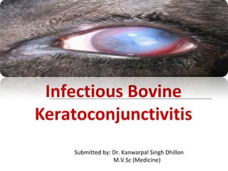
- 1. Infectious Bovine Keratoconjunctivitis Submitted by: Dr. Kanwarpal Singh Dhillon M.V.Sc (Medicine)
- 2. Pink eye or infectious bovine keratoconjunctivitis (IBK) is the most common & costly ocular disease of cattle. It is an inflammatory bacterial infection of the eye that can cause permanent blindness in severe cases. It is a highly contagious disease, causing inflammation of the cornea (the clear outer layer) and conjunctiva (the pink membrane lining the eyelids) of the eye. IBK is characterized by blepharospasm, conjunctivitis, lacrimation and varying degrees of corneal opacity and ulceration. The disease usually is acute and tends to spread rapidly The incidence of pinkeye increases in spring, peaks in the summer, and decreases in the fall/Autumn. Introduction
- 3. Etiology The primary infectious agent for pinkeye is the bacterium Moraxella bovis. The bacteria produce a toxin which attacks the surface of the eye (cornea) and conjuctiva, causing inflammation and ulceration. This bacterium is found in the eyes of many recovered and apparently normal cattle.
- 4. Predisposing Factors Infection with IBK or other microbes may increase the severity of infection with M bovis. Plant pollens, face flies, ultraviolet radiations, tall grass, dry and dusty environmental conditions and transportation stress. Flies (Musca spp.) can also serve as mechanical vectors for M. bovis. Eye irritation. Calves are more likely to develop the disease than adult cattle, as adult cattle appear to develop protective antibodies on the surface of the eye. Bull calves have a higher incidence of disease than heifer calves.
- 5. Clinical Signs Stage I Cattle have excessive tearing and increased sensitivity to light. They will blink frequently and there is redness along the eyelids. Cattle will often seek shade, which will decrease their grazing time. Pain associated with pinkeye also decreases their feed intake. Stage I will progress to a small ulcer in the centre of the cornea which appears as a small white spot. The cornea develops a slightly cloudy grey appearance due to inflammation. One or both eyes may be affected. Stage I
- 6. Stage II The clinical signs described in Stage I continue, but the ulcer spreads across the cornea. As more inflammation occurs, the cornea becomes increasingly cloudy. At this point, some of the dark color of the iris can still be seen. The blood vessels make the cornea appear pink, which is how the disease received its name. Stage II
- 7. Stage III The ulcer covers most of the cornea and the inflammation continues to spread into the inner parts of the eye. When this occurs, the inside of the eye fills with fibrin, which is a pus-like substance that gives the eye a yellow appearance versus the typical brown appearance. Stage III
- 8. Stage IV The ulcer extends completely through the cornea, and the iris may protrude through the ulcer. The iris will become stuck in the cornea even after healing. This may lead to glaucoma or persistent swelling of the eye. This eye will be partially or completely blind. The eye may go on to completely rupture, and will develop a shrunken appearance or enlarge if glaucoma (increased eye pressure) is present. This eye will be permanently blind. Stage IV
- 9. Early clinical signs are photophobia, blepharospasm and epiphora later the ocular discharge may become mucopurulent.
- 10. Severe corneal edema, corneal neovascularization and epiphora
- 11. Conjunctivitis, with or without varying degrees of keratitis, is usually present along with epiphora
- 12. Lesions In cattle, one or more small ulcers typically occur near the center of the cornea. Lesions may regress in the early stages or may continue to progress
- 13. Diagnosis Presumptive diagnosis is based on ocular signs and concurrent systemic disease. Microbial culture is important for confirmatory diagnosis. It is important to distinguish that the lesions are not due to some other cause like some systemic disease or foreign bodies or parasites.
- 14. Differential Diagnosis Traumatic conjunctivitis – evidence of phyical injury or presence of foreign material in the eye. Pasteurella multocida (capsular type A) – isolated from the eyes; outbreaks of severe keratitis with loss of corneal stroma. Mycoplasma bovis – isolated from the eyes; outbreaks characterized by severe conjunctivitis, corneal opacity, ulceration, & swelling of eyelids. Other diseases – Listeria monocytogenes iritis , Infectious bovine rhinotracheitis, Bovine malignant catarrh & Chalmydial keratoconjuctivitis & Thelaziasis
- 15. Treatment IBK is frequently a self-limiting disease. Topical therapy: Early, acute cases respond to Rx with ophthalmic ointments & solutions containing Ab. Administration of Benzathine cloxacillin 375mg (oil-based) is found to be effective. Two doses, 72 hrs. apart, are recommended. Effective antibiotics for topical ophthalmic use include triple antibiotic, Gentamicin, and a combination Oxytetracycline / Polymyxin B ointment.
- 16. Subconjuctival therapy: Procaine penicillin (3-6x10⁵ IU) is given through the skin of upper eyelid or under the bulbar conjunctiva Parental therapy: Sulfadimidine @100mg/kg BW. Long-acting oxytetracycline (2 injections @20 mg/kg, IM or SC, at a 48- to 72-hr interval) alone or with oral administration of OTT @2g/250kg BW for 10 days. Ancillary therapy: Animal should be placed in dark shelter out of direct sunlight. If housing is not possible, eye flap patches are glued on above the eye & can be flipped up for the medication.
- 17. Animals with substantial uveitis secondary to Keratoconjunctivitis that is particularly painful may benefit from topical ophthalmic application of 1% atropine ointment 1–3 times daily. Systemic NSAID may be used to provide relief from secondary uveitis.
- 20. Prevention and control Good management practices Separation of infected animals Ultraviolet radiation from sunlight may enhance the disease therefore affected animals should be provided with shade. Reduce the incidence of flies and subsequent spreading of bacteria with the application of pour-on treatments. Dust bags or insecticide-impregnated ear tags can be used to reduce the number of face flies
- 21. References: VETERINARY MEDICINE A textbook of the diseases of cattle, horses, sheep, pigs and goats by O. M. Radostits, C.C.Gay, K. W. Hinchcliff, P. D. Rebhun’s Diseases of dairy cattle by Thomas J. Divers & Simon F. Peek THANKS……..