2. amplification products, with sizes ranging from 550 bp
to 1.7 kb, identify each core OS type. Using this system,
we were able to define the core type of all 72 strains of
the E. coli ECOR collection, a collection of E. coli
strains that represents the genetic diversity of the
species.6
Our results show that 68% of the ECOR collec-
tion strains possess an R1 LPS core type, 12% are R2,
11% are R3, 3% are R4, and 6% are K-12. These data, in
addition to analyses of core OS types in bacteremic clin-
ical isolates of E. coli,7,8
confirm that R1 is the dominant
core type in clinical isolates of E. coli. Random, non-
verotoxigenic, E. coli isolates from cattle show a similar
predominance of the R1 core OS type but, in contrast,
verotoxigenic field isolates of serotypes O55, O111, and
152 Whitfield
BA
2
↑
1
GalΙΙΙ
3
↑
1
β-Glc
3
↑
1
KdoΙΙ-7←PEtN?HepΙΙΙ
1
↓
7
GlcN
1
↓
7
4
↑
P
↑
PEtN
lipidA
O-PS
waaC
waaTwaaGwaaO
waaF
waaWwaaV
waaL
4
↑
P
waaY
waaA
C
E.coliR4
Gal-1→2-Glc-1→3-Glc-1→3-HepΙΙ-
2
↑
1
GalII
4
↑
1
β-Gal
E.coliR3
3
↑
1
GlcNAc
Glc-1→2-Gal-1→3-Glc-1→3-HepΙΙ-
2
↑
1
Glc
E.coliR2
2
↑
1
GlcNAc
Glc-1→2-Glc-1→3-Glc-1→3-HepΙΙ-
6
↑
1
Gal
*
E.coliK-12
6
↑
1
Gal
Glc-1→2-Glc-1→3-Glc-1→3-HepΙΙ-
6
↑
1
Hep
7
↑
1
β-GlcNAc
2
↓
4
GalΙ-1→2-GlcΙΙ-1→3-GlcΙ-1→3-HepΙΙ-1→3-HepΙ-1→5-KdoΙ-2→
waaQ
1kbp
waaAwaaQwaaGwaaPwaaOwaaTwaaYwaaWwaaVwaaLwaaCwaaFgmhD
waaP
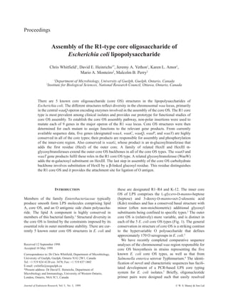
Fig.1.StructureofthecoreoligosaccharidesofE.coliandgeneticdeterminantsfortheassemblyoftheR1typecoreOS.(A)ShowsthestructureoftheR1coreOS,19
aswellastheligation
siteforOantigen(O-PS).4
TheGlcNattachedtoHepIIIhasbeenidentifiedasanon-stoichiometricsubstituentinstructuralstudies.19
Thenon-stoichiometricmodificationofKdoIIwith2-
aminoethylphosphateispredictedbycomparisonwithotherE.colicoreOSstructures.2
ThewaagenesencodingtheenzymescatalyzingeachtransferaregivenforresiduesintheR1coreOS
structure.(B)ShowsthevariationsinoutercoreOSstructureinE.coli.Amongtheseadditionalstructures,theO-PSattachmentsitehasonlybeenconfirmedfortheR2coreOStype22
andis
indicatedbytheasterisk.(C)ShowstheorganizationofthechromosomalwaaregionintheR1coretypeprototypestrain,E.coliF470.13,18
Filledarrowsindicategenesthatarevirtually
identicalinalloftheE.coliwaaloci.TheupstreampartsofthegmhDoperonthatterminateswithwaaChasnotbeensequencedintheR1coretypeandthepositionsofgmhDandwaaFare
onlypredicted,baseduponconservedsequencesinE.coliK-12andSalmonella.Inthoseorganisms,waaLoccupiestheterminalpositioninthegmhDoperon.
06JER19Whitfield 10/9/99 10:52 am Page 152
3. O157 only contain the R3 core OS type.5
It is currently
unclear whether the presence of a particular core type
(i.e. R1 or R3) confers any selective advantage to clini-
cal E. coli isolates in specific situations. A more likely
explanation is that this is simply a reflection of ancestral
R1 or R3 core type strains gaining additional DNA unre-
lated to LPS synthesis that allowed them to cause partic-
ular diseases.
Salmonella enterica serovar Typhimurium LT2 and E.
coli K-12 have historically provided paradigms for
genetic and biosynthetic studies on the assembly of LPSs
and lipooligosaccharides. However, there remain a num-
ber of unanswered questions in core OS assembly in these
bacteria, and direct experimental data are not yet available
to define the precise mechanism of action of many identi-
fied gene products. Comparative analyses of predicted
products encoded by the five known E. coli core OS
biosynthesis regions shed light on the role of some of
these gene products.4
Here we summarize our recent
analyses of the R1-type core OS assembly system, and the
assignment of function to each of the genes within the
central operon of the R1 waa cluster. This information
greatly enhances our understanding of the gene-structure
relationships in the assembly of the E. coli core OS mole-
cule. Moreover, given that R1 is the most prevalent core
type among clinical E. coli isolates, unraveling of the
genetics of the R1 assembly system provides an appropri-
ate and improved paradigm for core OS assembly.
Organization of the waa locus for LPS core OS
biosynthesis in E. coli
The chromosomal waa locus (formerly rfa) contains the
major gene operons for core OS assembly. The first waa
region to be sequenced in its entirety was that of E. coli
K-12. Although some functions have been biochemi-
cally characterized in E. coli K-12, the majority of func-
tional assignments have relied heavily on LPS structure
and LPS-specific phage-receptor data for Salmonella
mutants, and on heterologous complementation of those
mutations. The E. coli and Salmonella waa loci share a
common organization, with each consisting of 3 operons
(Fig. 1). In E. coli K-12, the locus maps to 81 minutes on
the linkage map.
For the purpose of discussion, the operons are identi-
fied by the first gene of the transcriptional unit. The
long, central waaQ operon contains genes necessary for
the assembly of the outer core OS and for modification
of core OS. Our analysis of the waaQ operon is the focus
of this paper. This operon contains some genes con-
served among E. coli and Salmonella and others that are
core OS type-specific. The waaQ operon is flanked by
two additional operons encoding conserved functions. In
E. coli K-12, the gmhD gene is the first of a block of four
genes (gmhD-waaFCL) which form an operon where the
first 3 genes are required for biosynthesis of the heptose
precursor and transfer of the first two heptosyl residues to
the nascent lipid A-core acceptor.1,4,9
With the exception
of the waaC gene, the sequence of the gmhD operon in the
R1 through R4 core OS types is unknown. The sequence
upstream of gmhD in E. coli K-12 indicates that the gmhD
operon is regulated by a heat shock promoter (reviewed
by Raetz et al1
). However, sequence diversity in the corre-
sponding upstream region in Salmonella suggests poten-
tial variations in regulation between different members of
the Enterobacteriaceae.10
The waaA transcript contains
the structural gene (waaA) for the bifunctional Kdo trans-
ferase,1
and a gene (designated orf18 or kdtB) encoding a
polypeptide of unknown function. The waaQ and waaA
operons are divergently transcribed and are separated by
400–500 bp of intervening DNA. This non-coding region
harbors the JUMPStart and ops sequences which co-oper-
ate with the RfaH protein to facilitate transcriptional
antitermination in the central waaQ operon.11,12
While the general organizational features of the core
OS biosynthesis region are conserved in E. coli, there
are differences in the gene content of the central operons
from E. coli strains representing the different core types.
One striking difference is that the clinically predominant
R1 (as well as the R4) core-type strains have their waaL
gene as the terminal gene of the waaQ operon. In con-
trast, this gene is the terminal gene of the gmhD operon
in the core OS biosynthesis regions of Salmonella and of
E. coli K-12, R2, and R3 core types.4,13
The waaL gene
product is required for attachment of various cell surface
polysaccharides (including O antigen) to lipid A-core
and is often simply referred to as ligase.14
It is currently
unclear whether there is any biological significance to
the different locations of waaL in operons that appear to
have differences in their regulation.
Inner core OS assembly
As might be predicted by the common structure of the
inner core OS in E. coli (Fig. 1), enzymes responsible for
its formation are highly conserved. In E. coli, the Kdo
transferase (WaaA) is bifunctional (or trifunctional),
adding two (or possibly three) Kdo residues.15
The
acceptor for the Kdo residues is the tetra-acyl lipid A
intermediate known as lipid IVA
.1
WaaA enzymes are
highly conserved, and the amino acid sequences of the 5
homologs from E. coli share greater than 96% identity.
Following addition of the final two acyl chains onto
Kdo2
-lipid IVA
, the heptose region is assembled. The
waaC gene product has been identified as the HepI
transferase16,17
and available evidence strongly supports
assignment of WaaF as the HepII transferase.17
Assembly of the R1-type core oligosaccharide of E. coli LPS 153
06JER19Whitfield 10/9/99 10:52 am Page 153
4. In E. coli, the heptose region of the core OS backbone
is modified by heptosyl and phosphoryl residues (Fig.
1). The gene products involved in these modifications
have only recently been resolved.18
By analyzing the
core OS structure of E. coli F470 (R1 core OS) mutants
carrying non-polar insertions in the relevant genes, we
were able to determine that: (i) waaP encodes a putative
kinase that phosphorylates HepI; (ii) the HepIII trans-
ferase is WaaQ; and (iii) waaY encodes an additional
kinase that catalyzes modification of HepII. Analysis of
the mutant phenotypes is complicated by the require-
ment that these modifications proceed in a specific order
(WaaP-Q-Y) with each step dependent on those preced-
ing it. Contrary to previous reports, WaaPQY-mediated
modifications are not essential for core completion,18
or
for addition of O polysaccharide (J. Yethon, D.E.
Heinrichs and C. Whitfield, unpublished results).
However, it is still unknown at what phase in the assem-
bly process these modifications occur in growing cells.
They may occur before completion of the remainder of
the core OS, or could occur once the core OS chain
extension is complete. An additional unresolved issue is
the identity of the enzyme(s) that contribute to the partial
substitution of HepI with either 2-aminoethyl diphos-
phate or phosphate (Fig. 1). WaaP could be involved in
transfer of either substituent or, alternatively, it may only
transfer phosphate groups with an additional unknown
enzyme catalyzing a subsequent transfer of phospho-
ethanolamine, completing the modification in a subset of
molecules.
The precise structure of the inner core OS plays a cru-
cial role in outer membrane stability in E. coli. Mutants
defective in waaP give rise to the phenotype known as
‘deep-rough’. Among the pleiotropic effects associated
with the ‘deep rough’ phenotype are: (i) an increase in
phospholipid:protein ratios and a depletion of outer
membrane porins; (ii) hypersensitivity to hydrophobic
compounds such as detergents, dyes, and some antibi-
otics; (iii) a ‘leaky’ outer membrane which releases con-
siderable amounts of periplasmic enzymes into the
medium; (iv) induction of colanic acid exopolysaccha-
ride synthesis; (v) reduced flagella expression; (vi)
secretion of an inactive form of secreted hemolysin; and
(vii) increased susceptibility to attack by lysosomal frac-
tions of polymorphonuclear leukocytes and to phago-
cytosis by macrophages (reviewed elsewhere1,4,9
). The
membrane perturbations resulting from ‘deep rough’
mutations are correlated with the absence of phosphory-
lation of HepI, as mutations in either waaQ or waaY
alone do not result in the ‘deep rough’ characteristics.18
While individual enzymes have been assigned to most
of the reactions in inner core OS assembly, there remain
some open questions. For example, in the R1 core OS
structure, HepIII is partially substituted with α(1→7)-
linked glucosamine.19
The corresponding gene for this
modification is not found within the sequenced chromoso-
mal waa gene region, indicating that, in the R1 system,
currently unmapped genes encode at least some core OS
biosynthesis enzymes. In addition, most, if not all, of the
core OS structures from E. coli and Salmonella carry 2-
aminoethyl phosphate residues on KdoII and a variable
amount of 2-aminoethyl diphosphate on HepI. The genes
and enzymology that underlie these substitutions are
currently under investigation.
Assembly of the R1 outer core OS
Our studies of the core OS assembly system from the E.
coli R1 core-type strain, F470, represent the only example
where precise, non-polar insertions have been constructed
in all of the genes that encode outer core glycosyltrans-
ferases. By determining the precise structure of the core
OS in each mutant, functional assignments have been
made for all of the glycosyltransferases (Fig. 1).13
The
core OS backbone is synthesized by the sequential
action of: (i) UDP-glucose:(heptosyl) lipopolysac-
charide α1,3-glucosyltransferase (WaaG; HexI addi-
tion); (ii) UDP-glucose:(glucosyl) lipopolysaccharide
α1,3-glucosyltransferase (WaaO; HexII addition); and
(iii) UDP-galactose:(glucosyl) lipopolysaccharide α1,2-
galactosyltransferase (WaaT; HexIII addition). The HexI
transferase, WaaG, is conserved among the different core
OS types in E. coli and Salmonella. The WaaO and WaaT
enzymes belong to a family of structurally related glyco-
syltransferases, known as the WaaIJ family. WaaIJ family
members share sequence motifs and some domains of
higher-order structure that are identified by hydrophobic
cluster analysis.13,20
In a glycosyltransferase classification
system developed by Breton et al.,21
the WaaT protein
would fall into family B. This is a family of galactosyl-
transferases that is made up of mostly bacterial, and few
eukaryotic, members. Although the WaaO, WaaT, and
WaaG enzymes all transfer a hexose from an UDP-hexose
precursor to LPS acceptors, WaaG is not a member of the
WaaIJ family.4
The α-galactosyl substituent on HexIII is added by the
WaaW enzyme, another member of the WaaIJ family of
glycosyltransferases.13
Perhaps surprisingly, the waaW
mutant core OS lacks substitution at both HexII and
HexIII. This is likely due to the fact that the terminal step
in R1 core OS assembly involves the addition of the β-
linked glucosyl residue at HexII by WaaV, and this step
requires the prior modification of HexIII catalyzed by the
WaaW transferase.13
These results could reflect the com-
plex structural requirements in the acceptor for individual
glycosyltransferases. Alternatively, essential protein:pro-
tein interactions in a putative assembly complex could be
lost due to the absence of a single component enzyme.
154 Whitfield
06JER19Whitfield 10/9/99 10:52 am Page 154
5. Ligation of O antigen to lipid A-core acceptor
Current data predict that the ligation reaction, which joins
newly synthesized O antigen to lipid A-core, occurs at the
periplasmic face of the plasma membrane (reviewed by
Whitfield et al.14
). The mechanism involved has not been
resolved, and the waaL gene product is the only enzyme
known to be required for ligation. The ligation-deficient
phenotype of a waaL mutant was first established in
Salmonella but has been confirmed in E. coli K-12,14
R1,13
and R222
core types. The ligase enzyme, or enzyme com-
plex, is essentially a glycosyltransferase with a complex
substrate requirement, but WaaL homologs contain no
features currently identified in known glycosyltrans-
ferases. This is perhaps expected, since their substrates are
undecaprenyl pyrophosphate-linked O antigens rather
than nucleotide diphosphosugars. The WaaL primary
sequences offer no obvious clues as to function and the 6
known WaaL proteins from E. coli and Salmonella collec-
tively share only low levels of similarity in their primary
sequences.4
However, their higher-order structures have
common features. All of the WaaL proteins are predicted
to be integral membrane proteins with 8 or more mem-
brane-spanning domains and their hydropathy profiles are
virtually identical. In contrast, sequence data for outer
core OS glycosyltransferases predict them to be periph-
eral membrane proteins. These features presumably
reflect differences in their substrate specificities.
Mutation of waaV (encoding the transferase which
adds the β-Glc side branch) eliminates ligation of O anti-
gen to type R1 lipid A-core.13
This and additional struc-
tural analysis identified the HexII (GlcII in E. coli R1)
side-branch substituent as the attachment site for O anti-
gen. This was quite unexpected as the only other systems
for which the attachment site had been determined
(Salmonella and E. coli R2) use the terminal (HexIII)
sugar of the core OS backbone as the ligation site. Such
differences in the acceptor for ligase activity begin to
explain the lack of primary sequence similarity among
the WaaL homologues. We are currently attempting to
resolve the structural requirements for ligation as well as
its mechanism of action.
Relationships between the waa regions of strains with
R1 and R4 core OSs
The R1 and R4 core OSs are identical with respect to
backbone structure and the α1,2-Gal substituent on
HexIII and, as would be expected, the relevant gene prod-
ucts all show > 95% identity. These two structures only
differ in the substituent at GlcII (Fig. 1). The R4 core type
lacks the β-glucosyltransferase (WaaV) but has a gene at
the same location within the waaQ operon that encodes a
β-galactosyltransferase (WaaX). This provides the only
molecular basis for the differences between the outer
core OS structure in types R1 and R4.13
The waaV and
waaX genes share little homology. Although the pre-
dicted WaaV and WaaX proteins show consensus
sequence features that are characteristic of β-glycosyl-
transferases, they are representatives of two distinct
enzyme families. When the sequence dissimilarities in
WaaV/X and the WaaL homologues are considered
together, acquisition and replacement (lateral gene trans-
fer) of a block of DNA that included waaV/waaX
together with waaL is the most likely explanation. A
change in ligase structure is expected given that the
attachment site residue must differ in the R1 and R4 core
OS structures.
CONCLUSIONS
With the assignment of function to the genes whose
products assemble an R1 type core OS molecule, many
of the ambiguities of core OS assembly have been
resolved. Comparing and contrasting homologous genes
(and predicted gene products) within each of the core OS
biosynthesis regions, along with the known chemical
structures of the core OS molecules resulting from the
activities of these enzymes, allows tentative assignment
of function to many other genes.4
This information can
be used to direct efforts towards definitively identifying
function by direct biochemical assays. Moreover, with
the identification of strictly conserved amino acid
residues within the growing list of known glycosyltrans-
ferase sequences, and through the use of site-directed
mutagenesis, the contribution of these critical residues to
the mechanism of action of glycosyltransferase enzymes
can be specifically addressed.
ACKNOWLEDGEMENTS
This work was supported by funding from the Natural
Sciences and Engineering Research Council (NSERC)
and the Canadian Bacterial Diseases Network (NCE pro-
gram) awarded to CW. DEH gratefully acknowledges
receipt of postdoctoral fellowships from NSERC and the
Medical Research Council. JAY is the recipient of a
postgraduate scholarship from NSERC and the Medical
Research Council of Canada.
Note added in proof:
Recent data indicate that the Orf18 gene encodes a phos-
phopantetheine adenyltransferase enzyme required for
coenzyme A biosynthesis (Izard T, Geerlof A. EMBO J
1999; 18: 2021–2030). The Orf18 enzyme is therefore
not involved directly in LPS biosynthesis as originally
thought.
Assembly of the R1-type core oligosaccharide of E. coli LPS 155
06JER19Whitfield 10/9/99 10:52 am Page 155
6. REFERENCES
1. Raetz CRH. Bacterial lipopolysaccharides: a remarkable family
of bioactive macroamphiphiles. In: Niedhardt FC, ed.
Escherichia coli and Salmonella. Cellular and Molecular
Biology. Washington, DC: American Society for Microbiology.
1996; 1035–1063.
2. Holst O, Brade H. Chemical structure of the core region of
lipopolysaccharides. In: Morrison DC, Ryan JL, eds. Bacterial
Endotoxic Lipopolysaccharides. Boca Raton: CRC Press, 1992;
171–205.
3. Hull S. Escherichia coli lipopolysaccharide in pathogenesis and
virulence. In: Sussman M, ed. Escherichia coli: Mechanisms of
Virulence. Cambridge, UK: Cambridge University Press, 1997;
145–167.
4. Heinrichs DE, Yethon JA, Whitfield C. Molecular basis for
structural diversity in the core regions of the lipopolysaccharides
of Escherichia coli and Salmonella enterica. Mol Microbiol
1998; 30: 221–232.
5. Amor K, Heinrichs DE, Johnson R, Ziebell K, Whitfield C. A
PCR-based system to determine the core oligosaccharide type in
Escherichia coli: application to the ECOR collection and VTEC
isolates. 99th Annual Meeting of the American Society for
Microbiology. Chicago, Illinois. 1999; Abstract #B/D41.
6. Ochman H, Selander RK. Standard reference strains of
Escherichia coli from natural populations. J Bacteriol 1984; 157:
690–693.
7. Appelmelk BJ, An Y-Q, Hekker TAM, Thijs LG, MacLaren DM,
deGraaf J. Frequencies of lipopolysaccharide core types in
Escherichia coli strains from bacteraemic patients. Microbiology
1994; 140: 1119–1124.
8. Gibb AR, Barclay GR, Poxton IR, di Padova F. Frequencies of
lipopolysaccharide core types among clinical isolates of
Escherichia coli defined with monoclonal antibodies. J Infect Dis
1992; 166: 1051–1057.
9. Schnaitman CA, Klena JD. Genetics of lipopolysaccharide
biosynthesis in enteric bacteria. Microbiol Rev 1993; 57: 655–682.
10. Sirisena DM, MacLachlan PR, Liu S-L, Hessel A, Sanderson KE.
Molecular analysis of the rfaD gene, for heptose synthesis, and
the rfaF gene, for heptose transfer, in lipopolysaccharide
synthesis in Salmonella typhimurium. J Bacteriol 1994; 176:
2379–2385.
11. Bailey MJ, Hughes C, Koronakis V. RfaH and the ops element:
components of a novel system controlling bacterial transcription
elongation. Mol Microbiol 1997; 26: 845–851.
12. Marolda CL, Valvano MA. Promoter region of the Escherichia
coli O7-specific lipopolysaccharide gene cluster: structural and
functional characterization of an upstream untranslated mRNA
sequence. J Bacteriol 1998; 180: 3070–3079.
13. Heinrichs DE, Yethon JA, Amor PA, Whitfield C. The assembly
system for the outer core portion of R1 and R4-type
lipopolysaccharides of Escherichia coli. The R1 core-specific β-
glucosyltransferase provides a novel attachment site for O
polysaccharides. J Biol Chem 1998; 273: 29497–29505.
14. Whitfield C, Amor PA, Köplin R. Modulation of surface
architecture of Gram-negative bacteria by the action of surface
polymer:lipid A-core ligase and by determinants of polymer
chain length. Mol Microbiol 1997; 23: 629–638.
15. Belunis CJ, Raetz CR. Biosynthesis of endotoxins. Purification
and catalytic properties of 3-deoxy-D-manno-octulosonic acid
transferase from Escherichia coli. J Biol Chem 1992; 267:
9988–9997.
16. Kadrmas JL, Raetz CR. Enzymatic synthesis of
lipopolysaccharide in Escherichia coli. Purification and
properties of heptosyltransferase i. J Biol Chem 1998; 273:
2799–2807.
17. Sirisena DM, Brozek KA, MacLachlan PR, Sanderson KE, Raetz
CR. The rfaC gene of Salmonella typhimurium. Cloning,
sequencing, and enzymatic function in heptose transfer to
lipopolysaccharide. J Biol Chem 1992; 267: 18874–18884.
18. Yethon JA, Heinrichs DE, Monteiro MA, Perry MB, Whitfield C.
Involvement of waaY, waaQ, and waaP in the modification of
Escherichia coli lipopolysaccharide, and their role in the
formation of a stable outer membrane. J Biol Chem 1998; 273:
26310–26316.
19. Vinogradov EV, van der Drift K, Thomas-Oates JE, Meshkov S,
Brade H, Holst O. The structures of the carbohydrate backbones
of the lipopolysaccharides from Escherichia coli rough mutants
F470 (R1 core type) and F576 (R2 core type). Eur J Biochem
1999; Submitted.
20. Shibayama K, Ohsuka S, Tanaka T, Arakawa Y, Ohta M.
Conserved structural regions involved in the catalytic mechanism
of Escherichia coli K-12 WaaO (RfaI). J Bacteriol 1998; 180:
5313–5318.
21. Breton C, Bettler E, Joziasse DH, Geremia RA, Imberty A.
Sequence-function relationships of prokaryotic and eukaryotic
galactosyltransferases. J Biochem (Tokyo) 1998; 123: 1000–1009.
22. Heinrichs DE, Monteiro MA, Perry MB, Whitfield C. The
assembly system for the lipopolysaccharide R2 core-type of
Escherichia coli is a hybrid of those found in Escherichia coli K-
12 and Salmonella enterica. Structure and function of the R2
WaaK and WaaL homologs. J Biol Chem 1998; 273: 8849–8859.
156 Whitfield
06JER19Whitfield 10/9/99 10:52 am Page 156