SlideShare une entreprise Scribd logo
1  sur  40
Télécharger pour lire hors ligne
Abstract
Tauopathies are diseases related to abnormal filamentous inclusions of microtubule-
associated protein tau commonly found across a spectrum of neurodegenerative disorders.
The dissociation of monomeric tau protein from microtubule bindings sites results in the
formation of aggregates with a high affinity for self-assembly. Aggregates propagate
interneuronally in a prion-like fashion, resulting in widespread neuronal damage, cognitive
decline and cell death. tau’s co-association with amyloid-beta in Alzheimer’s diseases
makes it a potential target for therapeutic intervention. More recently, immunotherapy in
passive and active forms has been shown to ameliorate amyloid load and substantially
rescue cognitive impairment. Antibody-mediated tau aggregation and clearance of tau
aggregates has been described in previous studies though none have evaluated the
effectiveness of antibodies at substochiometric concentrations sufficient to simulate the
exclusivity of the blood brain barrier. We found that substochoichiometric concentrations of
antibody binding to the C-terminus of tau exacerbated aggregation as measured by ThT
fluorescence. Fibril seeds formed in the presence of antibody decreased seeding efficiency
relative to non-antibody treated seeds, and end products of antibody treated seeds were
substantially more structured relative to their unseeded and normally seeded counterparts,
suggesting the potential formation of novel “strains”. Antibody in the presence of normal
seeds and monomeric tau lowered seeding efficiency in a concentration dependent
manner, indicating a therapeutically optimal concentration of antibody adequate enough to
decrease deplete seeding efficiency without overtly intensifying monomeric aggregation.
Relevant Vocabulary
Fibrils/Neurofibrillary Tangles/NFT Pathogenic clumps of tau protein
Aggregation The formation of fibrils from tau
Lag Phase
In-vitro: The time difference
between the beginning of an
aggregation assay and the onset of
elongation (fibril formation).
Elongation Rate
In-vitro: ThT fluorescence increase
per unit of time. Corresponds to
fibrils formed to unit of time.
Seeding The use of pre-formed fibrils as
templates to spur aggregation.
Fluorescence Emission of light
Circular Dichroism
The difference in the absorption of
left-circularly polarized light and
circularly right-polarized light that
occurs when sample contains chiral
centers (carbon with four different
groups attached to each of its four
bonds). This difference can help
determine secondary structure of
the protein and in other cases show
different structural states under
varying conditions.
Epitope
A region where an antibody binds.
Amyloid/Amyloidogenic
Capable of forming aggregates.
Disordered Protein
Protein lacking specific structure.
Alzheimer’s Disease
Alzheimer’s disease is characterized by progressive neurocognitive decline associated with
widespread propagation of amyloid-beta and tau protein fibrils. Early stages are
asymptomatic though the onset of cognitive debility and subsequent dementia emerges
with the prion-like propagation of amyloid deposits and tau neurofibrillary tangles, resulting
in pervasive neuronal death and white matter atrophy.
The Biochemical foundations of Alzheimer’s Disease
Multiple theories have been established to explain the physiological cascade involved in the
onset of Alzheimer’s disease. Oldest among these theories is the cholinergic hypothesis,
which arose during a particularly research-intensive era in the field of neurochemistry and
anatomy [1]. Findings from this two-decade period from the mid-1960s to the mid-1980s
established a foundation upon which the molecular basis of neurodegenerative diseases
could be closely examined. Chief among these neurophysiological mediators are
cholinergic receptors, which play an important role in a wide spectrum of homeostatic
functions. Consequently, the manifold nature of these receptors gives way to a broad
range of neurological disease states upon their dysfunction [2.3.4], including those found in
Alzheimer’s disease [5]. Amyloid beta deposits have been found to form extracellular
amyloid clumps known as plaques, leading to nueromodulating effects that can occur at
picomolar concentrations, irrespective of the neurotoxic state of amyloid beta [6]. The role
of acetylcholine in memory recall was demonstrated by the use of receptor antagonists in
monkeys and rats. Subjects receiving infusions in the perirhinal cortex showed marked
decline in the ability to recognize stimuli [7.8]. Subsequent studies demonstrated that
various degrees of cognitive impairment arise from region-specific application of receptor
antagonists [9,10]. Post-mortem examinations of AD brains revealed depleted levels of
cholinergic activity, particularly choline acetyltransferase, a transferase responsible for
acetylcholine synthesis, and acetylcholinesterase, a hydrolase that breaks down
acetylcholine in the neuromuscular junction and neural synapses, in the cerebral cortex
[11]. In Alzheimer’s patients, frontal and temporal regions of the brain responsible for
memory and cognition were especially depleted with respect to cholinergic receptors
[12.13].
Much of the criticism levied against this hypothesis stems from confounding factors that
show a natural decline of cholinergic activity in healthy rat brains [14, 15], as well as a
broad spectrum of neurodegenerative disease [16]. These revelations point to the more
general phenomenon of cholinergic decline as a symptom, rather than impetus, of
neurodegeneration.
The pivotal role of amyloid-beta in the progression of Alzheimer’s disease pins the peptide
as the central tenet of the amyloid cascade hypothesis. Upon observation of Auguste
Deter’s brain (who would later become the first patient to be formally diagnosed with
Alzheimer’s disease) Alois Alzheimer, the physician credited with the first published clinical
observation of AD dementia, noted “numerous small miliary foci are found in the superior
layers…[that] are determined by the storage of a peculiar material in the cortex”. Indeed,
Alzheimer would go on to conflate these plaques with “the most serious form of dementia”,
adding that “the plaques were excessively numerous and almost one-third of the [patient’s]
cortical cells had died off” [17]. These extracellular plaques would eventually come to be
known as abnormal accumulations of peptide amyloid-beta. The description of amyloid-
beta pathology as a “cascade” implies its central role as a vanguard of AD progression,
postulating the formation of amyloid plaques as the prerequisites for neurofibrillary tangle
formations of tau protein. Amyloid-beta’s precursor, amyloid precursor protein (APP), is a
transmembrane protein which has been found to influence synaptogenesis and, most
recently, protein synthesis in dividing human cells [18] among other processes. Its
abundance in interneuronal ER and Golgi [19] membranes contributes to its involvement in
AD pathogenesis, whereby the sequentially cleavage of APP by either α or β (BACE-1) and
γ-secretase enzymes, respectively, produces plaque forming and non-plaque forming
variants of free-floating amyloid-beta peptide in the neuronal interstitium. In the event of
primary cleavage by α-secretase, soluble APP (sAPPα) is secreted, leaving behind a 83-
residue membrane-bound fragment (CTFα) [20]. Conversely, initial cleavage with β-
secretase produces a 99 amino-acid transmembrane peptide (CTFβ). In both instances,
the membrane bound peptides are next cleaved by γ-secretase to yield amyloid-beta from
CTFβ and a small protein (P3) from CTFα. The amyloidogenic potential of cleavage
products is determined by the location of γ-secretase proteolysis; in the event of cleavage
of amyloid-beta valine-40, Ab-40, a 40 amino-acid variant, is secreted. In the event of
cleavage at alanine-42, ab 42, the 42 amino-acid variant, is secreted. While Ab-40 has
been determined to be a natural component of cerebrospinal fluid and plasma [21], even
potentially possessing neuroprotective properties, it’s counterpart, Ab-42 has been
implicated as the pathological trigger of plaque formation [22].
In a post-mortem examination of AD patients, Alois Alzheimer’s had also described
“peculiar, deeply stained bundles of neurofibrils” colocalized with dead cortical cells.
Unbeknownst to him, he was describing one of the two neuropathological findings
consistent with Alzheimer’s disease — tau neurofibrillary tangles. Distinguished in its
ubiquity across a spectrum of neurodegenerative disorders, tauopathies are not a unique to
Alzheimer’s disease, however tau fibrillation subsequent to amyloidosis is a hallmark sign.
As a major microtubule stabilizing protein in the central nervous system, tau maintains
cytoskeletal stability through polymerizing and depolymerization of tubulin subunits [23].
It’s affinity for tubulin is modulated by kinases and phosphatases [24]. In the event of
hyperphosphorylation, tau dissociates from its cytoskeletal origin in the form of free-floating
tau monomers. Consequently, these monomers self-assemble to form oligomeric
structures which serve as scaffolds for the development of larger, pathogenic neurofibrillary
tangles capable of propagating interneuronally, whereupon exogenous tau fibrils can
induce tauopathies in neighboring cells in a prion-like manner known as seeding [25].
The duality of amyloid plaques and tau fibrils in the pathophysiology of Alzheimer’s disease
lend credence to two of the later aforementioned theories. AD-associated tauopathies can
seldom form without the presence of amyloid plaques [26], however extracellular amyloid
deposition is not sufficient to elicit neurodegeneration [27,28]. The tau hypothesis is
therefore the most concise understanding of the biochemical underpinnings of Alzheimer’s
disease [29]. The molecular intersect between the two processes remains unclear,
however recent studies have shown that oxidative stress stemming from the presence of
toxic amyloid-beta upregulates a regulator (RCAN1) of calcineurine, a phosphatase of tau,
and glycogen-synthase kinase-3β (GSK3β), a tau kinase. Concomitantly, the imbalance
between an increase in phosphorylation activity and a decrease in dephosphorylation of tau
results in the formation of tau fibrils, thus providing a coherent link between amyloidosis
and fibrillation [30]. This link implies that the mitochondria invariably plays a part in AD
etiology, giving birth to a relatively novel theory in which the cellular powerhouse forms the
crux of the disease. The mitochondrial cascade hypothesis posits the formation of amyloid
plaques on the genetic resiliency of the mitochondrial electron transport chain. Over time,
the propensity of the mitochondria to regulate damage via reactive oxygen intermediates,
along with its ability to generate ATP via oxidative phosphorylation, declines [31]. Age-
related physiological changes in mitochondrial function result in compensatory responses,
among them the secretion of amyloid-beta. Indeed, studies have found an association
between mitochondrial amyloid-beta levels and the degree of cognitive impairment in
transgenic mice [32]. Moreover, rat neurons treated with electron transport chain inhibitors
have been found to enhance tau pathology [33,34] while cytochrome oxidase inhibitors,
which function to impede the reduction potential of the final link in the ETC, cause
substantial alterations in the cleavage of APP towards its toxic amyloid-beta descendant
[35]. This theory helps bridge the discrepancy between genetics and sporadic onset of
Alzheimer’s disease otherwise not explained by allelic variants that induce amyloidosis.
Microtubule Associated Protein tau
Microtubule associated protein tau is a seminal component in the maintenance of structural
integrity of neurons. Located on the 17th
chromosome, tau transcripts in the central
nervous system are composed of 16 exons, three of which (2,3, and 10) are alternatively
spliced to produce six potential isoforms expressed differentially throughout development,
with exon 1 serving as an untranslated transcriptional prom. These isoforms are
characterized by the presence of three or four repeat tubulin binding regions at the C-
terminus and the presence, or lack thereof, of additional inserts at the N-terminus. The
presence and absence of exon 10 in the modified tau transcript gives rise to four and three
repeat regions, respectively. Irrespective of the presence of exon 10, the repeat regions
3R (R1-R3) or 4R (R1-R4) are also encoded by exons 9,11, and 12 [36]. The largest of
these isoforms contains exon 4A (an intermediate region between exon 4 and 5) and is
unique in its localization to regions of the peripheral nervous system such as the spinal
cord and the retina.
Exon Size Isoform Repeat Domain
2,3,10 441 Adult 4 repeat
2,3 410 Adult 3 repeat
2,10 412 Adult 4 repeat
2 381 Adult 3 repeat
10 383 Adult 4 repeat
- 352 Fetal -
The importance of the N-terminus as a projection domain is maintained by a highly acidic
character capable of interacting with cellular components such as the plasma membrane
[37], mitochondria and serving as a key intermediate in the maintenance of structural
rigidity [38], axonal growth [39] and diameter [40]. Conversely, the C-terminus is
characterized as a positively charged, basic region connected to the N-terminus via a
proline-rich mediator [40]. This region is directly bound to cytoskeletal tubulin and
facilitates polymerization events conducive to cytoskeletal alterations (cite). It is important
to note that while 4R and 3R variants of tau bind microtubules, additional repeat regions
have been shown to enhance binding affinity while simultaneously contributing to
nucleation rates among dissociated tau [40].
Post-Translational Modification of tau
Post-translational modifications of tau have been proposed as key drivers of Alzheimer’s
pathology, among them glycosylation [41], acetylation [42] and phosphorylation [43]. N-
linked glycosylation (attachment of the oligosaccharide to the amide nitrogen of the
asparagine or arginine residue of a protein), targeting asparagine or arginine residues of
the tau, was found in non-hyperphosphorylated tau from Alzheimer diseased brain but not
in normal brain samples. Similarly, hyperphosphorylated neurofibrillary tangles and paired
helical filaments were found to be extensively glycosylated relative to microtubule
associated tau [44]. Moreover, the N-glycosylated tau functioned as a better substrate for
cAMP-dependent protein kinases compared to its deglycosylated counterpart [45].
Conversely, O-glycosylation (the reaction of a carbohydrate to a hydroxyl moiety), targeting
serine, threonine or tyrosine, was found to be inversely related to the level of tau
phosphorylation [41].
Acetylation (the attachment of an acetyl group to an amino moiety) of lysine residues
impairs the microtubule binding affinity of tau. Indeed, acetylation of Lys280 was only
found in hyperphosphorylated AD fibrils of mouse brain lysates [42], and deletion of SIRT1,
a protein deacetylase, intensified levels of phospho-tau [46].
The hyperphosphorylation of tau protein is a common factor among all aforementioned
scenarios [43]. As such, the phosphorylation state of tau has thus far been the main
determinant of tau pathology and the balance between kinase/phosphatase activity takes
center stage. Full length tau (441 aa) has been found to have a total of 80
serine/threonine, along with 5 threonine phosphorylation sites [47], each corresponding to
various severities of cytopathology in Alzheimer’s disease [48]. Most of these
phosphorylation sites lie in the proline-rich region connecting the projecting N-terminus with
the microtubule binding C-terminal region [40]. Similarly, tau serves as an intermediary
between phosphatases, enzymes that dephosphorylate targeted substrates, and
microtubule stability [49].
Structural and Mechanistic Features of tau Fibril Formation
Dissociation of protein tau from microtubule binding sites is the neuropathogenic foundation
of tauopathy in Alzheimer’s disease. Subsequent to detachment, monomeric tau assumes
an unstructured configuration, which can be attributed to its positive charge low
hydrophobic character at physiological pH levels and [50]. The lack of hydrophobic
residues precludes sufficient hydrophobic forces to sustain a secondary structure, and
phosphorylation events contribute to a chance in electrostatic character, disassociation and
self-assmbely [51]. These amyloid regions, narrowed down to hexapeptide sequences
275
(VQIINK)280
and 306
(VQIVYK)311
are sufficient for the growth and propagation of tau fibrils,
among other amyloid derivatives [52,53]. While a significant portion of tau retains its
random-coil structure even within fibrils, constituent regions of the amyloid core retain the
beta-sheet rich motifs remain [53]. This is also demonstrated by the aggregation of tau in
the presence of anionic compounds such as heparin [54] and arachidonic acid [55].
Spectroscopic studies using FRET and hydrogen/deuterium mass spec examinations have
proposed an ‘S’ shaped model for monomeric tau, whereby contact is maintained between
the N-terminus and the proline-rich region and the C-terminus and amylodigenic regions of
tau [56]. Interactions between tau hydrophobic regions or polyanionic substances results
in a conformational change from unstructured random-coils to beta-sheets, a pervasive
feature of amyloids [52].
tau monomer interactions result in the formation of parallel “stacks” of tau beta-strands
connected via intermolecular hydrogen bonds, similar to structures of amyloid-beta [57]
and alpha-synuclein deposits [58] in Parkinson’s Disease. Outer regions of tau filaments
exhibit exposed hydrogen bond donors and acceptors [59], features that promote further
aggregation and are absent in natural beta-sheet proteins to avoid aggregation [60]. In
this way, tau dimers are able to attract proximal monomers and grow in an unimpeded
stacking fashion.
Seeding and Intercellular Propagation of Tau
The presence of preformed tau aggregates potentiates fibrillation of endogenous tau by
enhancing recruitment of dissociated monomers and oligomers [61]. This facet of
tauopathies allows tau fibrils to propagate in a pathogenic, prion-like fashion whereby
exogenous fibrils or oligomers serve as “seeds”, or molecular scaffolds, for monomeric tau
in adjacent cells. Indeed, transgenic mice expressing P301L human mutant tau localized to
the entorhinal cortex demonstrated hierarchical propagation of fibrils to adjacent regions
[62]. Cultured cell experiments demonstrate cellular ability to uptake tau oligomers, but not
monomers, via endocytosis [63]. This seeding potential is determined by its structural
conformation. In these instances, deletion of motifs (275)VQIINK(280) and
(306)VQIVYK(311) eliminates the capacity of full-length tau to seed [64]. Currently, there
are two potential models to explain seeding: the oligomer-nucleated conformation induction
and template-assisted growth [65]. The major difference between these two models is the
structural component(s) of tau that influence fibril formation. Oligomer-nucleated
conformational induction establishes a high-energy scaffolds which attracts monomeric tau
that binds in succession to lower energy and form oligomers [66]. Unlike the template-
assisted growth model, fibrils do not integrate dissociated monomers, but are rather formed
only after the formation of oligomers [67]. Dimeric, trimeric and oligomeric intermediates
between monomer and fibril formation have been established in aggregation studies
involving other fibrillation prone agents [68] and AD peptide amyloid-beta [69]. Toxicology
comparisons between neurofibrillary tangles and tau oligomers injected into mouse brains
found that oligomer-infused brains showed diffuse tau pathology into neighboring brain
regions, whereas NFT-treated cells displayed localized deposits, implicating oligomers as
the component most responsible for intercellular tauopathies [70].
Braak Staging and the Prion-like Propagation of tau
The entorhinal region receives input from the neocortex and is involved in higher cognitive
functions and the limbic system, as well as in the formation of memories and emotions.
Intracellular tau deposits first appear in an area adjacent to the entorhinal region called the
transentorhinal region, which functions as a relay between the neocortex and the entorhinal
region. The manner of neurofibrillary tangle propagation is closely associated with the
degree of cognitive decline [71]. The limbic stages consist of minimal NFT presence in the
neocortex, with the fibrils concentration localized to the entorhinal and transentorhinal
regions, concomitant with noticeable cognitive impairment. End stage AD presents with
widespread damage to the neocortical areas, resulting in extensive cognitive impairment
and advanced dementia.
Figure 1:
The Blood-Brain Barrier: An Obstinate Foe
One of the major obstacles to immunotherapy against neurodegenerative disorders is the
human blood brain barrier (BBB), a restrictive vasculature of endothelial cells exhibiting
high electrical resistance. In healthy individuals, the BBB functions as a selective
safeguard against potential antigens and neurotoxins, impeding the entry of large or
hydrophilic molecules, while facilitating transport of metabolically essential nutrients and
molecules. The innate bulkiness of immunoglobulins poses a major obstacle to developing
effective therapeutic measures for combating neurodegenerative disorders. Indeed,
radioimmunoassays have found that approximately 0.1% of circulating IgGs, the most
common of the 5 immunoglobulin classes (A,G,M,E, and D), can be detected in the central
nervous system [72]. However, the efficacy of the BBB can be severely compromised
during neurological disorders such as multiple sclerosis, viral meningitis and tumors [73].
Inflammatory events in AD have also contributed to increased BBB permeability and the
pathological spread of amyloid plaques. Given the rapid turnover of cerebrospinal fluid into
the bloodstream, intrathecal injections directly into the CSF are equivalent to prolonged
intravenous injections, amounting to limited therapeutic efficacy [74]. Moreover, a
logarithmic decrease in drug distribution throughout the brain has been shown in bulk-flow
delivery of drugs directly into brain tissue [75]. As such, antibody delivery for neuro-
immune therapy is a popular research topic. Three major approaches to this problem
include the application of lipid-mediator molecules, which can passively diffuse through the
BBB, carrier mediated transport (CMT) of small water-soluble molecules, and the
exploitation of receptor-mediated transport (RMT) . Theranostics, the use of molecular
platforms for drug delivery and diagnostics, relies on lipid or water-based carriers to
transport antibodies across the membrane [76]. These platforms have been used in the
delivery of AB-antibody fragments via synthetic liposomal elements such as polyethylene
glycol polymer chains (PEG) [77]. Advances in RMT take advantage of metabolic
receptors mediating BBB access to transport bound antibodies into the brain parenchyma.
Insulin and transferrin receptor ligand-bound AB-antibodies have been shown to effectively
cross the BBB through receptor-mediated transcytosis, enhancing brain exposure 55-fold in
some instances [78,79].
Origins of Immunotherapy in Alzheimer’s Disease
As one of the hallmark pathologies of Alzheimer’s disease, aggregates of amyloid beta
have been one of the primary immune targets of AD therapies for quite some time. Mice
immunizedx with Aβ1-42, an alloform associated with toxic oligomers, showed reduced
plaque burdens and retained cognitive functions relative to their non-immunized
counterparts [80]. Subsequent human trials were halted after a subset of patients
developed encephalitis post-immunization, likely due to the extensive activation of CD8+
cytotoxicity [81,82]. Nevertheless, post-mortem autopsies indicated clearance of amyloid
with retention of tau pathology [83]. In another study, the clearance of extracellular
amyloid plaques was accompanied by the reduction of early tau pathology but retention of
hyperphosphorylated neurofibrillary tangles [84,85]. Conversely, tau antibody treatment did
not affect amyloid load, indicating that amyloid deposits serve as a precursor to tauopathy,
though analysis of cognitively normal individuals has shown tangle formation in the
temporal lobe without the presence of amyloid plaques.
Passive and active immunization of targeting tau fibrils has also become a mainstay in AD
immune-therapy. Studies exhibiting clearance after antibody treatment were either
targeted at tau phosphoepitopes or fibril specific conformations. In these cases,
phosphorylation of tau was reduced and fibril load significantly decreased [86] establishing
a correlation betweentau antibody titer count, fibril load and cognitive performance [87]. In
other cases, passive immunization of phosphorylated tau was found to significantly
decreased NFT burden while increasing microglial activity [88].
Mechanism of Antibody Mediated tau Therapy
Although the efficacy of tau antibodies against pathogenic aggregates has been well
documented, the mechanism by which this phenomenon occurs is obscure. Chief among
several theories is that antibodies directly inhibit the fibrillation or even work to reverse the
process altogether [89]. This theory is corroborated by the clearance amyloid-beta
aggregates in in-vitro studies. Indeed, studies have found that, similar to their amyloid-beta
counterparts, tau antibodies cross the neuronal membranes via clathrin-mediated
endocytosis and co-localize with intercellular fibrils [90]. Additionally, antibodies have been
found to interfere with the prion-like interneuronal propagation of tau by directly interacting
with extracellular tau seeds [91].
Due to the neuroinflammatory nature of tauopathies, microglial clearance has been found
to be a major form of fibril clearance [92]. However, mouse models studies for anti-
amyloid-beta antibodies have also shown that clearance can occur in a non-Fc-mediated
fashion with the use of antibodies lacking fragment crystallizable regions essential for the
interaction of immune system components, such as microglia, with pathogens [93].
Molecular Dynamics of Thioflavin T Binding
Thioflavin T, a benzothiazole dye, has been employed in numerous studies to detect and
analyze the extent of fibrilation in aggregation-prone proteins. When bound to fibrous
structures, the ThT maximum fluorescence wavelength increases substantially, producing a
discernible signal unique to amyloid fibrils. Immobilization of the benzylamine and
benzathiole rings on the molecule, producing a perpetually excited state that corresponds
to a manifold increase in fluorescence [94].
Methods
Protein Aggregation Assay
In vitro aggregation of tau protein was performed via 0.1 ml triplicates in a 96 well-plate in
the presence of 5 µM ThT. Each well was outfitted with 2 polyethylene balls (2.38-mm
diameter, Precision Ball, Reno, PA), the plate covered with a Mylar septum sheet (Thermo)
and shaken at 280 rpm in an Infinite M200 PRO Microplate Reader (Tecan, Austria).
Kinetic measurements were taken in 5 minute intervals using a 444-nm excitation and 485-
nm emission filter. Monomeric tau (15 µM, 0.6 mg/ml) was incubated in the presence of 5
µM heparin with the addition of 10 mM Hepes (pH 7.5), 1 mM EDTA, 100 mM NaCl, and 5
mM DTT in a 320 µL eppendorf tube and distributed in 0.1 mL aliquots into 3 triplicates.
Antibody concentrations varied from 5 nM – 125 nM.
Seeding assays were run in similar fashion with the addition of 0.06-0.5% of either non-
antibody treated (normal) tau fibril seeds or tau fibril seeds formed in the presence of 20 or
125 nM of antibody (abnormal).
Curve Fitting
The four parameter sigmoid curve was used to fit fluorescence data using Sigma Plot
software. Data was plotted in accordance with the formula
where Yo is the initial level of ThT fluorescence, a is the difference between the final and
initial level of ThT fluorescence, Xo is the midpoint of transition and 1/b is the rate constant
for fibril formation. The lag-time of fibril formation was determined by the formula Xo-2b and
the initiation rate was determined by the inverse of lag time.
[118]
Figure 2: Kinetic model of protein aggregation: the nucleation phase (lag phase) results in
minimal fluorescence until the concentration of oligomers reaches a critical mass resulting
in an exponential growth in fibrils (elongation phase). Incubation with seeds tends to
increase decrease lag phase and increase the rate at which fibrils are grown
Circular Dichroism
Far-UV CD spectra were measured using JASCO J-820 spectropolarimeter at 25 C. The
aforementioned solution of aggregated tau (15 µM [0.6 mg/ml]) was placed into a 0.2-mm
pathlength cell. Five spectra were averaged for each sample and acquired at high
sensitivity with 20 nm/min scan speed at 0.2 nm step size/1.0 nm bandwidth under constant
purging with nitrogen.
Determination of Stability
Aggregated tau samples (8 µl) were suspended in Hepes buffer (10 mM, pH 7.5) containing
various concentrations of guanidine thiocyanate (0.2-6.0M) in a total volume of 20 µl,
incubated for 1 hour at room temperature and then diluted to 300 µl with the concentration
of guanidinium thiocyanate adjusted to 0.2 M in the presence of 10 µM ThT. ThT
fluorescence was measured. An excitation wavelength of 442 nm and an emission
spectrum range of 470-500 nm was used. Excitation and emission slit values were 2.5 nm
and 5 nm, respectively. All measurements were performed in triplicate and averaged for
each sample.
Electron Microscopy
Five microliter volumes of post-aggregation protein solution was adsorbed onto prewashed
200-mesh formvar/carbon-coated nickel grids for 5 minutes. The grid was washed with 10
µl water, stained with 10 µl of 2% uranyl acetate for 2 minutes and washed with water prior
to drying at room temperature. Samples were analyzed with JEM-1400 transmission
electron microscope (JEOL) operated at 80 kV.
Results
Characterization of tau
Competent BL21(DE3) E.coli cells transformed with histone-tagged 0N4R four-repeat tau
(383 aa) cDNA were harvested for protein. Histone-modified tau was purified via nickel
column and dialyzed with TEV protease. Further purification was conducted using fast
protein liquid chromatography
Figure 3: tau after protease cleavage (3rd
row from left) shows bottom band at the 17 kD
mark, corresponding to his-tag cleavage and 55 kD mark corresponding to tau (40-60 kD).
Experimentation (I):
15 µM tau was incubated in 100 µl aliquots with various concentrations (5-125 nM) of
antibody 181, 231, 396, A10 and tau 13 in the presence of heparin sulfate. The samples
were placed in a plate reader with 5 µM ThT and shaken at 37 C degrees. Lag phase and
elongation rate were derived from fitting fluorescence data to a 4-parameter sigmoid curve.
Epitope Mapping
10 20 30 40 50
MAEPRQEFEV MEDHAGTYGL GDRKDQGGYT MHQDQEGDTD AGLKAEEAGI
60 70 80 90 100
GDTPSLEDEA AGHVTQARMV SKSKDGTGSD DKKAKGADGK TKIATPRGAA
110 120 130 140 150
PPGQKGQANA TRIPAKTPPA PKTPPSSGEP PKSGDRSGYS SPGSPGTPGS
160 170 180 190 200
RSRTPSLPTP PTREPKKVAV VRTPPKSPSS AKSRLQTAPV PMPDLKNVKS
210 220 230 240 250
KIGSTENLKH QPGGGKVQII NKKLDLSNVQ SKCGSKDNIK HVPGGGSVQI
260 270 280 290 300
VYKPVDLSKV TSKCGSLGNI HHKPGGGQVE VKSEKLDFKD RVQSKIGSLD
310 320 330 340 350
NITHVPGGGN KKIETHKLTF RENAKAKTDH GAEIVYKSPV VSGDTSPRHL
360 370 380
SNVSSTGSID MVDSPQLATL ADEVSASLAK QGL
tau 181
tau 231
tau 396
tau A10
tau 13
Amyloid Region
Figure 4: Five monoclonal antibodies were selected for kinetic studies. Three antibodies
(tau 13, 181, and 231) targeted epitopes upstream of the amyloid region and 2 targeted
epitopes downstream of the amyloid region. Two of the antibodies (tau 181 and 231)
bound to sequences in the proline-rich region, one targeted the N-terminus (tau 13) and
two targeted the C-terminus (tau A10 and 396).
Tau Antibodies Influence Fibril Formation at Substoichiometric Concentrations
Figure 5
A. B.
C. D.
.
!
E.
Figure 5 ThT fluorescence data of 15 µM monomeric tau aggregation in the presence of 5
µM heparin and antibody 181 (A), 231 (B), A10 (C), tau13 (D) and 396 (E) at 0 nM (red), 65
nM (orange) and 125 nM (yellow) concentrations. Anibtody 396 (E) showed significant
reduction in lag phase and increase in elongation rate compared to the control.
Analysis of Kinetic Parameters of Antibody 396 in Two Sequential Assays
Figure 7: First kinetic aggregation assay with antibody 396 showed influence on lag phase
across concentrations 20, 65, 95 and 125 nM. Elongation rate increase is only significant
for 20 nM antibody concentrations.
R
Figure 8: To avoid error, we ran the assay a second time in order to replicate the data.
Lag phase and elongation rate decreased and increased, respectively. Only tau monomers
treated with 125 nM antibody 396 showed a significant difference, in terms of elongation
rate and lag phase, from the control (p < 0.05).
Figure 9: Aggregates formed in the presence of antibody 396 were more numerous and
branched relative to the control. Fibril interconnectivity and thickness did not seem to vary
with antibody concentration. Different morphologies appeared within treatment groups. 5
nM antibody 396 treatment produced extensive branching and fibrillation with thick,
overlapping end-products. 20 nM treatments produced thin, defuse truncated wisps. 125
nM treatments resulted in thick, blotchy fibrils with pervasive overlap.
Experimentation (II):
15 µM tau was incubated in 100 µl aliquots with various concentrations (0.0625%-0.05%) of
seeding fibrils formed in the presence of 20 and 125 nM antibody 396. The samples were
placed in a plate reader with 5 µM ThT and shaken at 37 C degrees. Lag phase and
elongation rate were derived from fitting fluorescence data to a 4-parameter sigmoid curve.
Tau Fibrils Formed with Antibody 396 Have Lower Seeding Efficiency
Figure 10: Kinetic aggregation assays were conducted using normal fibrils seeded with 15
µM tau (red) or fibrils grown in the presence of 20 (orange) and 125 nM (yellow) antibody
396. Analysis of variance (ANOVA) shows that 0.25% is a sufficient concentration for
seeding with normal fibrils (red) to take place and decrease the lag phase (p < 0.05) and
increase elongation rate (p < 0.05) substantially. Seeds treated with 20 nM increase lag
phase and elongation rate to rates similar to non-seeded samples, differing significantly
from normally seeded samples at concentrations of 0.25% and 0.50%. Surprisingly, seeds
formed in a concentration of 125 nM antibody significantly differed from normal seeds at
only higher concentrations of 0.50%, whereas 20 nM seeds were also effective at 0.25%.
Overall, seeds harvested in the presence of antibody were far less effective in decreasing
the lag phase.
Antibody Treated Seeds Produce Structurally Distinct End Products
Figure 11: Circular dichromism indicates more structured end products with aggregates
formed in the presence of abnormal fibril seeds treated with 125 nM antibody 396 (green).
Aggregates formed in the presence of normal fibril seeds (orange) are more structured than
non-seeded aggregates (red) and similar to aggregates formed in the presence of
abnormal fibril seeds treated with 2 nM antibody 396 (yellow).
Electron Microscopy of End Products
Figure 12: Electron microscopy reveals some retention of oligomeric character among
normally seeded fibrils (top left,right) as opposed to a more pronounced branching and
fibrillary structure of aggregates formed in the presence of abnormal fibril seeds treated
with 125 nM antibody 396. Abnormally seeded end-products (125 nM) correspond to the
more structured spectra in figure 11, compared to a
Fibril Stability Assay
Figure 13: Guanidine thiocyanate was used to denature end products of tau aggregation in
the presence of normal and abnormal seeds. ThT fluorescence was used to measure fibril
denaturation at increasing concentrations of guanidine thiocyanate. The stability profile of
aggregates formed in the presence of normal seeds (½C: 1.91 M) did not significantly differ
from stability profile of aggregates formed in the presence of abnormal seeds treated with
125 nM Ab 396 (½C: 2.01 M). [½C: concentration of guanidine thiocyanate at which ThT
fluorescence of fibrils is half of the highest value].
Experimentation (III):
15 µM tau was incubated in 100 µl aliquots with various concentrations (5-125 nM) of
antibody 396 and normal fibril seeds in the presence of heparin sodium. The samples were
placed in a plate reader with 5 µM ThT and shaken at 37 C degrees. Lag phase and
elongation rate were derived from fitting fluorescence data to a 4-parameter sigmoid curve.
Antibody 396 Lowers Seeding Efficiency in Normally Seeded Aggregation in a
Concentration Dependent Manner
Figure 14: Kinetic Data
Figure 14: Kinetic aggregation assays were conducted using normal fibrils grown with 15
µM tau in the presence and absence of antibodies at various concentrations (5-125 nM).
Analysis of variance (ANOVA) indicates presence of antibody increases the lag phase and
decreases elongation rate in a concentration dependent manner. Surprisingly, the most
significant deviations from the control, in terms of lag phase and elongation rate, occur at
lower antibody concentrations and steadily decline as antibody concentration increases.
Figure 15: Electron Microscopy of End Products
Figure 15: Electron microscopy reveals fragmentation of normally seeded fibrils formed in
the presence of 125 nM antibody 396. Normally seeded end products appear thicker with
extensive branching compared to fibrils formed in the presence of 125 nM antibody 396.
Aggregation in the presence of seed and antibody produced truncated, thin filaments
similar to 20 nM treatment of antibody 396 in figure 9.
Seeding in the Presence of Normal Seeds Incubated with Antibody 396 Produces the
Most Structured Fibrils
Figure 16: Far-UV circular dichroism of end products indicates tau seeded with abnormal
seeds (125 nM) and normal seeds in the presence of antibody form more structured
products than fibrils formed from 15 µM tau treated with antibody 396.
Discussion
Active and passive immune targeting of neurodegenerative elements has become a major
therapeutic trend in biomedical research [96-99]. In Alzheimer’s disease, amyloid beta has
served as the leading focus in multiple clinical trials [100]. Immunological interventions
against tau fibril formations are far less common and remain in preclinical testing [87]. tau
antibodies have been shown in multiple studies to reduce interneuronal spread, seeding of
fibrils [in a concentration dependent manner] and reduce fibrillary load [101, 102, 103]]. In
this study, we have confirmed that tau antibodies interfere with seeding and may do so at
very low concentrations. Moreover, the ability of antibody 396 to influence kinetic
parameters of monomeric tau in the presence and absence of seeds in an opposing
manner indicates that it is capable of interacting with both monomers and fibrils.
The Relevance of Epitope “396”
Antibody 396 binds to a region located in the C-terminus of 4-repeat 383 tau isoform,
between amino acid 336 and 340. Truncation and phosphorylation studies of the C-
terminus determined the region plays a role in the inhibition of fibrillation and C-terminal
modification are critical to the induction of the process in tau and alpha-synuclein [104,
105]. It’s also important to note that prediction (Figure 17) of protein binding region
algorithm (ANCHOR) indicates a high probability that epitope “396” constitutes a
microtubule binding region. This is evidenced by the fact that serine “396”— 338 in isoform
D — is an essential phosphorylation site for the dissociation of tau from microtubules [119].
Though 336-340 is not the predicted amyloid region of isoform D (Figure 4), antibody
binding may potentially change the electrostatic character of tau, influencing the manner in
which it aggregates.
Figure 17: Anchoring prediction of tau epitope 396 (highlighted in black).
Seeding Efficiency
Seeding efficiency for antibody-treated seeds and normal seeds in the presence of
antibody 396 was markedly lower with respect to lag phase and elongation rate. In the
presence of 0.25% and 0.5% seeds treated with 20 nM antibody, lag phase is significantly
lower compared to non-seeded samples, but significantly higher compared to normally
seeded samples. Surprisingly, 125 nM antibody treated seeds were observed to have a
similar effect, but only at a concentration of 0.50%.
The lag phase and elongation rates for monomeric tau seeded with 0.50% normal seeds in
the presence of antibody 396 differed significantly from seeded tau in the absence of
antibody. Unexpectedly, inhibition of seeding was lowest at lower antibody concentration
and increased with increasing concentration. The ability of antibody 396 to minimize
seeding efficiency while simultaneously increase the propensity for monomeric tau to form
aggregates indicates a dual affinity for both tau fibrils and monomers. At lower
concentrations, the antibody may bind fibrils preferentially and thus lower seeding
efficiency. As concentration of antibody increases, tau monomer-immunoglobulin
interactions increase, thus contributing to the decrease in lag phase and increase in
!
elongation rate. This corresponds to previous studies in which Tau antibodies were found
to block tau aggregation seeding in vitro and decrease pathology in vivo [102]
Concentration as a Determinant of Antibody Efficacy
The lag phase and elongation rates of 15 µM tau aggregation in the presence of antibody
396 (experiment I) seem to indicate that the effect of immunoglobulin binding may be
concentration dependent. Specifically, lag phases and elongation rates of normally seeded
tau in the presence of antibody 396 deviate most significantly (experiment III) from the
control at lower concentrations and equalize with increasing concentration. This may
suggest the existence of a therapeutically optimal threshold concentration at which the
antibody lowers seeding efficiency without significantly lowering the lag phase of
monomeric tau aggregates.
Antibody 396 may Form Variable Conformational Strains of tau Fibrils
Conformational states are key determinants of seeding potency of tau aggregates [64].
Our data showed higher concentration of antibody 396, in seeding experiments involving
abnormal seeds and normal seeds in the presence of antibodies, produced more structured
end-products at higher concentration of antibody. Previous studies have documented tau
antibody-mediated seeding inhibition in-vitro and cognitive improvement in-vivo [103].
Experiments with prion protein have determined that conformers are predictive of the
incubation period in and seeding efficiencies in prionopathies [106, 107]. More specifically,
variations in the structure and stability differences within the amyloid core could give way to
novel strains of prion protein [108-110]. Similarly, distinct strains of tau have been found to
vary in seeding efficiency, structure and the extent of intercellular propagation (toxicity),
capable of producing different pathological states in-vivo [111]. These strains exhibit
distinct CD spectra and morphologies upon observation via EM [112].
While the amyloid cores of fibrils form beta structures, the configuration of the fibril, in
particular the largely disordered N and C-termini, remains disordered. Structural studies
have discovered a “fuzzy coat” ensemble that resembles a dual layered polyelectroylate
brush composed of N and C-termini components protruding from the core [113]. At
physiological levels, these extensions are thought to be negatively charged and possess
properties capable of interactions among neurofibrillary tangles in both a repulsive [114]
and adhesive manner [115]. These interactions are modified by changes in pH, electrolyte
concentration and posttranslational modifications such as phosphorylation [115,116]. The
propensity of antibody 396 to bind to the C-terminus may indicate a potential role for the
“fuzzy coat” of the tau fibril in the formation of potentially novel strains of the neurofibrillary
tangles.
Caveat
Relative to their tissue derived counterparts, heparin-induced tau fibrils formed in-vitro
possess greater chemical stability and a more defined secondary structure. In the
presence of heparin, monomeric recombinant tau seeded with brain-derived paired helical
filaments produced a spectrum of fibril confirmations distinct from their brain-derived
counterparts [117]. These findings present highlight potential drawbacks to the use of in-
vitro designs in structural modeling.
Conclusion
We have discovered a potentially novel mechanism of action for Tau antibody which has
not been previously determined by other laboratories. Fibrils produced in the presence of
these antibodies are less efficient at seeding and more structured than their untreated
counterparts.
References
1. The history of the cholinergic hypothesis. Contestabile A.Behavioural brain
research 221(2):334-40 (2011)
2. Schizophrenia and nicotinic receptors. Freedman R., Adler L. E., Bickford P.,
Byerley W., Coon H., Cullum C. M., Griffith J. M., Harris J. G., Leonard S. and Miller
C.Harvard review of psychiatry 2(4):179-92 (1994)
3. Neuromuscular junction in myasthenia gravis: decreased acetylcholine
receptors. Fambrough D. M., Drachman D. B. and Satyamurti S.Science
182(4109):293-5 (1973)
4. Nicotinic acetylcholine receptors in health and disease. Lindstrom J.Molecular
neurobiology 15(2):193-222 (1997)
5. Alzheimer's disease and acetylcholine receptors. Kihara T. and Shimohama
S.Acta neurobiologiae experimentalis 64(1):99-106 (2004)
6. Beta-amyloid peptides as direct cholinergic neuromodulators: a missing link?
Auld D. S., Kar S. and Quirion R.Trends Neurosci 21(1):43-9 (1998)
7. Effects of muscarinic blockade in perirhinal cortex during visual recognition.
Tang Y., Mishkin M. and Aigner T. G.Proc Natl Acad Sci U S A 94(23):12667-9
(1997)
8. Removal of cholinergic input to perirhinal cortex disrupts object recognition
but not spatial working memory in the rat. Winters B. D. and Bussey T.
J.European Journal of Neuroscience 21(8):2263-70 (2005)
9. Intraseptal muscarinic ligands and galanin: influence on hippocampal
acetylcholine and cognition. Elvander E., Schött P. A., Sandin J., Bjelke B., Kehr
J., Yoshitake T. and Ogren S. O.Neuroscience 126(3):541-57 (2004)
10. Intraseptal Infusion of the Cholinergic Agonist Carbachol Impairs Delayed-
non-match-to-sample Radial Arm Maze Performance in the Rat. Bunce, Jamie
G, Helen R Sabolek, and James J Chrobak. "." Hippocampus 14.4 (2004): 450-9.
Print.
11.Selective loss of central cholinergic neurons in Alzheimer's disease. Davies P.
and Maloney A. J. F.The Lancet 308(8000):1403 (1976)
12.Differential correlation between neurochemical deficits, neuropathology, and
cognitive status in Alzheimer's disease. Dournaud P., Delaere P., Hauw J. J. and
Epelbaum J.Neurobiol Aging 16(5):817-23 (1995)
13.Progressive decline in spatial learning and integrity of forebrain cholinergic
neurons in rats during aging. Fischer W., Chen K. S., Gage F. H. and Björklund
A.Neurobiol Aging 13(1):9-23 (1992)
14.Aging causes a preferential loss of cholinergic innervation of characterized
neocortical pyramidal neurons. Casu M. A., Wong T. P., De Koninck Y., Ribeiro-
da-Silva A. and Cuello A. C.Cereb Cortex 12(3):329-37 (2002)
15.Cholinergic receptors in cognitive disorders. Perry E. K., Perry R. H., Smith C.
J., Purohit D., Bonham J., Dick D. J., Candy J. M., Edwardson J. A. and Fairbairn
A.The Canadian journal of neurological sciences. Le journal canadien des sciences
neurologiques 13(4 Suppl):521-7 (1986)
16.Auguste D and Alzheimer's disease. Maurer K., Volk S. and Gerbaldo H.The
Lancet 349(9064):1546-9 (1997)
17.Amyloid precursor protein (APP) affects global protein synthesis in dividing
human cells. Sobol A., Galluzzo P., Liang S., Rambo B., Skucha S., Weber M. J.,
Alani S. and Bocchetta M.J Cell Physiol 230(5):1064-74 (2015)
18. Amyloid precursor protein (APP) affects global protein synthesis in dividing
human cells. Sobol A., Galluzzo P., Liang S., Rambo B., Skucha S., Weber M. J.,
Alani S. and Bocchetta M.J Cell Physiol 230(5):1064-74 (2015)
19.Endoplasmic reticulum and trans-Golgi network generate distinct populations
of Alzheimer beta-amyloid peptides. Greenfield J. P., Tsai J., Gouras G. K., Hai
B., Thinakaran G., Checler F., Sisodia S. S., Greengard P. and Xu H.Proc Natl Acad
Sci U S A 96(2):742-7 (1999)
20.Functions of Aβ, sAPPα and sAPPβ : similarities and differences.
Chasseigneaux S. and Allinquant B.J Neurochem 120 Suppl 1:99-108 (2012)
21.Isolation and quantification of soluble Alzheimer's beta-peptide from
biological fluids. Seubert P., Vigo-Pelfrey C., Esch F., Lee M., Dovey H., Davis D.,
Sinha S., Schlossmacher M., Whaley J. and Swindlehurst C.Nature 359(6393):325-7
(1992)
22.Intraneuronal Aβ42 accumulation in human brain. Gouras G. K., Tsai J., Naslund
J., Vincent B., Edgar M., Checler F., Greenfield J. P., Haroutunian V., Buxbaum J. D.
and Xu H.The American journal of pathology 156(1):15-20 (2000)
23.A protein factor essential for microtubule assembly. Weingarten M. D.,
Lockwood A. H., Hwo S. Y. and Kirschner M. W.Proc Natl Acad Sci U S A
72(5):1858-62 (1975)
24.tau domains, phosphorylation, and interactions with microtubules. Mandelkow
E. -M., Biernat J., Drewes G., Gustke N., Trinczek B. and Mandelkow
E.Neurobiology of aging 16(3):355-62 (1995)
25.Alzheimer neurofibrillary degeneration: significance, etiopathogenesis,
therapeutics and prevention. Iqbal K. and Grundke-Iqbal I.J Cell Mol Med
12(1):38-55 (2008)
26.The generation of a 17 kDa neurotoxic fragment: an alternative mechanism by
which tau mediates beta-amyloid-induced neurodegeneration. Park S. Y. and
Ferreira A.J Neurosci 25(22):5365-75 (2005)
27.Reducing endogenous tau ameliorates amyloid ß-induced deficits in an
Alzheimer's disease mouse model. Roberson E. D., Scearce-Levie K., Palop J. J.,
Yan F., Cheng I. H., Wu T., Gerstein H., Yu G. -Q. and Mucke L.Science
316(5825):750-4 (2007)
28.tau is essential to β-amyloid-induced neurotoxicity. Rapoport M., Dawson H. N.,
Binder L. I., Vitek M. P. and Ferreira A.Proceedings of the National Academy of
Sciences 99(9):6364-9 (2002)
29.tau-dependent microtubule disassembly initiated by prefibrillar beta-amyloid.
King M. E., Kan H. M., Baas P. W., Erisir A., Glabe C. G. and Bloom G. S.J Cell Biol
175(4):541-6 (2006)
30.Amyloid-β toxicity and tau hyperphosphorylation are linked via RCAN1 in
Alzheimer's disease. Lloret A., Badia M. C., Giraldo E., Ermak G., Alonso M. D.,
Pallardó F. V., Davies K. J. and Viña J.J Alzheimers Dis 27(4):701-9 (2011)
31.The mitochondrial energy transduction system and the aging process. Navarro
A. and Boveris A.American Journal of Physiology-Cell Physiology 292(2):C670-86
(2007)
32.Mitochondrial amyloid-beta levels are associated with the extent of
mitochondrial dysfunction in different brain regions and the degree of
cognitive impairment in Alzheimer's transgenic mice. Dragicevic N., Mamcarz
M., Zhu Y., Buzzeo R., Tan J., Arendash G. W. and Bradshaw P. C.J Alzheimers Dis
20 Suppl 2:S535-50 (2010)
33.Annonacin, a natural mitochondrial complex I inhibitor, causes tau pathology
in cultured neurons. Escobar-Khondiker M., Höllerhage M., Muriel M. P., Champy
P., Bach A., Depienne C., Respondek G., Yamada E. S., Lannuzel A., Yagi T.,
Hirsch E. C., Oertel W. H., Jacob R., Michel P. P., Ruberg M. and Höglinger G. U.J
Neurosci 27(29):7827-37 (2007)
34.The mitochondrial complex I inhibitor rotenone triggers a cerebral tauopathy.
Höglinger G. U., Lannuzel A., Khondiker M. E., Michel P. P., Duyckaerts C., Féger
J., Champy P., Prigent A., Medja F., Lombes A., Oertel W. H., Ruberg M. and Hirsch
E. C.J Neurochem 95(4):930-9 (2005)
35.Inhibition of energy metabolism alters the processing of amyloid precursor
protein and induces a potentially amyloidogenic derivative. Gabuzda D.,
Busciglio J., Chen L. B., Matsudaira P. and Yankner B. A.J Biol Chem
269(18):13623-8 (1994) amyloidosis in Alzheimer's disease
36.The microtubule binding domain of tau protein. Lee G., Neve R. L. and Kosik K.
S.Neuron 2(6):1615-24 (1989)
37.tau gene alternative splicing: expression patterns, regulation and modulation
of function in normal brain and neurodegenerative diseases. Andreadis
A.Biochim Biophys Acta 1739(2-3):91-103 (2005)
38.Interaction of tau with the neural plasma membrane mediated by tau's amino-
terminal projection domain. Brandt R., Léger J. and Lee G.J Cell Biol 131(5):1327-
40 (1995)
39.Domains of neuronal microtubule-associated proteins and flexural rigidity of
microtubules. Felgner H., Frank R., Biernat J., Mandelkow E. M., Mandelkow E.,
Ludin B., Matus A. and Schliwa M.J Cell Biol 138(5):1067-75 (1997)
40.Defects in axonal elongation and neuronal migration in mice with disrupted
tau and map1b genes. Takei Y., Teng J., Harada A. and Hirokawa N.J Cell Biol
150(5):989-1000 (2000)
41.tau protein as a differential biomarker of tauopathies. Sergeant N., Delacourte
A. and Buée L.Biochimica et Biophysica Acta (BBA)-Molecular Basis of Disease
1739(2):179-97 (2005)
42.The potential role of tau protein O-glycosylation in Alzheimer's disease.
Robertson L. A., Moya K. L. and Breen K. C.J Alzheimers Dis 6(5):489-95 (2004)
43.The acetylation of tau inhibits its function and promotes pathological tau
aggregation. Cohen T. J., Guo J. L., Hurtado D. E., Kwong L. K., Mills I. P.,
Trojanowski J. Q. and Lee V. M.Nature communications 2:252 (2011)
44.tau phosphorylation in neuronal cell function and dysfunction. Johnson G. V.
and Stoothoff W. H.Journal of cell science 117(24):5721-9 (2004)
45.Glycosylation of microtubule-associated protein tau in Alzheimers disease
brain. Takahashi M., Tsujioka Y., Yamada T., Tsuboi Y., Okada H., Yamamoto T.
and Liposits Z.Acta neuropathologica 97(6):635-41 (1999)
46.Role of glycosylation in hyperphosphorylation of tau in Alzheimer s disease.
Liu F., Zaidi T., Iqbal K., Grundke-Iqbal I., Merkle R. K. and Gong C. -X.FEBS letters
512(1):101-6 (2002)
47.Acetylation of tau inhibits its degradation and contributes to tauopathy. Min S.
-W., Cho S. -H., Zhou Y., Schroeder S., Haroutunian V., Seeley W. W., Huang E. J.,
Shen Y., Masliah E. and Mukherjee C.Neuron 67(6):953-66 (2010)
48.Abnormal hyperphosphorylation of tau: sites, regulation, and molecular
mechanism of neurofibrillary degeneration. Wang J. Z., Xia Y. Y., Grundke-Iqbal
I. and Iqbal K.J Alzheimers Dis 33 Suppl 1:S123-39 (2013)
49.Specific tau phosphorylation sites correlate with severity of neuronal
cytopathology in Alzheimer's disease. Augustinack J. C., Schneider A.,
Mandelkow E. M. and Hyman B. T.Acta Neuropathol 103(1):26-35 (2002)
50.Protein phosphatase 1 is targeted to microtubules by the microtubule-
associated protein tau. Liao H., Li Y., Brautigan D. L. and Gundersen G. G.J Biol
Chem 273(34):21901-8 (1998)
51.Why are "natively unfolded" proteins unstructured under physiologic
conditions? Uversky V. N., Gillespie J. R. and Fink A. L.Proteins 41(3):415-27
(2000
52.tau aggregation is driven by a transition from random coil to beta sheet
structure. von Bergen M., Barghorn S., Biernat J., Mandelkow E. M. and
Mandelkow E.Biochim Biophys Acta 1739(2-3):158-66 (2005)
53.Assembly of tau protein into Alzheimer paired helical filaments depends on a
local sequence motif ((306)VQIVYK(311)) forming beta structure. von Bergen
M., Friedhoff P., Biernat J., Heberle J., Mandelkow E. M. and Mandelkow E.Proc
Natl Acad Sci U S A 97(10):5129-34 (2000)
54.Assembly of microtubule-associated protein tau into Alzheimer-like filaments
induced by sulphated glycosaminoglycans. Goedert M., Jakes R., Spillantini M.
G., Hasegawa M., Smith M. J. and Crowther R. A.Nature 383(6600):550-3 (1996)
55.Differential assembly of human tau isoforms in the presence of arachidonic
acid. King M. E., Gamblin T. C., Kuret J. and Binder L. I.J Neurochem 74(4):1749-57
(2000)
56.Hyperphosphorylation of intrinsically disordered tau protein induces an
amyloidogenic shift in its conformational ensemble. Zhu S., Shala A., Bezginov
A., Sljoka A., Audette G. and Wilson D. J.PLoS One 10(3):e0120416 (2015)
57. "Structural and Dynamic Features of Alzheimer's Abeta Peptide in Amyloid
Fibrils Studied by Site-directed Spin Labeling. Török, Marianna, Saskia Milton,
Rakez Kayed, Peng Wu, Theresa McIntire, Charles G Glabe, and Ralf Langen.." J
Biol Chem 277.43 (October 25, 2002): 40810-5. Print.
58.Structural organization of alpha-synuclein fibrils studied by site-directed spin
labeling.Der-Sarkissian A., Jao C. C., Chen J. and Langen R.J Biol Chem
278(39):37530-5 (2003)
59.Template-assisted filament growth by parallel stacking of tau. Margittai M. and
Langen R.Proc Natl Acad Sci U S A 101(28):10278-83 (2004)
60.Natural beta-sheet proteins use negative design to avoid edge-to-edge
aggregation. Richardson J. S. and Richardson D. C.Proc Natl Acad Sci U S A
99(5):2754-9 (2002)
61.Seeding of normal tau by pathological tau conformers drives pathogenesis of
Alzheimer-like tangles. Guo J. L. and Lee V. M.J Biol Chem 286(17):15317-31
(2011)
62.Propagation of tau pathology in a model of early Alzheimer's disease. de
Calignon A., Polydoro M., Suárez-Calvet M., William C., Adamowicz D. H.,
Kopeikina K. J., Pitstick R., Sahara N., Ashe K. H. and Carlson G. A.Neuron
73(4):685-97 (2012)
63.Propagation of tau misfolding from the outside to the inside of a cell. Frost B.,
Jacks R. L. and Diamond M. I.J Biol Chem 284(19):12845-52 (2009)
64.Conformation determines the seeding potencies of native and recombinant tau
aggregates. Falcon B., Cavallini A., Angers R., Glover S., Murray T. K., Barnham L.,
Jackson S., O'Neill M. J., Isaacs A. M., Hutton M. L., Szekeres P. G., Goedert M.
and Bose S.J Biol Chem 290(2):1049-65 (2015)
65. Formation and propagation of tau oligomeric seedsGerson J. E. and Kayed
R.Front Neurol 4:93 (2013)
66.The role of prefibrillar structures in the assembly of a peptide amyloid.
Ruschak A. M. and Miranker A. D.Journal of molecular biology 393(1):214-26 (2009)
67.Preparation and characterization of neurotoxic tau oligomers. Lasagna-Reeves
C. A., Castillo-Carranza D. L., Guerrero-Muoz M. J., Jackson G. R. and Kayed
R.Biochemistry 49(47):10039-41 (2010)
68.Early events in the fibrillation of monomeric insulin. Ahmad A., Uversky V. N.,
Hong D. and Fink A. L.J Biol Chem 280(52):42669-75 (2005)
69.Amyloid-β protein oligomerization and the importance of tetramers and
dodecamers in the aetiology of Alzheimer's disease. Bernstein S. L., Dupuis N.
F., Lazo N. D., Wyttenbach T., Condron M. M., Bitan G., Teplow D. B., Shea J. E.,
Ruotolo B. T., Robinson C. V. and Bowers M. T.Nat Chem 1(4):326-31 (2009)
70.Alzheimer brain-derived tau oligomers propagate pathology from endogenous
tau. Lasagna-Reeves C. A., Castillo-Carranza D. L., Sengupta U., Guerrero-Munoz
M. J., Kiritoshi T., Neugebauer V., Jackson G. R. and Kayed R.Sci Rep 2:700 (2012)
71.Staging of Alzheimer's disease-related neurofibrillary changes. Braak H. and
Braak E.Neurobiology of aging 16(3):271-8 (1995)
72.Radioimmunoassays for Ig classes G, A, M, D, and E in spinal fluids: normal
values of different age groups. Nerenberg S. T. and Prasad R.The Journal of
laboratory and clinical medicine 86(5):887-98 (1975)
73.Cerebrospinal fluid IgG, IgA, IgM, IgD, and IgE levels in central nervous
system disorders. Nerenberg S. T., Prasad R. and Rothman M. E.Neurology
28(10):988-90 (1978)
74.Fate of adrenal cortical steroids following intrathecal injection. FISHMAN R. A.
and CHRISTY N. P.Neurology 15:1-6 (1965)
75.Point source concentration of GDNF may explain failure of phase II clinical
trial. Salvatore M. F., Ai Y., Fischer B., Zhang A. M., Grondin R. C., Zhang Z.,
Gerhardt G. A. and Gash D. M.Exp Neurol 202(2):497-505 (2006)
76.Liposomes and nanotechnology in drug development: focus on neurological
targets. Ramos-Cabrer P. and Campos F.Int J Nanomedicine 8:951-60 (2013)
77.Enhanced glutathione PEGylated liposomal brain delivery of an anti-amyloid
single domain antibody fragment in a mouse model for Alzheimer's disease.
Rotman M., Welling M. M., Bunschoten A., de Backer M. E., Rip J., Nabuurs R. J.,
Gaillard P. J., van Buchem M. A., van der Maarel S. M. and van der Weerd L.Journal
of Controlled Release 203:40-50 (2015)
78.Fusion antibody for Alzheimer's disease with bidirectional transport across
the blood-brain barrier and abeta fibril disaggregation. Boado R. J., Zhang Y.,
Zhang Y., Xia C. F. and Pardridge W. M.Bioconjug Chem 18(2):447-55 (2007)
79.Increased brain penetration and potency of a therapeutic antibody using a
monovalent molecular shuttle. Niewoehner J., Bohrmann B., Collin L., Urich E.,
Sade H., Maier P., Rueger P., Stracke J. O., Lau W., Tissot A. C., Loetscher H.,
Ghosh A. and Freskgård P. O.Neuron 81(1):49-60 (2014)
80.Immunization with amyloid-beta attenuates Alzheimer-disease-like pathology
in the PDAPP mouse. Schenk D., Barbour R., Dunn W., Gordon G., Grajeda H.,
Guido T., Hu K., Huang J., Johnson-Wood K., Khan K., Kholodenko D., Lee M., Liao
Z., Lieberburg I., Motter R., Mutter L., Soriano F., Shopp G., Vasquez N., Vandevert
C., Walker S., Wogulis M., Yednock T., Games D. and Seubert P.Nature
400(6740):173-7 (1999)
81.Subacute meningoencephalitis in a subset of patients with AD after Aβ42
immunization. Orgogozo J. -M., Gilman S., Dartigues J. -F., Laurent B., Puel M.,
Kirby L. C., Jouanny P., Dubois B., Eisner L. and Flitman S.Neurology 61(1):46-54
(2003)
82.Neuropathology and pathogenesis of encephalitis following amyloid β
immunization in Alzheimer's disease. Ferrer I., Rovira M. B., Guerra M. L. S., Rey
M. J. and Costa-Jussá F.Brain Pathology 14(1):11-20 (2004)
83.Neuropathology of human Alzheimer disease after immunization with amyloid-
beta peptide: a case report. Nicoll J. A., Wilkinson D., Holmes C., Steart P.,
Markham H. and Weller R. O.Nat Med 9(4):448-52 (2003)
84.Aβ immunotherapy leads to clearance of early, but not late,
hyperphosphorylated tau aggregates via the proteasome. Oddo S., Billings L.,
Kesslak J. P., Cribbs D. H. and LaFerla F. M.Neuron 43(3):321-32 (2004)
85.Passive immunization with anti-tau antibodies in two transgenic models:
reduction of tau pathology and delay of disease progression. Chai X., Wu S.,
Murray T. K., Kinley R., Cella C. V., Sims H., Buckner N., Hanmer J., Davies P.,
O'Neill M. J., Hutton M. L. and Citron M.J Biol Chem 286(39):34457-67 (2011)
86.tau-targeted immunization impedes progression of neurofibrillary
histopathology in aged P301L tau transgenic mice. Bi M., Ittner A., Ke Y. D.,
Götz J. and Ittner L. M.PLoS One 6(12):e26860 (2011)
87.Immunotherapy targeting pathological tau prevents cognitive decline in a new
tangle mouse model. Boutajangout A., Quartermain D. and Sigurdsson E. M.J
Neurosci 30(49):16559-66 (2010)
88.Efficacy and safety of immunization with phosphorylated tau against
neurofibrillary tangles in mice. Boimel M., Grigoriadis N., Lourbopoulos A., Haber
E., Abramsky O. and Rosenmann H.Exp Neurol 224(2):472-85 (2010)
89.Disaggregation of Alzheimer beta-amyloid by site-directed mAb. Solomon B.,
Koppel R., Frankel D. and Hanan-Aharon E.Proc Natl Acad Sci U S A 94(8):4109-12
(1997)
90.Antibody uptake into neurons occurs primarily via clathrin-dependent Fcγ
receptor endocytosis and is a prerequisite for acute tau protein clearance.
Congdon E. E., Gu J., Sait H. B. and Sigurdsson E. M.J Biol Chem 288(49):35452-
65 (2013)
91.Proteopathic tau seeding predicts tauopathy in vivo. Holmes B. B., Furman J. L.,
Mahan T. E., Yamasaki T. R., Mirbaha H., Eades W. C., Belaygorod L., Cairns N. J.,
Holtzman D. M. and Diamond M. I.Proc Natl Acad Sci U S A 111(41):E4376-85
(2014)
92.tau oligomers and fibrils induce activation of microglial cells. Morales I.,
Jiménez J. M., Mancilla M. and Maccioni R. B.J Alzheimers Dis 37(4):849-56 (2013)
93.Non-Fc-mediated mechanisms are involved in clearance of amyloid-beta in
vivo by immunotherapy. Bacskai B. J., Kajdasz S. T., McLellan M. E., Games D.,
Seubert P., Schenk D. and Hyman B. T.J Neurosci 22(18):7873-8 (2002)
94.Molecular mechanism of Thioflavin-T binding to amyloid fibrils. Biancalana M.
and Koide S.Biochim Biophys Acta 1804(7):1405-12 (2010)
95.Circular dichroism spectroscopy as a tool for monitoring aggregation in
monoclonal antibody therapeutics. Joshi V., Shivach T., Yadav N. and Rathore A.
S.Anal Chem 86(23):11606-13 (2014)
96.Active and passive immunotherapy for neurodegenerative disorders. Brody D.
L. and Holtzman D. M.Annu Rev Neurosci 31:175-93 (2008)
97.Immunotherapy for Alzheimer's disease: hoops and hurdles. Lemere C. A.Mol
Neurodegener 8:36 (2013)
98.Can Alzheimer disease be prevented by amyloid-beta immunotherapy? Lemere
C. A. and Masliah E.Nat Rev Neurol 6(2):108-19 (2010)
99.Immunotherapy for Alzheimer's disease. Wisniewski T. and Goñi F.Biochem
Pharmacol 88(4):499-507 (2014)
100. Safety, tolerability, and antibody response of active Aβ immunotherapy
with CAD106 in patients with Alzheimer's disease: randomised, double-blind,
placebo-controlled, first-in-human study. Winblad B., Andreasen N., Minthon L.,
Floesser A., Imbert G., Dumortier T., Maguire R. P., Blennow K., Lundmark J.,
Staufenbiel M., Orgogozo J. M. and Graf A.Lancet Neurol 11(7):597-604 (2012)
101. First-in-man tau vaccine targeting structural determinants essential for
pathological tau-tau interaction reduces tau oligomerisation and
neurofibrillary degeneration in an Alzheimer's disease model. Kontsekova E.,
Zilka N., Kovacech B., Novak P. and Novak M.Alzheimers Res Ther 6(4):44 (2014)
102. Anti-tau antibodies that block tau aggregate seeding in vitro markedly
decrease pathology and improve cognition in vivo. Yanamandra K., Kfoury N.,
Jiang H., Mahan T. E., Ma S., Maloney S. E., Wozniak D. F., Diamond M. I. and
Holtzman D. M.Neuron 80(2):402-14 (2013)
103. Anti-tau antibody reduces insoluble tau and decreases brain atrophy.
Yanamandra K., Jiang H., Mahan T. E., Maloney S. E., Wozniak D. F., Diamond M.
I. and Holtzman D. M.Ann Clin Transl Neurol 2(3):278-88 (20
104. C-terminal inhibition of tau assembly in vitro and in Alzheimer's disease.
Abraha A., Ghoshal N., Gamblin T. C., Cryns V., Berry R. W., Kuret J. and Binder L.
I.J Cell Sci 113 Pt 21:3737-45 (2000)
105. Aggregation promoting C-terminal truncation of alpha-synuclein is a
normal cellular process and is enhanced by the familial Parkinson's disease-
linked mutations. Li W., West N., Colla E., Pletnikova O., Troncoso J. C., Marsh L.,
Dawson T. M., Jäkälä P., Hartmann T., Price D. L. and Lee M. K.Proc Natl Acad Sci
U S A 102(6):2162-7 (2005)
106. "Small Protease Sensitive Oligomers of PrPSc in Distinct Human Prions
Determine Conversion Rate of PrP(C)." Kim, Chae, Tracy Haldiman, Krystyna
Surewicz, Yvonne Cohen, Wei Chen, Janis Blevins, Man-Sun Sy, Mark Cohen,
Qingzhong Kong, Glenn C Telling, Witold K Surewicz, and Jiri G Safar. PLoS
Pathog 8.8 (2012)
107. "Continuum of Prion Protein Structures Enciphers a Multitude of Prion
Isolate-specified Phenotypes." Legname, Giuseppe, Hoang-Oanh B Nguyen,
David Peretz, Fred E Cohen, Stephen J DeArmond, and Stanley B Prusiner. Proc
Natl Acad Sci U S A 103.50 (December 12, 2006): 19105-10.
108. Parallel in-register intermolecular β-sheet architectures for prion-seeded
prion protein (PrP) amyloids. Groveman B. R., Dolan M. A., taubner L. M., Kraus
A., Wickner R. B. and Caughey B.J Biol Chem 289(35):24129-42 (2014)
109. Distinct prion strains are defined by amyloid core structure and chaperone
binding site dynamics. Frederick K. K., Debelouchina G. T., Kayatekin C., Dorminy
T., Jacavone A. C., Griffin R. G. and Lindquist S.Chem Biol 21(2):295-305 (2014)
110. Conformational stability of mammalian prion protein amyloid fibrils is
dictated by a packing polymorphism within the core region. Cobb N. J., Apostol
M. I., Chen S., Smirnovas V. and Surewicz W. K.J Biol Chem 289(5):2643-50 (2014)
111. Distinct tau prion strains propagate in cells and mice and define different
tauopathies. Sanders D. W., Kaufman S. K., DeVos S. L., Sharma A. M., Mirbaha
H., Li A., Barker S. J., Foley A. C., Thorpe J. R., Serpell L. C., Miller T. M., Grinberg
L. T., Seeley W. W. and Diamond M. I.Neuron 82(6):1271-88 (2014)
112. Conformational diversity of wild-type tau fibrils specified by templated
conformation change. Frost B., Ollesch J., Wille H. and Diamond M. I.J Biol Chem
284(6):3546-51 (2009)
113. Structural characterization of the core of the paired helical filament of
Alzheimer disease. Wischik C. M., Novak M., Edwards P. C., Klug A., Tichelaar W.
and Crowther R. A.Proc Natl Acad Sci U S A 85(13):4884-8 (1988)
114. Entropic exclusion by neurofilament sidearms: a mechanism for
maintaining interfilament spacing. Brown H. G. and Hoh J. H.Biochemistry
36(49):15035-40 (1997)
115. The fuzzy coat of pathological human tau fibrils is a two-layered
polyelectrolyte brush. Wegmann S., Medalsy I. D., Mandelkow E. and Müller D.
J.Proc Natl Acad Sci U S A 110(4):E313-21 (2013)
116. Modulation of repulsive forces between neurofilaments by sidearm
phosphorylation. Kumar S. and Hoh J. H.Biochem Biophys Res Commun
324(2):489-96 (2004)
117. Conformational features of tau fibrils from Alzheimer's disease brain are
faithfully propagated by unmodified recombinant protein. Morozova O. A.,
March Z. M., Robinson A. S. and Colby D. W.Biochemistry 52(40):6960-7 (2013)
118. Phosphorylation of Amyloid Beta (Aβ) Peptides - a Trigger for Formation of
Toxic Aggregates in Alzheimer's Disease. Kumar, Sathish, and Jochen Walter.
Aging (Albany NY) 3.8 (August, 2011): 803-12. Print.
119. "Tau Phosphorylation at Serine 396 and Serine 404 by Human
Recombinant Tau Protein Kinase II Inhibits Tau's Ability to Promote
Microtubule Assembly." Evans, D B, K B Rank, K Bhattacharya, D R Thomsen, M
E Gurney, and S K Sharma. J Biol Chem 275.32 (August 11, 2000): 24977-83. Print.
!
!

Contenu connexe

Tendances

Review Nature 399 A15
Review Nature 399 A15Review Nature 399 A15
Review Nature 399 A15MADHU.MATHEWS
 
Sperm dna fragmentation
Sperm dna fragmentationSperm dna fragmentation
Sperm dna fragmentationWael Alhuleily
 
Seminar- recent advances in apoptosis
Seminar- recent advances in apoptosisSeminar- recent advances in apoptosis
Seminar- recent advances in apoptosisEkta Jajodia
 
Genetics of Mitochondrial disorders
Genetics of Mitochondrial disordersGenetics of Mitochondrial disorders
Genetics of Mitochondrial disordersPramod Krishnan
 
The nano skeleton in the closet
The nano skeleton in the closetThe nano skeleton in the closet
The nano skeleton in the closetAdonis Sfera, MD
 
Muthusamy_et_al-2015-Aging_Cell
Muthusamy_et_al-2015-Aging_CellMuthusamy_et_al-2015-Aging_Cell
Muthusamy_et_al-2015-Aging_CellLaura Sommerville
 
Apoptosis extrinsic mechanism
Apoptosis extrinsic mechanismApoptosis extrinsic mechanism
Apoptosis extrinsic mechanismYasha Bhardwaj
 
Cellular Senescence Presentation
Cellular Senescence PresentationCellular Senescence Presentation
Cellular Senescence PresentationStephen Liu
 
PIIS1552526009012771(1)(3)
PIIS1552526009012771(1)(3)PIIS1552526009012771(1)(3)
PIIS1552526009012771(1)(3)Marco Garza
 
Cell Death and Cancer
Cell Death and CancerCell Death and Cancer
Cell Death and CancerSiva Prasad
 
Apoptosis extrinsic pathway
Apoptosis extrinsic pathwayApoptosis extrinsic pathway
Apoptosis extrinsic pathwayLord Favourite
 
Mitochondial disorders
Mitochondial disordersMitochondial disorders
Mitochondial disordersaqsa ayoub
 

Tendances (20)

Protein misfolding
Protein misfoldingProtein misfolding
Protein misfolding
 
Review Nature 399 A15
Review Nature 399 A15Review Nature 399 A15
Review Nature 399 A15
 
BOC 134 lecture - 7 Cell Death
BOC 134 lecture - 7 Cell DeathBOC 134 lecture - 7 Cell Death
BOC 134 lecture - 7 Cell Death
 
Telomerase in psychiatry
Telomerase in psychiatryTelomerase in psychiatry
Telomerase in psychiatry
 
Apoptosis
ApoptosisApoptosis
Apoptosis
 
Sperm dna fragmentation
Sperm dna fragmentationSperm dna fragmentation
Sperm dna fragmentation
 
Apoptosis
ApoptosisApoptosis
Apoptosis
 
Regulation of Apoptosis by BCL-2 Family Members (Prof. Thomas Kaufmann)
Regulation of Apoptosis by BCL-2 Family Members (Prof. Thomas Kaufmann)Regulation of Apoptosis by BCL-2 Family Members (Prof. Thomas Kaufmann)
Regulation of Apoptosis by BCL-2 Family Members (Prof. Thomas Kaufmann)
 
Seminar- recent advances in apoptosis
Seminar- recent advances in apoptosisSeminar- recent advances in apoptosis
Seminar- recent advances in apoptosis
 
BOC lecture 7 cell death
BOC lecture 7 cell deathBOC lecture 7 cell death
BOC lecture 7 cell death
 
Genetics of Mitochondrial disorders
Genetics of Mitochondrial disordersGenetics of Mitochondrial disorders
Genetics of Mitochondrial disorders
 
The nano skeleton in the closet
The nano skeleton in the closetThe nano skeleton in the closet
The nano skeleton in the closet
 
Muthusamy_et_al-2015-Aging_Cell
Muthusamy_et_al-2015-Aging_CellMuthusamy_et_al-2015-Aging_Cell
Muthusamy_et_al-2015-Aging_Cell
 
Apoptosis extrinsic mechanism
Apoptosis extrinsic mechanismApoptosis extrinsic mechanism
Apoptosis extrinsic mechanism
 
Cellular Senescence Presentation
Cellular Senescence PresentationCellular Senescence Presentation
Cellular Senescence Presentation
 
PIIS1552526009012771(1)(3)
PIIS1552526009012771(1)(3)PIIS1552526009012771(1)(3)
PIIS1552526009012771(1)(3)
 
Cell Death and Cancer
Cell Death and CancerCell Death and Cancer
Cell Death and Cancer
 
Apoptosis extrinsic pathway
Apoptosis extrinsic pathwayApoptosis extrinsic pathway
Apoptosis extrinsic pathway
 
Mitochondial disorders
Mitochondial disordersMitochondial disorders
Mitochondial disorders
 
Cell senescence
Cell senescenceCell senescence
Cell senescence
 

Similaire à Konstantin Ravvin: Immune Therapy Targeting Tauopathies in Alzheimer's Disease

Neurobiological aspects-of-alzheimers-disease
Neurobiological aspects-of-alzheimers-disease                      Neurobiological aspects-of-alzheimers-disease
Neurobiological aspects-of-alzheimers-disease Lobna A.Mohamed
 
Development and implementation of a novel interactome platform in studies on ...
Development and implementation of a novel interactome platform in studies on ...Development and implementation of a novel interactome platform in studies on ...
Development and implementation of a novel interactome platform in studies on ...Ewelina Maliszewska-Cyna, PhD
 
Alzheimers presentation.docx
Alzheimers presentation.docxAlzheimers presentation.docx
Alzheimers presentation.docxHena Jawaid
 
Alzheimers presentation.docx
Alzheimers presentation.docxAlzheimers presentation.docx
Alzheimers presentation.docxteredeloscobos
 
Alzheimers presentation.docx (1)
Alzheimers presentation.docx (1)Alzheimers presentation.docx (1)
Alzheimers presentation.docx (1)Ashwani Kumar
 
ALZHEIMER DISEASE.pdf
ALZHEIMER DISEASE.pdfALZHEIMER DISEASE.pdf
ALZHEIMER DISEASE.pdfAmeena Kadar
 
Pathophysiology of alzeimers's disease (1)
Pathophysiology of alzeimers's disease (1)Pathophysiology of alzeimers's disease (1)
Pathophysiology of alzeimers's disease (1)ShifaTaj3
 
A New Perspective On Alzheimer S Disease As A Brain Expression Of A Complex M...
A New Perspective On Alzheimer S Disease As A Brain Expression Of A Complex M...A New Perspective On Alzheimer S Disease As A Brain Expression Of A Complex M...
A New Perspective On Alzheimer S Disease As A Brain Expression Of A Complex M...Tony Lisko
 
Alzheimers disease and its treatments
Alzheimers disease and its treatmentsAlzheimers disease and its treatments
Alzheimers disease and its treatmentsRavish Yadav
 
Alzheimers disease by Ritika soni
Alzheimers disease by Ritika soniAlzheimers disease by Ritika soni
Alzheimers disease by Ritika soniShimla
 
Neurodegenrtative disorders.pptx
Neurodegenrtative disorders.pptxNeurodegenrtative disorders.pptx
Neurodegenrtative disorders.pptxAyeshaSaleem423816
 

Similaire à Konstantin Ravvin: Immune Therapy Targeting Tauopathies in Alzheimer's Disease (20)

konstantinravvin89
konstantinravvin89konstantinravvin89
konstantinravvin89
 
Alzheimer disease
Alzheimer diseaseAlzheimer disease
Alzheimer disease
 
Neurobiological aspects-of-alzheimers-disease
Neurobiological aspects-of-alzheimers-disease                      Neurobiological aspects-of-alzheimers-disease
Neurobiological aspects-of-alzheimers-disease
 
Development and implementation of a novel interactome platform in studies on ...
Development and implementation of a novel interactome platform in studies on ...Development and implementation of a novel interactome platform in studies on ...
Development and implementation of a novel interactome platform in studies on ...
 
Alzheimers presentation.docx
Alzheimers presentation.docxAlzheimers presentation.docx
Alzheimers presentation.docx
 
Alzheimers presentation.docx
Alzheimers presentation.docxAlzheimers presentation.docx
Alzheimers presentation.docx
 
Neurodegenerative disorders
Neurodegenerative disordersNeurodegenerative disorders
Neurodegenerative disorders
 
Alzheimer disease (ad)
Alzheimer disease (ad)Alzheimer disease (ad)
Alzheimer disease (ad)
 
Cate buline are buburuza
Cate buline are buburuzaCate buline are buburuza
Cate buline are buburuza
 
aguzzi2010.pdf
aguzzi2010.pdfaguzzi2010.pdf
aguzzi2010.pdf
 
Alzheimers presentation.docx (1)
Alzheimers presentation.docx (1)Alzheimers presentation.docx (1)
Alzheimers presentation.docx (1)
 
Alzheimers disease
Alzheimers diseaseAlzheimers disease
Alzheimers disease
 
Lit Review
Lit ReviewLit Review
Lit Review
 
Alzheimer
AlzheimerAlzheimer
Alzheimer
 
ALZHEIMER DISEASE.pdf
ALZHEIMER DISEASE.pdfALZHEIMER DISEASE.pdf
ALZHEIMER DISEASE.pdf
 
Pathophysiology of alzeimers's disease (1)
Pathophysiology of alzeimers's disease (1)Pathophysiology of alzeimers's disease (1)
Pathophysiology of alzeimers's disease (1)
 
A New Perspective On Alzheimer S Disease As A Brain Expression Of A Complex M...
A New Perspective On Alzheimer S Disease As A Brain Expression Of A Complex M...A New Perspective On Alzheimer S Disease As A Brain Expression Of A Complex M...
A New Perspective On Alzheimer S Disease As A Brain Expression Of A Complex M...
 
Alzheimers disease and its treatments
Alzheimers disease and its treatmentsAlzheimers disease and its treatments
Alzheimers disease and its treatments
 
Alzheimers disease by Ritika soni
Alzheimers disease by Ritika soniAlzheimers disease by Ritika soni
Alzheimers disease by Ritika soni
 
Neurodegenrtative disorders.pptx
Neurodegenrtative disorders.pptxNeurodegenrtative disorders.pptx
Neurodegenrtative disorders.pptx
 

Dernier

9999266834 Call Girls In Noida Sector 22 (Delhi) Call Girl Service
9999266834 Call Girls In Noida Sector 22 (Delhi) Call Girl Service9999266834 Call Girls In Noida Sector 22 (Delhi) Call Girl Service
9999266834 Call Girls In Noida Sector 22 (Delhi) Call Girl Servicenishacall1
 
Conjugation, transduction and transformation
Conjugation, transduction and transformationConjugation, transduction and transformation
Conjugation, transduction and transformationAreesha Ahmad
 
chemical bonding Essentials of Physical Chemistry2.pdf
chemical bonding Essentials of Physical Chemistry2.pdfchemical bonding Essentials of Physical Chemistry2.pdf
chemical bonding Essentials of Physical Chemistry2.pdfTukamushabaBismark
 
FAIRSpectra - Enabling the FAIRification of Analytical Science
FAIRSpectra - Enabling the FAIRification of Analytical ScienceFAIRSpectra - Enabling the FAIRification of Analytical Science
FAIRSpectra - Enabling the FAIRification of Analytical ScienceAlex Henderson
 
Module for Grade 9 for Asynchronous/Distance learning
Module for Grade 9 for Asynchronous/Distance learningModule for Grade 9 for Asynchronous/Distance learning
Module for Grade 9 for Asynchronous/Distance learninglevieagacer
 
COST ESTIMATION FOR A RESEARCH PROJECT.pptx
COST ESTIMATION FOR A RESEARCH PROJECT.pptxCOST ESTIMATION FOR A RESEARCH PROJECT.pptx
COST ESTIMATION FOR A RESEARCH PROJECT.pptxFarihaAbdulRasheed
 
Introduction to Viruses
Introduction to VirusesIntroduction to Viruses
Introduction to VirusesAreesha Ahmad
 
Digital Dentistry.Digital Dentistryvv.pptx
Digital Dentistry.Digital Dentistryvv.pptxDigital Dentistry.Digital Dentistryvv.pptx
Digital Dentistry.Digital Dentistryvv.pptxMohamedFarag457087
 
❤Jammu Kashmir Call Girls 8617697112 Personal Whatsapp Number 💦✅.
❤Jammu Kashmir Call Girls 8617697112 Personal Whatsapp Number 💦✅.❤Jammu Kashmir Call Girls 8617697112 Personal Whatsapp Number 💦✅.
❤Jammu Kashmir Call Girls 8617697112 Personal Whatsapp Number 💦✅.Nitya salvi
 
Pests of cotton_Borer_Pests_Binomics_Dr.UPR.pdf
Pests of cotton_Borer_Pests_Binomics_Dr.UPR.pdfPests of cotton_Borer_Pests_Binomics_Dr.UPR.pdf
Pests of cotton_Borer_Pests_Binomics_Dr.UPR.pdfPirithiRaju
 
Forensic Biology & Its biological significance.pdf
Forensic Biology & Its biological significance.pdfForensic Biology & Its biological significance.pdf
Forensic Biology & Its biological significance.pdfrohankumarsinghrore1
 
Vip profile Call Girls In Lonavala 9748763073 For Genuine Sex Service At Just...
Vip profile Call Girls In Lonavala 9748763073 For Genuine Sex Service At Just...Vip profile Call Girls In Lonavala 9748763073 For Genuine Sex Service At Just...
Vip profile Call Girls In Lonavala 9748763073 For Genuine Sex Service At Just...Monika Rani
 
The Mariana Trench remarkable geological features on Earth.pptx
The Mariana Trench remarkable geological features on Earth.pptxThe Mariana Trench remarkable geological features on Earth.pptx
The Mariana Trench remarkable geological features on Earth.pptxseri bangash
 
Human & Veterinary Respiratory Physilogy_DR.E.Muralinath_Associate Professor....
Human & Veterinary Respiratory Physilogy_DR.E.Muralinath_Associate Professor....Human & Veterinary Respiratory Physilogy_DR.E.Muralinath_Associate Professor....
Human & Veterinary Respiratory Physilogy_DR.E.Muralinath_Associate Professor....muralinath2
 
pumpkin fruit fly, water melon fruit fly, cucumber fruit fly
pumpkin fruit fly, water melon fruit fly, cucumber fruit flypumpkin fruit fly, water melon fruit fly, cucumber fruit fly
pumpkin fruit fly, water melon fruit fly, cucumber fruit flyPRADYUMMAURYA1
 
Asymmetry in the atmosphere of the ultra-hot Jupiter WASP-76 b
Asymmetry in the atmosphere of the ultra-hot Jupiter WASP-76 bAsymmetry in the atmosphere of the ultra-hot Jupiter WASP-76 b
Asymmetry in the atmosphere of the ultra-hot Jupiter WASP-76 bSérgio Sacani
 
GBSN - Microbiology (Unit 2)
GBSN - Microbiology (Unit 2)GBSN - Microbiology (Unit 2)
GBSN - Microbiology (Unit 2)Areesha Ahmad
 
module for grade 9 for distance learning
module for grade 9 for distance learningmodule for grade 9 for distance learning
module for grade 9 for distance learninglevieagacer
 
GBSN - Microbiology (Unit 3)
GBSN - Microbiology (Unit 3)GBSN - Microbiology (Unit 3)
GBSN - Microbiology (Unit 3)Areesha Ahmad
 

Dernier (20)

9999266834 Call Girls In Noida Sector 22 (Delhi) Call Girl Service
9999266834 Call Girls In Noida Sector 22 (Delhi) Call Girl Service9999266834 Call Girls In Noida Sector 22 (Delhi) Call Girl Service
9999266834 Call Girls In Noida Sector 22 (Delhi) Call Girl Service
 
Conjugation, transduction and transformation
Conjugation, transduction and transformationConjugation, transduction and transformation
Conjugation, transduction and transformation
 
+971581248768>> SAFE AND ORIGINAL ABORTION PILLS FOR SALE IN DUBAI AND ABUDHA...
+971581248768>> SAFE AND ORIGINAL ABORTION PILLS FOR SALE IN DUBAI AND ABUDHA...+971581248768>> SAFE AND ORIGINAL ABORTION PILLS FOR SALE IN DUBAI AND ABUDHA...
+971581248768>> SAFE AND ORIGINAL ABORTION PILLS FOR SALE IN DUBAI AND ABUDHA...
 
chemical bonding Essentials of Physical Chemistry2.pdf
chemical bonding Essentials of Physical Chemistry2.pdfchemical bonding Essentials of Physical Chemistry2.pdf
chemical bonding Essentials of Physical Chemistry2.pdf
 
FAIRSpectra - Enabling the FAIRification of Analytical Science
FAIRSpectra - Enabling the FAIRification of Analytical ScienceFAIRSpectra - Enabling the FAIRification of Analytical Science
FAIRSpectra - Enabling the FAIRification of Analytical Science
 
Module for Grade 9 for Asynchronous/Distance learning
Module for Grade 9 for Asynchronous/Distance learningModule for Grade 9 for Asynchronous/Distance learning
Module for Grade 9 for Asynchronous/Distance learning
 
COST ESTIMATION FOR A RESEARCH PROJECT.pptx
COST ESTIMATION FOR A RESEARCH PROJECT.pptxCOST ESTIMATION FOR A RESEARCH PROJECT.pptx
COST ESTIMATION FOR A RESEARCH PROJECT.pptx
 
Introduction to Viruses
Introduction to VirusesIntroduction to Viruses
Introduction to Viruses
 
Digital Dentistry.Digital Dentistryvv.pptx
Digital Dentistry.Digital Dentistryvv.pptxDigital Dentistry.Digital Dentistryvv.pptx
Digital Dentistry.Digital Dentistryvv.pptx
 
❤Jammu Kashmir Call Girls 8617697112 Personal Whatsapp Number 💦✅.
❤Jammu Kashmir Call Girls 8617697112 Personal Whatsapp Number 💦✅.❤Jammu Kashmir Call Girls 8617697112 Personal Whatsapp Number 💦✅.
❤Jammu Kashmir Call Girls 8617697112 Personal Whatsapp Number 💦✅.
 
Pests of cotton_Borer_Pests_Binomics_Dr.UPR.pdf
Pests of cotton_Borer_Pests_Binomics_Dr.UPR.pdfPests of cotton_Borer_Pests_Binomics_Dr.UPR.pdf
Pests of cotton_Borer_Pests_Binomics_Dr.UPR.pdf
 
Forensic Biology & Its biological significance.pdf
Forensic Biology & Its biological significance.pdfForensic Biology & Its biological significance.pdf
Forensic Biology & Its biological significance.pdf
 
Vip profile Call Girls In Lonavala 9748763073 For Genuine Sex Service At Just...
Vip profile Call Girls In Lonavala 9748763073 For Genuine Sex Service At Just...Vip profile Call Girls In Lonavala 9748763073 For Genuine Sex Service At Just...
Vip profile Call Girls In Lonavala 9748763073 For Genuine Sex Service At Just...
 
The Mariana Trench remarkable geological features on Earth.pptx
The Mariana Trench remarkable geological features on Earth.pptxThe Mariana Trench remarkable geological features on Earth.pptx
The Mariana Trench remarkable geological features on Earth.pptx
 
Human & Veterinary Respiratory Physilogy_DR.E.Muralinath_Associate Professor....
Human & Veterinary Respiratory Physilogy_DR.E.Muralinath_Associate Professor....Human & Veterinary Respiratory Physilogy_DR.E.Muralinath_Associate Professor....
Human & Veterinary Respiratory Physilogy_DR.E.Muralinath_Associate Professor....
 
pumpkin fruit fly, water melon fruit fly, cucumber fruit fly
pumpkin fruit fly, water melon fruit fly, cucumber fruit flypumpkin fruit fly, water melon fruit fly, cucumber fruit fly
pumpkin fruit fly, water melon fruit fly, cucumber fruit fly
 
Asymmetry in the atmosphere of the ultra-hot Jupiter WASP-76 b
Asymmetry in the atmosphere of the ultra-hot Jupiter WASP-76 bAsymmetry in the atmosphere of the ultra-hot Jupiter WASP-76 b
Asymmetry in the atmosphere of the ultra-hot Jupiter WASP-76 b
 
GBSN - Microbiology (Unit 2)
GBSN - Microbiology (Unit 2)GBSN - Microbiology (Unit 2)
GBSN - Microbiology (Unit 2)
 
module for grade 9 for distance learning
module for grade 9 for distance learningmodule for grade 9 for distance learning
module for grade 9 for distance learning
 
GBSN - Microbiology (Unit 3)
GBSN - Microbiology (Unit 3)GBSN - Microbiology (Unit 3)
GBSN - Microbiology (Unit 3)
 

Konstantin Ravvin: Immune Therapy Targeting Tauopathies in Alzheimer's Disease

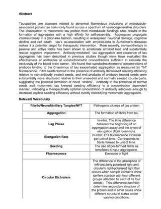
  • 1. Abstract Tauopathies are diseases related to abnormal filamentous inclusions of microtubule- associated protein tau commonly found across a spectrum of neurodegenerative disorders. The dissociation of monomeric tau protein from microtubule bindings sites results in the formation of aggregates with a high affinity for self-assembly. Aggregates propagate interneuronally in a prion-like fashion, resulting in widespread neuronal damage, cognitive decline and cell death. tau’s co-association with amyloid-beta in Alzheimer’s diseases makes it a potential target for therapeutic intervention. More recently, immunotherapy in passive and active forms has been shown to ameliorate amyloid load and substantially rescue cognitive impairment. Antibody-mediated tau aggregation and clearance of tau aggregates has been described in previous studies though none have evaluated the effectiveness of antibodies at substochiometric concentrations sufficient to simulate the exclusivity of the blood brain barrier. We found that substochoichiometric concentrations of antibody binding to the C-terminus of tau exacerbated aggregation as measured by ThT fluorescence. Fibril seeds formed in the presence of antibody decreased seeding efficiency relative to non-antibody treated seeds, and end products of antibody treated seeds were substantially more structured relative to their unseeded and normally seeded counterparts, suggesting the potential formation of novel “strains”. Antibody in the presence of normal seeds and monomeric tau lowered seeding efficiency in a concentration dependent manner, indicating a therapeutically optimal concentration of antibody adequate enough to decrease deplete seeding efficiency without overtly intensifying monomeric aggregation. Relevant Vocabulary Fibrils/Neurofibrillary Tangles/NFT Pathogenic clumps of tau protein Aggregation The formation of fibrils from tau Lag Phase In-vitro: The time difference between the beginning of an aggregation assay and the onset of elongation (fibril formation). Elongation Rate In-vitro: ThT fluorescence increase per unit of time. Corresponds to fibrils formed to unit of time. Seeding The use of pre-formed fibrils as templates to spur aggregation. Fluorescence Emission of light Circular Dichroism The difference in the absorption of left-circularly polarized light and circularly right-polarized light that occurs when sample contains chiral centers (carbon with four different groups attached to each of its four bonds). This difference can help determine secondary structure of the protein and in other cases show different structural states under varying conditions.
  • 2. Epitope A region where an antibody binds. Amyloid/Amyloidogenic Capable of forming aggregates. Disordered Protein Protein lacking specific structure. Alzheimer’s Disease Alzheimer’s disease is characterized by progressive neurocognitive decline associated with widespread propagation of amyloid-beta and tau protein fibrils. Early stages are asymptomatic though the onset of cognitive debility and subsequent dementia emerges with the prion-like propagation of amyloid deposits and tau neurofibrillary tangles, resulting in pervasive neuronal death and white matter atrophy. The Biochemical foundations of Alzheimer’s Disease Multiple theories have been established to explain the physiological cascade involved in the onset of Alzheimer’s disease. Oldest among these theories is the cholinergic hypothesis, which arose during a particularly research-intensive era in the field of neurochemistry and anatomy [1]. Findings from this two-decade period from the mid-1960s to the mid-1980s established a foundation upon which the molecular basis of neurodegenerative diseases could be closely examined. Chief among these neurophysiological mediators are cholinergic receptors, which play an important role in a wide spectrum of homeostatic functions. Consequently, the manifold nature of these receptors gives way to a broad range of neurological disease states upon their dysfunction [2.3.4], including those found in Alzheimer’s disease [5]. Amyloid beta deposits have been found to form extracellular amyloid clumps known as plaques, leading to nueromodulating effects that can occur at picomolar concentrations, irrespective of the neurotoxic state of amyloid beta [6]. The role of acetylcholine in memory recall was demonstrated by the use of receptor antagonists in monkeys and rats. Subjects receiving infusions in the perirhinal cortex showed marked
  • 3. decline in the ability to recognize stimuli [7.8]. Subsequent studies demonstrated that various degrees of cognitive impairment arise from region-specific application of receptor antagonists [9,10]. Post-mortem examinations of AD brains revealed depleted levels of cholinergic activity, particularly choline acetyltransferase, a transferase responsible for acetylcholine synthesis, and acetylcholinesterase, a hydrolase that breaks down acetylcholine in the neuromuscular junction and neural synapses, in the cerebral cortex [11]. In Alzheimer’s patients, frontal and temporal regions of the brain responsible for memory and cognition were especially depleted with respect to cholinergic receptors [12.13]. Much of the criticism levied against this hypothesis stems from confounding factors that show a natural decline of cholinergic activity in healthy rat brains [14, 15], as well as a broad spectrum of neurodegenerative disease [16]. These revelations point to the more general phenomenon of cholinergic decline as a symptom, rather than impetus, of neurodegeneration. The pivotal role of amyloid-beta in the progression of Alzheimer’s disease pins the peptide as the central tenet of the amyloid cascade hypothesis. Upon observation of Auguste Deter’s brain (who would later become the first patient to be formally diagnosed with Alzheimer’s disease) Alois Alzheimer, the physician credited with the first published clinical observation of AD dementia, noted “numerous small miliary foci are found in the superior layers…[that] are determined by the storage of a peculiar material in the cortex”. Indeed, Alzheimer would go on to conflate these plaques with “the most serious form of dementia”, adding that “the plaques were excessively numerous and almost one-third of the [patient’s] cortical cells had died off” [17]. These extracellular plaques would eventually come to be known as abnormal accumulations of peptide amyloid-beta. The description of amyloid- beta pathology as a “cascade” implies its central role as a vanguard of AD progression,
  • 4. postulating the formation of amyloid plaques as the prerequisites for neurofibrillary tangle formations of tau protein. Amyloid-beta’s precursor, amyloid precursor protein (APP), is a transmembrane protein which has been found to influence synaptogenesis and, most recently, protein synthesis in dividing human cells [18] among other processes. Its abundance in interneuronal ER and Golgi [19] membranes contributes to its involvement in AD pathogenesis, whereby the sequentially cleavage of APP by either α or β (BACE-1) and γ-secretase enzymes, respectively, produces plaque forming and non-plaque forming variants of free-floating amyloid-beta peptide in the neuronal interstitium. In the event of primary cleavage by α-secretase, soluble APP (sAPPα) is secreted, leaving behind a 83- residue membrane-bound fragment (CTFα) [20]. Conversely, initial cleavage with β- secretase produces a 99 amino-acid transmembrane peptide (CTFβ). In both instances, the membrane bound peptides are next cleaved by γ-secretase to yield amyloid-beta from CTFβ and a small protein (P3) from CTFα. The amyloidogenic potential of cleavage products is determined by the location of γ-secretase proteolysis; in the event of cleavage of amyloid-beta valine-40, Ab-40, a 40 amino-acid variant, is secreted. In the event of cleavage at alanine-42, ab 42, the 42 amino-acid variant, is secreted. While Ab-40 has been determined to be a natural component of cerebrospinal fluid and plasma [21], even potentially possessing neuroprotective properties, it’s counterpart, Ab-42 has been implicated as the pathological trigger of plaque formation [22]. In a post-mortem examination of AD patients, Alois Alzheimer’s had also described “peculiar, deeply stained bundles of neurofibrils” colocalized with dead cortical cells. Unbeknownst to him, he was describing one of the two neuropathological findings consistent with Alzheimer’s disease — tau neurofibrillary tangles. Distinguished in its ubiquity across a spectrum of neurodegenerative disorders, tauopathies are not a unique to Alzheimer’s disease, however tau fibrillation subsequent to amyloidosis is a hallmark sign.
  • 5. As a major microtubule stabilizing protein in the central nervous system, tau maintains cytoskeletal stability through polymerizing and depolymerization of tubulin subunits [23]. It’s affinity for tubulin is modulated by kinases and phosphatases [24]. In the event of hyperphosphorylation, tau dissociates from its cytoskeletal origin in the form of free-floating tau monomers. Consequently, these monomers self-assemble to form oligomeric structures which serve as scaffolds for the development of larger, pathogenic neurofibrillary tangles capable of propagating interneuronally, whereupon exogenous tau fibrils can induce tauopathies in neighboring cells in a prion-like manner known as seeding [25]. The duality of amyloid plaques and tau fibrils in the pathophysiology of Alzheimer’s disease lend credence to two of the later aforementioned theories. AD-associated tauopathies can seldom form without the presence of amyloid plaques [26], however extracellular amyloid deposition is not sufficient to elicit neurodegeneration [27,28]. The tau hypothesis is therefore the most concise understanding of the biochemical underpinnings of Alzheimer’s disease [29]. The molecular intersect between the two processes remains unclear, however recent studies have shown that oxidative stress stemming from the presence of toxic amyloid-beta upregulates a regulator (RCAN1) of calcineurine, a phosphatase of tau, and glycogen-synthase kinase-3β (GSK3β), a tau kinase. Concomitantly, the imbalance between an increase in phosphorylation activity and a decrease in dephosphorylation of tau results in the formation of tau fibrils, thus providing a coherent link between amyloidosis and fibrillation [30]. This link implies that the mitochondria invariably plays a part in AD etiology, giving birth to a relatively novel theory in which the cellular powerhouse forms the crux of the disease. The mitochondrial cascade hypothesis posits the formation of amyloid plaques on the genetic resiliency of the mitochondrial electron transport chain. Over time, the propensity of the mitochondria to regulate damage via reactive oxygen intermediates, along with its ability to generate ATP via oxidative phosphorylation, declines [31]. Age-
  • 6. related physiological changes in mitochondrial function result in compensatory responses, among them the secretion of amyloid-beta. Indeed, studies have found an association between mitochondrial amyloid-beta levels and the degree of cognitive impairment in transgenic mice [32]. Moreover, rat neurons treated with electron transport chain inhibitors have been found to enhance tau pathology [33,34] while cytochrome oxidase inhibitors, which function to impede the reduction potential of the final link in the ETC, cause substantial alterations in the cleavage of APP towards its toxic amyloid-beta descendant [35]. This theory helps bridge the discrepancy between genetics and sporadic onset of Alzheimer’s disease otherwise not explained by allelic variants that induce amyloidosis. Microtubule Associated Protein tau Microtubule associated protein tau is a seminal component in the maintenance of structural integrity of neurons. Located on the 17th chromosome, tau transcripts in the central nervous system are composed of 16 exons, three of which (2,3, and 10) are alternatively spliced to produce six potential isoforms expressed differentially throughout development, with exon 1 serving as an untranslated transcriptional prom. These isoforms are characterized by the presence of three or four repeat tubulin binding regions at the C- terminus and the presence, or lack thereof, of additional inserts at the N-terminus. The presence and absence of exon 10 in the modified tau transcript gives rise to four and three repeat regions, respectively. Irrespective of the presence of exon 10, the repeat regions 3R (R1-R3) or 4R (R1-R4) are also encoded by exons 9,11, and 12 [36]. The largest of these isoforms contains exon 4A (an intermediate region between exon 4 and 5) and is unique in its localization to regions of the peripheral nervous system such as the spinal cord and the retina.
  • 7. Exon Size Isoform Repeat Domain 2,3,10 441 Adult 4 repeat 2,3 410 Adult 3 repeat 2,10 412 Adult 4 repeat 2 381 Adult 3 repeat 10 383 Adult 4 repeat - 352 Fetal - The importance of the N-terminus as a projection domain is maintained by a highly acidic character capable of interacting with cellular components such as the plasma membrane [37], mitochondria and serving as a key intermediate in the maintenance of structural rigidity [38], axonal growth [39] and diameter [40]. Conversely, the C-terminus is characterized as a positively charged, basic region connected to the N-terminus via a proline-rich mediator [40]. This region is directly bound to cytoskeletal tubulin and facilitates polymerization events conducive to cytoskeletal alterations (cite). It is important to note that while 4R and 3R variants of tau bind microtubules, additional repeat regions have been shown to enhance binding affinity while simultaneously contributing to nucleation rates among dissociated tau [40]. Post-Translational Modification of tau Post-translational modifications of tau have been proposed as key drivers of Alzheimer’s pathology, among them glycosylation [41], acetylation [42] and phosphorylation [43]. N- linked glycosylation (attachment of the oligosaccharide to the amide nitrogen of the asparagine or arginine residue of a protein), targeting asparagine or arginine residues of the tau, was found in non-hyperphosphorylated tau from Alzheimer diseased brain but not in normal brain samples. Similarly, hyperphosphorylated neurofibrillary tangles and paired helical filaments were found to be extensively glycosylated relative to microtubule associated tau [44]. Moreover, the N-glycosylated tau functioned as a better substrate for cAMP-dependent protein kinases compared to its deglycosylated counterpart [45].
  • 8. Conversely, O-glycosylation (the reaction of a carbohydrate to a hydroxyl moiety), targeting serine, threonine or tyrosine, was found to be inversely related to the level of tau phosphorylation [41]. Acetylation (the attachment of an acetyl group to an amino moiety) of lysine residues impairs the microtubule binding affinity of tau. Indeed, acetylation of Lys280 was only found in hyperphosphorylated AD fibrils of mouse brain lysates [42], and deletion of SIRT1, a protein deacetylase, intensified levels of phospho-tau [46]. The hyperphosphorylation of tau protein is a common factor among all aforementioned scenarios [43]. As such, the phosphorylation state of tau has thus far been the main determinant of tau pathology and the balance between kinase/phosphatase activity takes center stage. Full length tau (441 aa) has been found to have a total of 80 serine/threonine, along with 5 threonine phosphorylation sites [47], each corresponding to various severities of cytopathology in Alzheimer’s disease [48]. Most of these phosphorylation sites lie in the proline-rich region connecting the projecting N-terminus with the microtubule binding C-terminal region [40]. Similarly, tau serves as an intermediary between phosphatases, enzymes that dephosphorylate targeted substrates, and microtubule stability [49]. Structural and Mechanistic Features of tau Fibril Formation Dissociation of protein tau from microtubule binding sites is the neuropathogenic foundation of tauopathy in Alzheimer’s disease. Subsequent to detachment, monomeric tau assumes an unstructured configuration, which can be attributed to its positive charge low hydrophobic character at physiological pH levels and [50]. The lack of hydrophobic residues precludes sufficient hydrophobic forces to sustain a secondary structure, and phosphorylation events contribute to a chance in electrostatic character, disassociation and
  • 9. self-assmbely [51]. These amyloid regions, narrowed down to hexapeptide sequences 275 (VQIINK)280 and 306 (VQIVYK)311 are sufficient for the growth and propagation of tau fibrils, among other amyloid derivatives [52,53]. While a significant portion of tau retains its random-coil structure even within fibrils, constituent regions of the amyloid core retain the beta-sheet rich motifs remain [53]. This is also demonstrated by the aggregation of tau in the presence of anionic compounds such as heparin [54] and arachidonic acid [55]. Spectroscopic studies using FRET and hydrogen/deuterium mass spec examinations have proposed an ‘S’ shaped model for monomeric tau, whereby contact is maintained between the N-terminus and the proline-rich region and the C-terminus and amylodigenic regions of tau [56]. Interactions between tau hydrophobic regions or polyanionic substances results in a conformational change from unstructured random-coils to beta-sheets, a pervasive feature of amyloids [52]. tau monomer interactions result in the formation of parallel “stacks” of tau beta-strands connected via intermolecular hydrogen bonds, similar to structures of amyloid-beta [57] and alpha-synuclein deposits [58] in Parkinson’s Disease. Outer regions of tau filaments exhibit exposed hydrogen bond donors and acceptors [59], features that promote further aggregation and are absent in natural beta-sheet proteins to avoid aggregation [60]. In this way, tau dimers are able to attract proximal monomers and grow in an unimpeded stacking fashion. Seeding and Intercellular Propagation of Tau The presence of preformed tau aggregates potentiates fibrillation of endogenous tau by enhancing recruitment of dissociated monomers and oligomers [61]. This facet of tauopathies allows tau fibrils to propagate in a pathogenic, prion-like fashion whereby exogenous fibrils or oligomers serve as “seeds”, or molecular scaffolds, for monomeric tau in adjacent cells. Indeed, transgenic mice expressing P301L human mutant tau localized to
  • 10. the entorhinal cortex demonstrated hierarchical propagation of fibrils to adjacent regions [62]. Cultured cell experiments demonstrate cellular ability to uptake tau oligomers, but not monomers, via endocytosis [63]. This seeding potential is determined by its structural conformation. In these instances, deletion of motifs (275)VQIINK(280) and (306)VQIVYK(311) eliminates the capacity of full-length tau to seed [64]. Currently, there are two potential models to explain seeding: the oligomer-nucleated conformation induction and template-assisted growth [65]. The major difference between these two models is the structural component(s) of tau that influence fibril formation. Oligomer-nucleated conformational induction establishes a high-energy scaffolds which attracts monomeric tau that binds in succession to lower energy and form oligomers [66]. Unlike the template- assisted growth model, fibrils do not integrate dissociated monomers, but are rather formed only after the formation of oligomers [67]. Dimeric, trimeric and oligomeric intermediates between monomer and fibril formation have been established in aggregation studies involving other fibrillation prone agents [68] and AD peptide amyloid-beta [69]. Toxicology comparisons between neurofibrillary tangles and tau oligomers injected into mouse brains found that oligomer-infused brains showed diffuse tau pathology into neighboring brain regions, whereas NFT-treated cells displayed localized deposits, implicating oligomers as the component most responsible for intercellular tauopathies [70]. Braak Staging and the Prion-like Propagation of tau The entorhinal region receives input from the neocortex and is involved in higher cognitive functions and the limbic system, as well as in the formation of memories and emotions. Intracellular tau deposits first appear in an area adjacent to the entorhinal region called the transentorhinal region, which functions as a relay between the neocortex and the entorhinal region. The manner of neurofibrillary tangle propagation is closely associated with the degree of cognitive decline [71]. The limbic stages consist of minimal NFT presence in the
  • 11. neocortex, with the fibrils concentration localized to the entorhinal and transentorhinal regions, concomitant with noticeable cognitive impairment. End stage AD presents with widespread damage to the neocortical areas, resulting in extensive cognitive impairment and advanced dementia. Figure 1: The Blood-Brain Barrier: An Obstinate Foe One of the major obstacles to immunotherapy against neurodegenerative disorders is the human blood brain barrier (BBB), a restrictive vasculature of endothelial cells exhibiting high electrical resistance. In healthy individuals, the BBB functions as a selective safeguard against potential antigens and neurotoxins, impeding the entry of large or hydrophilic molecules, while facilitating transport of metabolically essential nutrients and molecules. The innate bulkiness of immunoglobulins poses a major obstacle to developing effective therapeutic measures for combating neurodegenerative disorders. Indeed, radioimmunoassays have found that approximately 0.1% of circulating IgGs, the most common of the 5 immunoglobulin classes (A,G,M,E, and D), can be detected in the central
  • 12. nervous system [72]. However, the efficacy of the BBB can be severely compromised during neurological disorders such as multiple sclerosis, viral meningitis and tumors [73]. Inflammatory events in AD have also contributed to increased BBB permeability and the pathological spread of amyloid plaques. Given the rapid turnover of cerebrospinal fluid into the bloodstream, intrathecal injections directly into the CSF are equivalent to prolonged intravenous injections, amounting to limited therapeutic efficacy [74]. Moreover, a logarithmic decrease in drug distribution throughout the brain has been shown in bulk-flow delivery of drugs directly into brain tissue [75]. As such, antibody delivery for neuro- immune therapy is a popular research topic. Three major approaches to this problem include the application of lipid-mediator molecules, which can passively diffuse through the BBB, carrier mediated transport (CMT) of small water-soluble molecules, and the exploitation of receptor-mediated transport (RMT) . Theranostics, the use of molecular platforms for drug delivery and diagnostics, relies on lipid or water-based carriers to transport antibodies across the membrane [76]. These platforms have been used in the delivery of AB-antibody fragments via synthetic liposomal elements such as polyethylene glycol polymer chains (PEG) [77]. Advances in RMT take advantage of metabolic receptors mediating BBB access to transport bound antibodies into the brain parenchyma. Insulin and transferrin receptor ligand-bound AB-antibodies have been shown to effectively cross the BBB through receptor-mediated transcytosis, enhancing brain exposure 55-fold in some instances [78,79]. Origins of Immunotherapy in Alzheimer’s Disease As one of the hallmark pathologies of Alzheimer’s disease, aggregates of amyloid beta have been one of the primary immune targets of AD therapies for quite some time. Mice immunizedx with Aβ1-42, an alloform associated with toxic oligomers, showed reduced plaque burdens and retained cognitive functions relative to their non-immunized
  • 13. counterparts [80]. Subsequent human trials were halted after a subset of patients developed encephalitis post-immunization, likely due to the extensive activation of CD8+ cytotoxicity [81,82]. Nevertheless, post-mortem autopsies indicated clearance of amyloid with retention of tau pathology [83]. In another study, the clearance of extracellular amyloid plaques was accompanied by the reduction of early tau pathology but retention of hyperphosphorylated neurofibrillary tangles [84,85]. Conversely, tau antibody treatment did not affect amyloid load, indicating that amyloid deposits serve as a precursor to tauopathy, though analysis of cognitively normal individuals has shown tangle formation in the temporal lobe without the presence of amyloid plaques. Passive and active immunization of targeting tau fibrils has also become a mainstay in AD immune-therapy. Studies exhibiting clearance after antibody treatment were either targeted at tau phosphoepitopes or fibril specific conformations. In these cases, phosphorylation of tau was reduced and fibril load significantly decreased [86] establishing a correlation betweentau antibody titer count, fibril load and cognitive performance [87]. In other cases, passive immunization of phosphorylated tau was found to significantly decreased NFT burden while increasing microglial activity [88]. Mechanism of Antibody Mediated tau Therapy Although the efficacy of tau antibodies against pathogenic aggregates has been well documented, the mechanism by which this phenomenon occurs is obscure. Chief among several theories is that antibodies directly inhibit the fibrillation or even work to reverse the process altogether [89]. This theory is corroborated by the clearance amyloid-beta aggregates in in-vitro studies. Indeed, studies have found that, similar to their amyloid-beta counterparts, tau antibodies cross the neuronal membranes via clathrin-mediated endocytosis and co-localize with intercellular fibrils [90]. Additionally, antibodies have been
  • 14. found to interfere with the prion-like interneuronal propagation of tau by directly interacting with extracellular tau seeds [91]. Due to the neuroinflammatory nature of tauopathies, microglial clearance has been found to be a major form of fibril clearance [92]. However, mouse models studies for anti- amyloid-beta antibodies have also shown that clearance can occur in a non-Fc-mediated fashion with the use of antibodies lacking fragment crystallizable regions essential for the interaction of immune system components, such as microglia, with pathogens [93]. Molecular Dynamics of Thioflavin T Binding Thioflavin T, a benzothiazole dye, has been employed in numerous studies to detect and analyze the extent of fibrilation in aggregation-prone proteins. When bound to fibrous structures, the ThT maximum fluorescence wavelength increases substantially, producing a discernible signal unique to amyloid fibrils. Immobilization of the benzylamine and benzathiole rings on the molecule, producing a perpetually excited state that corresponds to a manifold increase in fluorescence [94]. Methods Protein Aggregation Assay In vitro aggregation of tau protein was performed via 0.1 ml triplicates in a 96 well-plate in the presence of 5 µM ThT. Each well was outfitted with 2 polyethylene balls (2.38-mm diameter, Precision Ball, Reno, PA), the plate covered with a Mylar septum sheet (Thermo) and shaken at 280 rpm in an Infinite M200 PRO Microplate Reader (Tecan, Austria). Kinetic measurements were taken in 5 minute intervals using a 444-nm excitation and 485- nm emission filter. Monomeric tau (15 µM, 0.6 mg/ml) was incubated in the presence of 5 µM heparin with the addition of 10 mM Hepes (pH 7.5), 1 mM EDTA, 100 mM NaCl, and 5
  • 15. mM DTT in a 320 µL eppendorf tube and distributed in 0.1 mL aliquots into 3 triplicates. Antibody concentrations varied from 5 nM – 125 nM. Seeding assays were run in similar fashion with the addition of 0.06-0.5% of either non- antibody treated (normal) tau fibril seeds or tau fibril seeds formed in the presence of 20 or 125 nM of antibody (abnormal). Curve Fitting The four parameter sigmoid curve was used to fit fluorescence data using Sigma Plot software. Data was plotted in accordance with the formula where Yo is the initial level of ThT fluorescence, a is the difference between the final and initial level of ThT fluorescence, Xo is the midpoint of transition and 1/b is the rate constant for fibril formation. The lag-time of fibril formation was determined by the formula Xo-2b and the initiation rate was determined by the inverse of lag time. [118]
  • 16. Figure 2: Kinetic model of protein aggregation: the nucleation phase (lag phase) results in minimal fluorescence until the concentration of oligomers reaches a critical mass resulting in an exponential growth in fibrils (elongation phase). Incubation with seeds tends to increase decrease lag phase and increase the rate at which fibrils are grown Circular Dichroism Far-UV CD spectra were measured using JASCO J-820 spectropolarimeter at 25 C. The aforementioned solution of aggregated tau (15 µM [0.6 mg/ml]) was placed into a 0.2-mm pathlength cell. Five spectra were averaged for each sample and acquired at high sensitivity with 20 nm/min scan speed at 0.2 nm step size/1.0 nm bandwidth under constant purging with nitrogen. Determination of Stability Aggregated tau samples (8 µl) were suspended in Hepes buffer (10 mM, pH 7.5) containing various concentrations of guanidine thiocyanate (0.2-6.0M) in a total volume of 20 µl, incubated for 1 hour at room temperature and then diluted to 300 µl with the concentration of guanidinium thiocyanate adjusted to 0.2 M in the presence of 10 µM ThT. ThT fluorescence was measured. An excitation wavelength of 442 nm and an emission spectrum range of 470-500 nm was used. Excitation and emission slit values were 2.5 nm and 5 nm, respectively. All measurements were performed in triplicate and averaged for each sample. Electron Microscopy Five microliter volumes of post-aggregation protein solution was adsorbed onto prewashed 200-mesh formvar/carbon-coated nickel grids for 5 minutes. The grid was washed with 10 µl water, stained with 10 µl of 2% uranyl acetate for 2 minutes and washed with water prior
  • 17. to drying at room temperature. Samples were analyzed with JEM-1400 transmission electron microscope (JEOL) operated at 80 kV. Results Characterization of tau Competent BL21(DE3) E.coli cells transformed with histone-tagged 0N4R four-repeat tau (383 aa) cDNA were harvested for protein. Histone-modified tau was purified via nickel column and dialyzed with TEV protease. Further purification was conducted using fast protein liquid chromatography Figure 3: tau after protease cleavage (3rd row from left) shows bottom band at the 17 kD mark, corresponding to his-tag cleavage and 55 kD mark corresponding to tau (40-60 kD). Experimentation (I): 15 µM tau was incubated in 100 µl aliquots with various concentrations (5-125 nM) of antibody 181, 231, 396, A10 and tau 13 in the presence of heparin sulfate. The samples were placed in a plate reader with 5 µM ThT and shaken at 37 C degrees. Lag phase and elongation rate were derived from fitting fluorescence data to a 4-parameter sigmoid curve.
  • 18. Epitope Mapping 10 20 30 40 50 MAEPRQEFEV MEDHAGTYGL GDRKDQGGYT MHQDQEGDTD AGLKAEEAGI 60 70 80 90 100 GDTPSLEDEA AGHVTQARMV SKSKDGTGSD DKKAKGADGK TKIATPRGAA 110 120 130 140 150 PPGQKGQANA TRIPAKTPPA PKTPPSSGEP PKSGDRSGYS SPGSPGTPGS 160 170 180 190 200 RSRTPSLPTP PTREPKKVAV VRTPPKSPSS AKSRLQTAPV PMPDLKNVKS 210 220 230 240 250 KIGSTENLKH QPGGGKVQII NKKLDLSNVQ SKCGSKDNIK HVPGGGSVQI 260 270 280 290 300 VYKPVDLSKV TSKCGSLGNI HHKPGGGQVE VKSEKLDFKD RVQSKIGSLD 310 320 330 340 350 NITHVPGGGN KKIETHKLTF RENAKAKTDH GAEIVYKSPV VSGDTSPRHL 360 370 380 SNVSSTGSID MVDSPQLATL ADEVSASLAK QGL tau 181 tau 231 tau 396 tau A10 tau 13 Amyloid Region Figure 4: Five monoclonal antibodies were selected for kinetic studies. Three antibodies (tau 13, 181, and 231) targeted epitopes upstream of the amyloid region and 2 targeted epitopes downstream of the amyloid region. Two of the antibodies (tau 181 and 231) bound to sequences in the proline-rich region, one targeted the N-terminus (tau 13) and two targeted the C-terminus (tau A10 and 396).
  • 19. Tau Antibodies Influence Fibril Formation at Substoichiometric Concentrations Figure 5 A. B. C. D. . ! E. Figure 5 ThT fluorescence data of 15 µM monomeric tau aggregation in the presence of 5 µM heparin and antibody 181 (A), 231 (B), A10 (C), tau13 (D) and 396 (E) at 0 nM (red), 65 nM (orange) and 125 nM (yellow) concentrations. Anibtody 396 (E) showed significant reduction in lag phase and increase in elongation rate compared to the control.
  • 20. Analysis of Kinetic Parameters of Antibody 396 in Two Sequential Assays Figure 7: First kinetic aggregation assay with antibody 396 showed influence on lag phase across concentrations 20, 65, 95 and 125 nM. Elongation rate increase is only significant for 20 nM antibody concentrations. R Figure 8: To avoid error, we ran the assay a second time in order to replicate the data. Lag phase and elongation rate decreased and increased, respectively. Only tau monomers treated with 125 nM antibody 396 showed a significant difference, in terms of elongation rate and lag phase, from the control (p < 0.05).
  • 21. Figure 9: Aggregates formed in the presence of antibody 396 were more numerous and branched relative to the control. Fibril interconnectivity and thickness did not seem to vary with antibody concentration. Different morphologies appeared within treatment groups. 5 nM antibody 396 treatment produced extensive branching and fibrillation with thick, overlapping end-products. 20 nM treatments produced thin, defuse truncated wisps. 125 nM treatments resulted in thick, blotchy fibrils with pervasive overlap.
  • 22. Experimentation (II): 15 µM tau was incubated in 100 µl aliquots with various concentrations (0.0625%-0.05%) of seeding fibrils formed in the presence of 20 and 125 nM antibody 396. The samples were placed in a plate reader with 5 µM ThT and shaken at 37 C degrees. Lag phase and elongation rate were derived from fitting fluorescence data to a 4-parameter sigmoid curve. Tau Fibrils Formed with Antibody 396 Have Lower Seeding Efficiency
  • 23. Figure 10: Kinetic aggregation assays were conducted using normal fibrils seeded with 15 µM tau (red) or fibrils grown in the presence of 20 (orange) and 125 nM (yellow) antibody 396. Analysis of variance (ANOVA) shows that 0.25% is a sufficient concentration for seeding with normal fibrils (red) to take place and decrease the lag phase (p < 0.05) and increase elongation rate (p < 0.05) substantially. Seeds treated with 20 nM increase lag phase and elongation rate to rates similar to non-seeded samples, differing significantly from normally seeded samples at concentrations of 0.25% and 0.50%. Surprisingly, seeds formed in a concentration of 125 nM antibody significantly differed from normal seeds at only higher concentrations of 0.50%, whereas 20 nM seeds were also effective at 0.25%. Overall, seeds harvested in the presence of antibody were far less effective in decreasing the lag phase. Antibody Treated Seeds Produce Structurally Distinct End Products
  • 24. Figure 11: Circular dichromism indicates more structured end products with aggregates formed in the presence of abnormal fibril seeds treated with 125 nM antibody 396 (green). Aggregates formed in the presence of normal fibril seeds (orange) are more structured than non-seeded aggregates (red) and similar to aggregates formed in the presence of abnormal fibril seeds treated with 2 nM antibody 396 (yellow). Electron Microscopy of End Products Figure 12: Electron microscopy reveals some retention of oligomeric character among normally seeded fibrils (top left,right) as opposed to a more pronounced branching and fibrillary structure of aggregates formed in the presence of abnormal fibril seeds treated with 125 nM antibody 396. Abnormally seeded end-products (125 nM) correspond to the more structured spectra in figure 11, compared to a
  • 25. Fibril Stability Assay Figure 13: Guanidine thiocyanate was used to denature end products of tau aggregation in the presence of normal and abnormal seeds. ThT fluorescence was used to measure fibril denaturation at increasing concentrations of guanidine thiocyanate. The stability profile of aggregates formed in the presence of normal seeds (½C: 1.91 M) did not significantly differ from stability profile of aggregates formed in the presence of abnormal seeds treated with 125 nM Ab 396 (½C: 2.01 M). [½C: concentration of guanidine thiocyanate at which ThT fluorescence of fibrils is half of the highest value].
  • 26. Experimentation (III): 15 µM tau was incubated in 100 µl aliquots with various concentrations (5-125 nM) of antibody 396 and normal fibril seeds in the presence of heparin sodium. The samples were placed in a plate reader with 5 µM ThT and shaken at 37 C degrees. Lag phase and elongation rate were derived from fitting fluorescence data to a 4-parameter sigmoid curve. Antibody 396 Lowers Seeding Efficiency in Normally Seeded Aggregation in a Concentration Dependent Manner Figure 14: Kinetic Data
  • 27. Figure 14: Kinetic aggregation assays were conducted using normal fibrils grown with 15 µM tau in the presence and absence of antibodies at various concentrations (5-125 nM). Analysis of variance (ANOVA) indicates presence of antibody increases the lag phase and decreases elongation rate in a concentration dependent manner. Surprisingly, the most significant deviations from the control, in terms of lag phase and elongation rate, occur at lower antibody concentrations and steadily decline as antibody concentration increases. Figure 15: Electron Microscopy of End Products Figure 15: Electron microscopy reveals fragmentation of normally seeded fibrils formed in the presence of 125 nM antibody 396. Normally seeded end products appear thicker with extensive branching compared to fibrils formed in the presence of 125 nM antibody 396. Aggregation in the presence of seed and antibody produced truncated, thin filaments similar to 20 nM treatment of antibody 396 in figure 9.
  • 28. Seeding in the Presence of Normal Seeds Incubated with Antibody 396 Produces the Most Structured Fibrils Figure 16: Far-UV circular dichroism of end products indicates tau seeded with abnormal seeds (125 nM) and normal seeds in the presence of antibody form more structured products than fibrils formed from 15 µM tau treated with antibody 396. Discussion
  • 29. Active and passive immune targeting of neurodegenerative elements has become a major therapeutic trend in biomedical research [96-99]. In Alzheimer’s disease, amyloid beta has served as the leading focus in multiple clinical trials [100]. Immunological interventions against tau fibril formations are far less common and remain in preclinical testing [87]. tau antibodies have been shown in multiple studies to reduce interneuronal spread, seeding of fibrils [in a concentration dependent manner] and reduce fibrillary load [101, 102, 103]]. In this study, we have confirmed that tau antibodies interfere with seeding and may do so at very low concentrations. Moreover, the ability of antibody 396 to influence kinetic parameters of monomeric tau in the presence and absence of seeds in an opposing manner indicates that it is capable of interacting with both monomers and fibrils. The Relevance of Epitope “396” Antibody 396 binds to a region located in the C-terminus of 4-repeat 383 tau isoform, between amino acid 336 and 340. Truncation and phosphorylation studies of the C- terminus determined the region plays a role in the inhibition of fibrillation and C-terminal modification are critical to the induction of the process in tau and alpha-synuclein [104, 105]. It’s also important to note that prediction (Figure 17) of protein binding region algorithm (ANCHOR) indicates a high probability that epitope “396” constitutes a microtubule binding region. This is evidenced by the fact that serine “396”— 338 in isoform D — is an essential phosphorylation site for the dissociation of tau from microtubules [119]. Though 336-340 is not the predicted amyloid region of isoform D (Figure 4), antibody binding may potentially change the electrostatic character of tau, influencing the manner in which it aggregates.
  • 30. Figure 17: Anchoring prediction of tau epitope 396 (highlighted in black). Seeding Efficiency Seeding efficiency for antibody-treated seeds and normal seeds in the presence of antibody 396 was markedly lower with respect to lag phase and elongation rate. In the presence of 0.25% and 0.5% seeds treated with 20 nM antibody, lag phase is significantly lower compared to non-seeded samples, but significantly higher compared to normally seeded samples. Surprisingly, 125 nM antibody treated seeds were observed to have a similar effect, but only at a concentration of 0.50%. The lag phase and elongation rates for monomeric tau seeded with 0.50% normal seeds in the presence of antibody 396 differed significantly from seeded tau in the absence of antibody. Unexpectedly, inhibition of seeding was lowest at lower antibody concentration and increased with increasing concentration. The ability of antibody 396 to minimize seeding efficiency while simultaneously increase the propensity for monomeric tau to form aggregates indicates a dual affinity for both tau fibrils and monomers. At lower concentrations, the antibody may bind fibrils preferentially and thus lower seeding efficiency. As concentration of antibody increases, tau monomer-immunoglobulin interactions increase, thus contributing to the decrease in lag phase and increase in !
  • 31. elongation rate. This corresponds to previous studies in which Tau antibodies were found to block tau aggregation seeding in vitro and decrease pathology in vivo [102] Concentration as a Determinant of Antibody Efficacy The lag phase and elongation rates of 15 µM tau aggregation in the presence of antibody 396 (experiment I) seem to indicate that the effect of immunoglobulin binding may be concentration dependent. Specifically, lag phases and elongation rates of normally seeded tau in the presence of antibody 396 deviate most significantly (experiment III) from the control at lower concentrations and equalize with increasing concentration. This may suggest the existence of a therapeutically optimal threshold concentration at which the antibody lowers seeding efficiency without significantly lowering the lag phase of monomeric tau aggregates. Antibody 396 may Form Variable Conformational Strains of tau Fibrils Conformational states are key determinants of seeding potency of tau aggregates [64]. Our data showed higher concentration of antibody 396, in seeding experiments involving abnormal seeds and normal seeds in the presence of antibodies, produced more structured end-products at higher concentration of antibody. Previous studies have documented tau antibody-mediated seeding inhibition in-vitro and cognitive improvement in-vivo [103]. Experiments with prion protein have determined that conformers are predictive of the incubation period in and seeding efficiencies in prionopathies [106, 107]. More specifically, variations in the structure and stability differences within the amyloid core could give way to novel strains of prion protein [108-110]. Similarly, distinct strains of tau have been found to vary in seeding efficiency, structure and the extent of intercellular propagation (toxicity), capable of producing different pathological states in-vivo [111]. These strains exhibit distinct CD spectra and morphologies upon observation via EM [112].
  • 32. While the amyloid cores of fibrils form beta structures, the configuration of the fibril, in particular the largely disordered N and C-termini, remains disordered. Structural studies have discovered a “fuzzy coat” ensemble that resembles a dual layered polyelectroylate brush composed of N and C-termini components protruding from the core [113]. At physiological levels, these extensions are thought to be negatively charged and possess properties capable of interactions among neurofibrillary tangles in both a repulsive [114] and adhesive manner [115]. These interactions are modified by changes in pH, electrolyte concentration and posttranslational modifications such as phosphorylation [115,116]. The propensity of antibody 396 to bind to the C-terminus may indicate a potential role for the “fuzzy coat” of the tau fibril in the formation of potentially novel strains of the neurofibrillary tangles. Caveat Relative to their tissue derived counterparts, heparin-induced tau fibrils formed in-vitro possess greater chemical stability and a more defined secondary structure. In the presence of heparin, monomeric recombinant tau seeded with brain-derived paired helical filaments produced a spectrum of fibril confirmations distinct from their brain-derived counterparts [117]. These findings present highlight potential drawbacks to the use of in- vitro designs in structural modeling. Conclusion We have discovered a potentially novel mechanism of action for Tau antibody which has not been previously determined by other laboratories. Fibrils produced in the presence of these antibodies are less efficient at seeding and more structured than their untreated counterparts.
  • 33. References 1. The history of the cholinergic hypothesis. Contestabile A.Behavioural brain research 221(2):334-40 (2011) 2. Schizophrenia and nicotinic receptors. Freedman R., Adler L. E., Bickford P., Byerley W., Coon H., Cullum C. M., Griffith J. M., Harris J. G., Leonard S. and Miller C.Harvard review of psychiatry 2(4):179-92 (1994) 3. Neuromuscular junction in myasthenia gravis: decreased acetylcholine receptors. Fambrough D. M., Drachman D. B. and Satyamurti S.Science 182(4109):293-5 (1973) 4. Nicotinic acetylcholine receptors in health and disease. Lindstrom J.Molecular neurobiology 15(2):193-222 (1997) 5. Alzheimer's disease and acetylcholine receptors. Kihara T. and Shimohama S.Acta neurobiologiae experimentalis 64(1):99-106 (2004) 6. Beta-amyloid peptides as direct cholinergic neuromodulators: a missing link? Auld D. S., Kar S. and Quirion R.Trends Neurosci 21(1):43-9 (1998) 7. Effects of muscarinic blockade in perirhinal cortex during visual recognition. Tang Y., Mishkin M. and Aigner T. G.Proc Natl Acad Sci U S A 94(23):12667-9 (1997) 8. Removal of cholinergic input to perirhinal cortex disrupts object recognition but not spatial working memory in the rat. Winters B. D. and Bussey T. J.European Journal of Neuroscience 21(8):2263-70 (2005) 9. Intraseptal muscarinic ligands and galanin: influence on hippocampal acetylcholine and cognition. Elvander E., Schött P. A., Sandin J., Bjelke B., Kehr J., Yoshitake T. and Ogren S. O.Neuroscience 126(3):541-57 (2004) 10. Intraseptal Infusion of the Cholinergic Agonist Carbachol Impairs Delayed- non-match-to-sample Radial Arm Maze Performance in the Rat. Bunce, Jamie G, Helen R Sabolek, and James J Chrobak. "." Hippocampus 14.4 (2004): 450-9. Print. 11.Selective loss of central cholinergic neurons in Alzheimer's disease. Davies P. and Maloney A. J. F.The Lancet 308(8000):1403 (1976) 12.Differential correlation between neurochemical deficits, neuropathology, and cognitive status in Alzheimer's disease. Dournaud P., Delaere P., Hauw J. J. and Epelbaum J.Neurobiol Aging 16(5):817-23 (1995) 13.Progressive decline in spatial learning and integrity of forebrain cholinergic neurons in rats during aging. Fischer W., Chen K. S., Gage F. H. and Björklund A.Neurobiol Aging 13(1):9-23 (1992) 14.Aging causes a preferential loss of cholinergic innervation of characterized neocortical pyramidal neurons. Casu M. A., Wong T. P., De Koninck Y., Ribeiro- da-Silva A. and Cuello A. C.Cereb Cortex 12(3):329-37 (2002) 15.Cholinergic receptors in cognitive disorders. Perry E. K., Perry R. H., Smith C. J., Purohit D., Bonham J., Dick D. J., Candy J. M., Edwardson J. A. and Fairbairn A.The Canadian journal of neurological sciences. Le journal canadien des sciences neurologiques 13(4 Suppl):521-7 (1986) 16.Auguste D and Alzheimer's disease. Maurer K., Volk S. and Gerbaldo H.The
  • 34. Lancet 349(9064):1546-9 (1997) 17.Amyloid precursor protein (APP) affects global protein synthesis in dividing human cells. Sobol A., Galluzzo P., Liang S., Rambo B., Skucha S., Weber M. J., Alani S. and Bocchetta M.J Cell Physiol 230(5):1064-74 (2015) 18. Amyloid precursor protein (APP) affects global protein synthesis in dividing human cells. Sobol A., Galluzzo P., Liang S., Rambo B., Skucha S., Weber M. J., Alani S. and Bocchetta M.J Cell Physiol 230(5):1064-74 (2015) 19.Endoplasmic reticulum and trans-Golgi network generate distinct populations of Alzheimer beta-amyloid peptides. Greenfield J. P., Tsai J., Gouras G. K., Hai B., Thinakaran G., Checler F., Sisodia S. S., Greengard P. and Xu H.Proc Natl Acad Sci U S A 96(2):742-7 (1999) 20.Functions of Aβ, sAPPα and sAPPβ : similarities and differences. Chasseigneaux S. and Allinquant B.J Neurochem 120 Suppl 1:99-108 (2012) 21.Isolation and quantification of soluble Alzheimer's beta-peptide from biological fluids. Seubert P., Vigo-Pelfrey C., Esch F., Lee M., Dovey H., Davis D., Sinha S., Schlossmacher M., Whaley J. and Swindlehurst C.Nature 359(6393):325-7 (1992) 22.Intraneuronal Aβ42 accumulation in human brain. Gouras G. K., Tsai J., Naslund J., Vincent B., Edgar M., Checler F., Greenfield J. P., Haroutunian V., Buxbaum J. D. and Xu H.The American journal of pathology 156(1):15-20 (2000) 23.A protein factor essential for microtubule assembly. Weingarten M. D., Lockwood A. H., Hwo S. Y. and Kirschner M. W.Proc Natl Acad Sci U S A 72(5):1858-62 (1975) 24.tau domains, phosphorylation, and interactions with microtubules. Mandelkow E. -M., Biernat J., Drewes G., Gustke N., Trinczek B. and Mandelkow E.Neurobiology of aging 16(3):355-62 (1995) 25.Alzheimer neurofibrillary degeneration: significance, etiopathogenesis, therapeutics and prevention. Iqbal K. and Grundke-Iqbal I.J Cell Mol Med 12(1):38-55 (2008) 26.The generation of a 17 kDa neurotoxic fragment: an alternative mechanism by which tau mediates beta-amyloid-induced neurodegeneration. Park S. Y. and Ferreira A.J Neurosci 25(22):5365-75 (2005) 27.Reducing endogenous tau ameliorates amyloid ß-induced deficits in an Alzheimer's disease mouse model. Roberson E. D., Scearce-Levie K., Palop J. J., Yan F., Cheng I. H., Wu T., Gerstein H., Yu G. -Q. and Mucke L.Science 316(5825):750-4 (2007) 28.tau is essential to β-amyloid-induced neurotoxicity. Rapoport M., Dawson H. N., Binder L. I., Vitek M. P. and Ferreira A.Proceedings of the National Academy of Sciences 99(9):6364-9 (2002) 29.tau-dependent microtubule disassembly initiated by prefibrillar beta-amyloid. King M. E., Kan H. M., Baas P. W., Erisir A., Glabe C. G. and Bloom G. S.J Cell Biol 175(4):541-6 (2006) 30.Amyloid-β toxicity and tau hyperphosphorylation are linked via RCAN1 in Alzheimer's disease. Lloret A., Badia M. C., Giraldo E., Ermak G., Alonso M. D., Pallardó F. V., Davies K. J. and Viña J.J Alzheimers Dis 27(4):701-9 (2011) 31.The mitochondrial energy transduction system and the aging process. Navarro A. and Boveris A.American Journal of Physiology-Cell Physiology 292(2):C670-86 (2007) 32.Mitochondrial amyloid-beta levels are associated with the extent of mitochondrial dysfunction in different brain regions and the degree of cognitive impairment in Alzheimer's transgenic mice. Dragicevic N., Mamcarz
  • 35. M., Zhu Y., Buzzeo R., Tan J., Arendash G. W. and Bradshaw P. C.J Alzheimers Dis 20 Suppl 2:S535-50 (2010) 33.Annonacin, a natural mitochondrial complex I inhibitor, causes tau pathology in cultured neurons. Escobar-Khondiker M., Höllerhage M., Muriel M. P., Champy P., Bach A., Depienne C., Respondek G., Yamada E. S., Lannuzel A., Yagi T., Hirsch E. C., Oertel W. H., Jacob R., Michel P. P., Ruberg M. and Höglinger G. U.J Neurosci 27(29):7827-37 (2007) 34.The mitochondrial complex I inhibitor rotenone triggers a cerebral tauopathy. Höglinger G. U., Lannuzel A., Khondiker M. E., Michel P. P., Duyckaerts C., Féger J., Champy P., Prigent A., Medja F., Lombes A., Oertel W. H., Ruberg M. and Hirsch E. C.J Neurochem 95(4):930-9 (2005) 35.Inhibition of energy metabolism alters the processing of amyloid precursor protein and induces a potentially amyloidogenic derivative. Gabuzda D., Busciglio J., Chen L. B., Matsudaira P. and Yankner B. A.J Biol Chem 269(18):13623-8 (1994) amyloidosis in Alzheimer's disease 36.The microtubule binding domain of tau protein. Lee G., Neve R. L. and Kosik K. S.Neuron 2(6):1615-24 (1989) 37.tau gene alternative splicing: expression patterns, regulation and modulation of function in normal brain and neurodegenerative diseases. Andreadis A.Biochim Biophys Acta 1739(2-3):91-103 (2005) 38.Interaction of tau with the neural plasma membrane mediated by tau's amino- terminal projection domain. Brandt R., Léger J. and Lee G.J Cell Biol 131(5):1327- 40 (1995) 39.Domains of neuronal microtubule-associated proteins and flexural rigidity of microtubules. Felgner H., Frank R., Biernat J., Mandelkow E. M., Mandelkow E., Ludin B., Matus A. and Schliwa M.J Cell Biol 138(5):1067-75 (1997) 40.Defects in axonal elongation and neuronal migration in mice with disrupted tau and map1b genes. Takei Y., Teng J., Harada A. and Hirokawa N.J Cell Biol 150(5):989-1000 (2000) 41.tau protein as a differential biomarker of tauopathies. Sergeant N., Delacourte A. and Buée L.Biochimica et Biophysica Acta (BBA)-Molecular Basis of Disease 1739(2):179-97 (2005) 42.The potential role of tau protein O-glycosylation in Alzheimer's disease. Robertson L. A., Moya K. L. and Breen K. C.J Alzheimers Dis 6(5):489-95 (2004) 43.The acetylation of tau inhibits its function and promotes pathological tau aggregation. Cohen T. J., Guo J. L., Hurtado D. E., Kwong L. K., Mills I. P., Trojanowski J. Q. and Lee V. M.Nature communications 2:252 (2011) 44.tau phosphorylation in neuronal cell function and dysfunction. Johnson G. V. and Stoothoff W. H.Journal of cell science 117(24):5721-9 (2004) 45.Glycosylation of microtubule-associated protein tau in Alzheimers disease brain. Takahashi M., Tsujioka Y., Yamada T., Tsuboi Y., Okada H., Yamamoto T. and Liposits Z.Acta neuropathologica 97(6):635-41 (1999) 46.Role of glycosylation in hyperphosphorylation of tau in Alzheimer s disease. Liu F., Zaidi T., Iqbal K., Grundke-Iqbal I., Merkle R. K. and Gong C. -X.FEBS letters 512(1):101-6 (2002) 47.Acetylation of tau inhibits its degradation and contributes to tauopathy. Min S. -W., Cho S. -H., Zhou Y., Schroeder S., Haroutunian V., Seeley W. W., Huang E. J., Shen Y., Masliah E. and Mukherjee C.Neuron 67(6):953-66 (2010) 48.Abnormal hyperphosphorylation of tau: sites, regulation, and molecular mechanism of neurofibrillary degeneration. Wang J. Z., Xia Y. Y., Grundke-Iqbal
  • 36. I. and Iqbal K.J Alzheimers Dis 33 Suppl 1:S123-39 (2013) 49.Specific tau phosphorylation sites correlate with severity of neuronal cytopathology in Alzheimer's disease. Augustinack J. C., Schneider A., Mandelkow E. M. and Hyman B. T.Acta Neuropathol 103(1):26-35 (2002) 50.Protein phosphatase 1 is targeted to microtubules by the microtubule- associated protein tau. Liao H., Li Y., Brautigan D. L. and Gundersen G. G.J Biol Chem 273(34):21901-8 (1998) 51.Why are "natively unfolded" proteins unstructured under physiologic conditions? Uversky V. N., Gillespie J. R. and Fink A. L.Proteins 41(3):415-27 (2000 52.tau aggregation is driven by a transition from random coil to beta sheet structure. von Bergen M., Barghorn S., Biernat J., Mandelkow E. M. and Mandelkow E.Biochim Biophys Acta 1739(2-3):158-66 (2005) 53.Assembly of tau protein into Alzheimer paired helical filaments depends on a local sequence motif ((306)VQIVYK(311)) forming beta structure. von Bergen M., Friedhoff P., Biernat J., Heberle J., Mandelkow E. M. and Mandelkow E.Proc Natl Acad Sci U S A 97(10):5129-34 (2000) 54.Assembly of microtubule-associated protein tau into Alzheimer-like filaments induced by sulphated glycosaminoglycans. Goedert M., Jakes R., Spillantini M. G., Hasegawa M., Smith M. J. and Crowther R. A.Nature 383(6600):550-3 (1996) 55.Differential assembly of human tau isoforms in the presence of arachidonic acid. King M. E., Gamblin T. C., Kuret J. and Binder L. I.J Neurochem 74(4):1749-57 (2000) 56.Hyperphosphorylation of intrinsically disordered tau protein induces an amyloidogenic shift in its conformational ensemble. Zhu S., Shala A., Bezginov A., Sljoka A., Audette G. and Wilson D. J.PLoS One 10(3):e0120416 (2015) 57. "Structural and Dynamic Features of Alzheimer's Abeta Peptide in Amyloid Fibrils Studied by Site-directed Spin Labeling. Török, Marianna, Saskia Milton, Rakez Kayed, Peng Wu, Theresa McIntire, Charles G Glabe, and Ralf Langen.." J Biol Chem 277.43 (October 25, 2002): 40810-5. Print. 58.Structural organization of alpha-synuclein fibrils studied by site-directed spin labeling.Der-Sarkissian A., Jao C. C., Chen J. and Langen R.J Biol Chem 278(39):37530-5 (2003) 59.Template-assisted filament growth by parallel stacking of tau. Margittai M. and Langen R.Proc Natl Acad Sci U S A 101(28):10278-83 (2004) 60.Natural beta-sheet proteins use negative design to avoid edge-to-edge aggregation. Richardson J. S. and Richardson D. C.Proc Natl Acad Sci U S A 99(5):2754-9 (2002) 61.Seeding of normal tau by pathological tau conformers drives pathogenesis of Alzheimer-like tangles. Guo J. L. and Lee V. M.J Biol Chem 286(17):15317-31 (2011) 62.Propagation of tau pathology in a model of early Alzheimer's disease. de Calignon A., Polydoro M., Suárez-Calvet M., William C., Adamowicz D. H., Kopeikina K. J., Pitstick R., Sahara N., Ashe K. H. and Carlson G. A.Neuron 73(4):685-97 (2012) 63.Propagation of tau misfolding from the outside to the inside of a cell. Frost B., Jacks R. L. and Diamond M. I.J Biol Chem 284(19):12845-52 (2009) 64.Conformation determines the seeding potencies of native and recombinant tau aggregates. Falcon B., Cavallini A., Angers R., Glover S., Murray T. K., Barnham L., Jackson S., O'Neill M. J., Isaacs A. M., Hutton M. L., Szekeres P. G., Goedert M.
  • 37. and Bose S.J Biol Chem 290(2):1049-65 (2015) 65. Formation and propagation of tau oligomeric seedsGerson J. E. and Kayed R.Front Neurol 4:93 (2013) 66.The role of prefibrillar structures in the assembly of a peptide amyloid. Ruschak A. M. and Miranker A. D.Journal of molecular biology 393(1):214-26 (2009) 67.Preparation and characterization of neurotoxic tau oligomers. Lasagna-Reeves C. A., Castillo-Carranza D. L., Guerrero-Muoz M. J., Jackson G. R. and Kayed R.Biochemistry 49(47):10039-41 (2010) 68.Early events in the fibrillation of monomeric insulin. Ahmad A., Uversky V. N., Hong D. and Fink A. L.J Biol Chem 280(52):42669-75 (2005) 69.Amyloid-β protein oligomerization and the importance of tetramers and dodecamers in the aetiology of Alzheimer's disease. Bernstein S. L., Dupuis N. F., Lazo N. D., Wyttenbach T., Condron M. M., Bitan G., Teplow D. B., Shea J. E., Ruotolo B. T., Robinson C. V. and Bowers M. T.Nat Chem 1(4):326-31 (2009) 70.Alzheimer brain-derived tau oligomers propagate pathology from endogenous tau. Lasagna-Reeves C. A., Castillo-Carranza D. L., Sengupta U., Guerrero-Munoz M. J., Kiritoshi T., Neugebauer V., Jackson G. R. and Kayed R.Sci Rep 2:700 (2012) 71.Staging of Alzheimer's disease-related neurofibrillary changes. Braak H. and Braak E.Neurobiology of aging 16(3):271-8 (1995) 72.Radioimmunoassays for Ig classes G, A, M, D, and E in spinal fluids: normal values of different age groups. Nerenberg S. T. and Prasad R.The Journal of laboratory and clinical medicine 86(5):887-98 (1975) 73.Cerebrospinal fluid IgG, IgA, IgM, IgD, and IgE levels in central nervous system disorders. Nerenberg S. T., Prasad R. and Rothman M. E.Neurology 28(10):988-90 (1978) 74.Fate of adrenal cortical steroids following intrathecal injection. FISHMAN R. A. and CHRISTY N. P.Neurology 15:1-6 (1965) 75.Point source concentration of GDNF may explain failure of phase II clinical trial. Salvatore M. F., Ai Y., Fischer B., Zhang A. M., Grondin R. C., Zhang Z., Gerhardt G. A. and Gash D. M.Exp Neurol 202(2):497-505 (2006) 76.Liposomes and nanotechnology in drug development: focus on neurological targets. Ramos-Cabrer P. and Campos F.Int J Nanomedicine 8:951-60 (2013) 77.Enhanced glutathione PEGylated liposomal brain delivery of an anti-amyloid single domain antibody fragment in a mouse model for Alzheimer's disease. Rotman M., Welling M. M., Bunschoten A., de Backer M. E., Rip J., Nabuurs R. J., Gaillard P. J., van Buchem M. A., van der Maarel S. M. and van der Weerd L.Journal of Controlled Release 203:40-50 (2015) 78.Fusion antibody for Alzheimer's disease with bidirectional transport across the blood-brain barrier and abeta fibril disaggregation. Boado R. J., Zhang Y., Zhang Y., Xia C. F. and Pardridge W. M.Bioconjug Chem 18(2):447-55 (2007) 79.Increased brain penetration and potency of a therapeutic antibody using a monovalent molecular shuttle. Niewoehner J., Bohrmann B., Collin L., Urich E., Sade H., Maier P., Rueger P., Stracke J. O., Lau W., Tissot A. C., Loetscher H., Ghosh A. and Freskgård P. O.Neuron 81(1):49-60 (2014) 80.Immunization with amyloid-beta attenuates Alzheimer-disease-like pathology in the PDAPP mouse. Schenk D., Barbour R., Dunn W., Gordon G., Grajeda H., Guido T., Hu K., Huang J., Johnson-Wood K., Khan K., Kholodenko D., Lee M., Liao Z., Lieberburg I., Motter R., Mutter L., Soriano F., Shopp G., Vasquez N., Vandevert C., Walker S., Wogulis M., Yednock T., Games D. and Seubert P.Nature 400(6740):173-7 (1999) 81.Subacute meningoencephalitis in a subset of patients with AD after Aβ42
  • 38. immunization. Orgogozo J. -M., Gilman S., Dartigues J. -F., Laurent B., Puel M., Kirby L. C., Jouanny P., Dubois B., Eisner L. and Flitman S.Neurology 61(1):46-54 (2003) 82.Neuropathology and pathogenesis of encephalitis following amyloid β immunization in Alzheimer's disease. Ferrer I., Rovira M. B., Guerra M. L. S., Rey M. J. and Costa-Jussá F.Brain Pathology 14(1):11-20 (2004) 83.Neuropathology of human Alzheimer disease after immunization with amyloid- beta peptide: a case report. Nicoll J. A., Wilkinson D., Holmes C., Steart P., Markham H. and Weller R. O.Nat Med 9(4):448-52 (2003) 84.Aβ immunotherapy leads to clearance of early, but not late, hyperphosphorylated tau aggregates via the proteasome. Oddo S., Billings L., Kesslak J. P., Cribbs D. H. and LaFerla F. M.Neuron 43(3):321-32 (2004) 85.Passive immunization with anti-tau antibodies in two transgenic models: reduction of tau pathology and delay of disease progression. Chai X., Wu S., Murray T. K., Kinley R., Cella C. V., Sims H., Buckner N., Hanmer J., Davies P., O'Neill M. J., Hutton M. L. and Citron M.J Biol Chem 286(39):34457-67 (2011) 86.tau-targeted immunization impedes progression of neurofibrillary histopathology in aged P301L tau transgenic mice. Bi M., Ittner A., Ke Y. D., Götz J. and Ittner L. M.PLoS One 6(12):e26860 (2011) 87.Immunotherapy targeting pathological tau prevents cognitive decline in a new tangle mouse model. Boutajangout A., Quartermain D. and Sigurdsson E. M.J Neurosci 30(49):16559-66 (2010) 88.Efficacy and safety of immunization with phosphorylated tau against neurofibrillary tangles in mice. Boimel M., Grigoriadis N., Lourbopoulos A., Haber E., Abramsky O. and Rosenmann H.Exp Neurol 224(2):472-85 (2010) 89.Disaggregation of Alzheimer beta-amyloid by site-directed mAb. Solomon B., Koppel R., Frankel D. and Hanan-Aharon E.Proc Natl Acad Sci U S A 94(8):4109-12 (1997) 90.Antibody uptake into neurons occurs primarily via clathrin-dependent Fcγ receptor endocytosis and is a prerequisite for acute tau protein clearance. Congdon E. E., Gu J., Sait H. B. and Sigurdsson E. M.J Biol Chem 288(49):35452- 65 (2013) 91.Proteopathic tau seeding predicts tauopathy in vivo. Holmes B. B., Furman J. L., Mahan T. E., Yamasaki T. R., Mirbaha H., Eades W. C., Belaygorod L., Cairns N. J., Holtzman D. M. and Diamond M. I.Proc Natl Acad Sci U S A 111(41):E4376-85 (2014) 92.tau oligomers and fibrils induce activation of microglial cells. Morales I., Jiménez J. M., Mancilla M. and Maccioni R. B.J Alzheimers Dis 37(4):849-56 (2013) 93.Non-Fc-mediated mechanisms are involved in clearance of amyloid-beta in vivo by immunotherapy. Bacskai B. J., Kajdasz S. T., McLellan M. E., Games D., Seubert P., Schenk D. and Hyman B. T.J Neurosci 22(18):7873-8 (2002) 94.Molecular mechanism of Thioflavin-T binding to amyloid fibrils. Biancalana M. and Koide S.Biochim Biophys Acta 1804(7):1405-12 (2010) 95.Circular dichroism spectroscopy as a tool for monitoring aggregation in monoclonal antibody therapeutics. Joshi V., Shivach T., Yadav N. and Rathore A. S.Anal Chem 86(23):11606-13 (2014) 96.Active and passive immunotherapy for neurodegenerative disorders. Brody D. L. and Holtzman D. M.Annu Rev Neurosci 31:175-93 (2008) 97.Immunotherapy for Alzheimer's disease: hoops and hurdles. Lemere C. A.Mol Neurodegener 8:36 (2013) 98.Can Alzheimer disease be prevented by amyloid-beta immunotherapy? Lemere
  • 39. C. A. and Masliah E.Nat Rev Neurol 6(2):108-19 (2010) 99.Immunotherapy for Alzheimer's disease. Wisniewski T. and Goñi F.Biochem Pharmacol 88(4):499-507 (2014) 100. Safety, tolerability, and antibody response of active Aβ immunotherapy with CAD106 in patients with Alzheimer's disease: randomised, double-blind, placebo-controlled, first-in-human study. Winblad B., Andreasen N., Minthon L., Floesser A., Imbert G., Dumortier T., Maguire R. P., Blennow K., Lundmark J., Staufenbiel M., Orgogozo J. M. and Graf A.Lancet Neurol 11(7):597-604 (2012) 101. First-in-man tau vaccine targeting structural determinants essential for pathological tau-tau interaction reduces tau oligomerisation and neurofibrillary degeneration in an Alzheimer's disease model. Kontsekova E., Zilka N., Kovacech B., Novak P. and Novak M.Alzheimers Res Ther 6(4):44 (2014) 102. Anti-tau antibodies that block tau aggregate seeding in vitro markedly decrease pathology and improve cognition in vivo. Yanamandra K., Kfoury N., Jiang H., Mahan T. E., Ma S., Maloney S. E., Wozniak D. F., Diamond M. I. and Holtzman D. M.Neuron 80(2):402-14 (2013) 103. Anti-tau antibody reduces insoluble tau and decreases brain atrophy. Yanamandra K., Jiang H., Mahan T. E., Maloney S. E., Wozniak D. F., Diamond M. I. and Holtzman D. M.Ann Clin Transl Neurol 2(3):278-88 (20 104. C-terminal inhibition of tau assembly in vitro and in Alzheimer's disease. Abraha A., Ghoshal N., Gamblin T. C., Cryns V., Berry R. W., Kuret J. and Binder L. I.J Cell Sci 113 Pt 21:3737-45 (2000) 105. Aggregation promoting C-terminal truncation of alpha-synuclein is a normal cellular process and is enhanced by the familial Parkinson's disease- linked mutations. Li W., West N., Colla E., Pletnikova O., Troncoso J. C., Marsh L., Dawson T. M., Jäkälä P., Hartmann T., Price D. L. and Lee M. K.Proc Natl Acad Sci U S A 102(6):2162-7 (2005) 106. "Small Protease Sensitive Oligomers of PrPSc in Distinct Human Prions Determine Conversion Rate of PrP(C)." Kim, Chae, Tracy Haldiman, Krystyna Surewicz, Yvonne Cohen, Wei Chen, Janis Blevins, Man-Sun Sy, Mark Cohen, Qingzhong Kong, Glenn C Telling, Witold K Surewicz, and Jiri G Safar. PLoS Pathog 8.8 (2012) 107. "Continuum of Prion Protein Structures Enciphers a Multitude of Prion Isolate-specified Phenotypes." Legname, Giuseppe, Hoang-Oanh B Nguyen, David Peretz, Fred E Cohen, Stephen J DeArmond, and Stanley B Prusiner. Proc Natl Acad Sci U S A 103.50 (December 12, 2006): 19105-10. 108. Parallel in-register intermolecular β-sheet architectures for prion-seeded prion protein (PrP) amyloids. Groveman B. R., Dolan M. A., taubner L. M., Kraus A., Wickner R. B. and Caughey B.J Biol Chem 289(35):24129-42 (2014) 109. Distinct prion strains are defined by amyloid core structure and chaperone binding site dynamics. Frederick K. K., Debelouchina G. T., Kayatekin C., Dorminy T., Jacavone A. C., Griffin R. G. and Lindquist S.Chem Biol 21(2):295-305 (2014) 110. Conformational stability of mammalian prion protein amyloid fibrils is dictated by a packing polymorphism within the core region. Cobb N. J., Apostol M. I., Chen S., Smirnovas V. and Surewicz W. K.J Biol Chem 289(5):2643-50 (2014) 111. Distinct tau prion strains propagate in cells and mice and define different tauopathies. Sanders D. W., Kaufman S. K., DeVos S. L., Sharma A. M., Mirbaha H., Li A., Barker S. J., Foley A. C., Thorpe J. R., Serpell L. C., Miller T. M., Grinberg L. T., Seeley W. W. and Diamond M. I.Neuron 82(6):1271-88 (2014) 112. Conformational diversity of wild-type tau fibrils specified by templated conformation change. Frost B., Ollesch J., Wille H. and Diamond M. I.J Biol Chem
  • 40. 284(6):3546-51 (2009) 113. Structural characterization of the core of the paired helical filament of Alzheimer disease. Wischik C. M., Novak M., Edwards P. C., Klug A., Tichelaar W. and Crowther R. A.Proc Natl Acad Sci U S A 85(13):4884-8 (1988) 114. Entropic exclusion by neurofilament sidearms: a mechanism for maintaining interfilament spacing. Brown H. G. and Hoh J. H.Biochemistry 36(49):15035-40 (1997) 115. The fuzzy coat of pathological human tau fibrils is a two-layered polyelectrolyte brush. Wegmann S., Medalsy I. D., Mandelkow E. and Müller D. J.Proc Natl Acad Sci U S A 110(4):E313-21 (2013) 116. Modulation of repulsive forces between neurofilaments by sidearm phosphorylation. Kumar S. and Hoh J. H.Biochem Biophys Res Commun 324(2):489-96 (2004) 117. Conformational features of tau fibrils from Alzheimer's disease brain are faithfully propagated by unmodified recombinant protein. Morozova O. A., March Z. M., Robinson A. S. and Colby D. W.Biochemistry 52(40):6960-7 (2013) 118. Phosphorylation of Amyloid Beta (Aβ) Peptides - a Trigger for Formation of Toxic Aggregates in Alzheimer's Disease. Kumar, Sathish, and Jochen Walter. Aging (Albany NY) 3.8 (August, 2011): 803-12. Print. 119. "Tau Phosphorylation at Serine 396 and Serine 404 by Human Recombinant Tau Protein Kinase II Inhibits Tau's Ability to Promote Microtubule Assembly." Evans, D B, K B Rank, K Bhattacharya, D R Thomsen, M E Gurney, and S K Sharma. J Biol Chem 275.32 (August 11, 2000): 24977-83. Print. ! !