2. 1993; Jacobs et al., 1997!. Paradoxically, b-lactam inhibitors of
b-lactamases can induce the production of the enzyme they are
meant to inhibit.
Class C ~type I! b-lactamases like AmpC ~Bush et al., 1995! are
among the most difficult to counter. Compounds that are suppos-
edly “b-lactamase stable,” like aztreonam, are hydrolyzed byAmpC,
and b-lactam–based inhibitors, such as clavulanic acid, do not
inhibit them at clinically relevant concentrations. Presumably driven
by an increased use of “b-lactamase stable” drugs and b-lactamase
inhibitors that are themselves b-lactams, AmpC-based resistance
to b-lactams has become increasingly prominent. The recent mo-
bilization of these enzymes on plasmids ~Gonzalez Leiza et al.,
1994; Koeck et al., 1997! and the isolation of mutant enzymes in
the wild ~Nukaga et al., 1995! threaten further spread of these
broad-spectrum b-lactamases.
We investigated non-b-lactam inhibitors of AmpC from Esche-
richia coli, reasoning that they would not be subject to the suite
of existing resistance mechanisms developed against b-lactams.
A non-b-lactam compound would avoid hydrolysis by mutant
b-lactamases, might not be affected by porin channel mutations,
and might not up-regulate b-lactamase production. Because such
inhibitors would not have been in the biosphere over evolutionary
time, bacteria might need to develop new resistance mechanisms,
rather than simply recruiting existing ones. The high sequence and
structural similarity among class C b-lactamases ~Usher et al.,
1998! suggests that an inhibitor of AmpC will inhibit class C
enzymes from other Gram-negative pathogens.
Boronic acids inhibit b-lactamases as transition-state analogs
~Beesley et al., 1983; Strynadka et al., 1996!. Using structure-
based methods, we identified a novel boronic acid inhibitor,
benzo~b!thiophene-2-boronic acid ~BZBTH2B!, with a Ki of
27 nM for AmpC ~Weston et al., 1998!. This inhibitor is 100-fold
more potent than boronic acid inhibitors previously described for
this enzyme and is specific for AmpC over mechanistically related
enzymes ~Weston et al., 1998!.
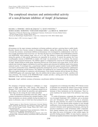
BZBTH2B does not resemble b-lactams ~Fig. 1!, but neverthe-
less inhibits AmpC b-lactamase potently and specifically. This
raises several questions. To what interactions does the high affinity
of this inhibitor owe? What does this inhibitor tell us about the
functional group recognition in AmpC? Can such non-b-lactam
inhibitors evade the classic b-lactam resistance mechanisms? To
address these questions, we determined the X-ray crystallographic
structure of the BZBTH2B0AmpC complex. We also investigated
the actions of BZBTH2B0b-lactam combinations against patho-
genic bacteria that complement AmpC expression with secondary
resistance mechanisms, increasing resistance. Both the crystallo-
graphic structure and antibacterial experiments reveal surprising
properties of BZBTH2B. The inhibitor makes unusual interactions
with conserved, catalytic residues in the AmpC active site, and it
has an antibiotic profile dramatically different from traditional
anti-b-lactamase compounds, such as clavulanic acid or aztreonam
~Fig. 1!.
Results
Crystallographic structure of BZBTH2B0AmpC complex
The X-ray crystallographic structure of BZBTH2B bound to AmpC
b-lactamase was determined to 2.25 Å resolution ~Table 1!. The
electron density for the inhibitor in this structure was well defined
~Fig. 2!. Following refinement of the complexed structure, a sim-
ulated annealing omit map of the area surrounding the inhibitor
was calculated and showed unambiguous positive difference den-
sity for the inhibitor when contoured at 3s. The position of the
sulfur atom of the thiophene ring of the inhibitor was verified by
calculating electron density maps for models with the sulfur atom
in each of two possible orientations. The two positions differed by
a 1808 rotation around the boron–carbon atom 1 bond of the in-
hibitor. Fo Ϫ Fc difference electron density maps were contoured at
3s and showed significant negative difference density surrounding
the sulfur atom when in the position 1808 from the one shown in
Figure 2. Overall, the structure of the BZBTH2B0AmpC complex
resembles the apo-enzyme, with a root-mean-square deviation
~RMSD! for all atoms of 0.76 Å. The RMSD between the com-
plexed and apo-enzymes for active site residues ~Ser64, Lys67,
Tyr150, Asn152, Tyr221, Lys315, and Ala318! in molecule 1 of the
A B
C D
Fig. 1. Comparison of b-lactamase ligands. A: Cephalothin, a cephalosporin substrate. The R1 and R2 side chains are labeled.
B: Clavulanic acid, a clinically used b-lactamase inhibitor. C: Aztreonam, a “b-lactamase stable” molecule. D: Benzo~b!thiophene-
2-boronic acid.
Structure of a novel b-lactamase inhibitor 2331
3. asymmetric unit is 0.28 Å. This rises to 1.06 Å if Gln120 is in-
cluded; this residue showed the most movement in the active site.
The two monomers of AmpC in the asymmetric unit of the com-
plex also resemble each other, differing by an RMSD of 0.84 Å for
all atoms. The quality of the model was evaluated with Procheck
~Laskowski et al., 1993!; 90.2% of the nonglycine and nonproline
residues were in the most favored regions of a Ramachandran plot
~9.6% were in the additionally allowed region!. The final R and
Rfree values of the refined structure were 16.7 and 22.4%, respec-
tively. The structure has been deposited with the PDB as 1C3B.
The inhibitor appears to form quadrupole–quadrupole inter-
actions with Tyr221 and quadrupole–dipole interactions withAsn152
~Fig. 3!. The distance between ring carbon atom 7 of the inhibitor
and the aromatic ring centroid of Tyr221 is 4.0 Å, and the distance
between the centroid of the second ring of BZBTH2B and the
amide nitrogen of Asn152 is 3.0 Å ~Table 2!. The angle between
the rings of the inhibitor and Tyr221 is 478, and the amide nitrogen
of Asn152 is perpendicular to the ring plane of the inhibitor. These
distances and angles are consistent with quadrupolar interactions
observed in other protein structures ~Thornton et al., 1988!.
An observed interaction with mechanistic implications is the
hydrogen bond between the boronic acid O2 atom and Tyr150
~2.8 Å, Fig. 3, Table 2!. In forming this new interaction, the cat-
alytic site has rearranged subtly. Lys67 and Tyr150, which usually
form a hydrogen bond ~Oefner et al., 1990; Lobkovsky et al., 1993,
1994; Usher et al., 1998!, have moved by ;0.7 and 0.4 Å, respec-
tively. Consequently, the interaction between them is lost; Lys67
now forms a hydrogen bond with the main-chain oxygen of Ala220
~not shown! while Tyr150 interacts with the boronic acid O2 atom
~Fig. 3!.
Fig. 2. 2Fo Ϫ Fc electron density ~in stereo! of the refined model, contoured at 1s. The area shown is the active site region with
BZBTH2B covalently bound to the catalytic residue Ser64 of AmpC b-lactamase. Residues shown are within 3.8 Å of the inhibitor
and are completely conserved in class C b-lactamases. Green spheres represent ordered water molecules. Carbon atoms are colored
gray, oxygen atoms red, and nitrogen atoms blue. This figure was generated using the programs MOLSCRIPT ~Kraulis, 1991! and
Raster3D ~Bacon & Anderson, 1988! and displayed using the program BOBSCRIPT ~Esnouf, 1997!.
Table 1. Data collection and refinement statistics
Cell constants ~Å; deg! a ϭ 118.86, b ϭ 78.01,
c ϭ 98.96; b ϭ 116.07
Resolution ~Å! 2.25
Unique reflections 33,738
Rmerge 9.4 ~13.7!a
Completeness ~%! 86.6 ~76.4!a
^I&0^sI& 11.4
Resolution range for refinement ~Å! 20–2.25 ~2.37–2.25 Å!a
Number of water molecules 94
RMSD bond lengths ~Å! 0.012
RMSD bond angles ~deg! 1.732
R-factor ~%! 16.7
Rfree ~%! 22.4b
Average B-factor, protein ~Å2
! 28.63c
Average B-factor, inhibitor ~Å2
! 25.42c
a
Values in parentheses are for the highest resolution shell used in
refinement.
b
Rfree was calculated with 10% of reflections set aside randomly.
c
Values cited were calculated for both molecules in the asymmetric unit.
Fig. 3. Key interactions ~Table 2! observed in the structure of BZBTH2B
in complex with AmpC. Dashed lines indicate hydrogen bonding ~yellow!
or quadrupolar interactions ~magenta!. Green spheres represent ordered
water molecules. The atoms are colored as in Figure 2. The figure was
generated with MidasPlus ~Ferrin et al., 1988!.
2332 R.A. Powers et al.
4. Two well-ordered water molecules appear in the active site,
making extensive interactions that involve the inhibitor and the
shifted Tyr150. The first water hydrogen bonds with the boronic
acid O2 atom and with the active site residue Thr316 ~Fig. 3;
Table 2!. The second water forms a hydrogen bond with the first
water molecule. This second water also interacts with catalytic
residues Asn346 and Arg349 ~Fig. 3; Table 2!. Additionally, the
boronic acid O1 atom hydrogen bonds with the backbone nitrogens
of Ser64 ~2.9 Å! and Ala318 ~2.7 Å!, and also with the carbonyl
oxygen of Ala318 ~2.9 Å! ~Table 2!.
Antimicrobial activity of BZBTH2B
The high affinity of BZBTH2B led us to investigate its ability
to potentiate the activity of b-lactams against resistant bacteria.
BZBTH2B increased the activity of the widely used third-generation
cephalosporin ceftazidime ~CAZ! for all bacterial strains over-
expressing AmpC b-lactamase against which it was tested
~Fig. 4A!. These included common hospital-acquired pathogens
such as E. coli, Enterobacter cloacae, Pseudomonas aeruginosa,
and Citrobacter freundii, which are often resistant to most b-lactams.
BZBTH2B increased the efficacy of CAZ up to 64-fold and re-
duced ~improved! its minimum inhibitory concentration ~MIC! to
as low as 2 mg0mL for some pathogens. BZBTH2B alone had no
measurable antibiotic activity at these concentrations.
Because BZBTH2B is not a b-lactam, we hypothesized that it
would be unaffected by traditional b-lactam resistance mecha-
nisms. To test this, we investigated how the inhibitor was affected
by porin channel mutants that are known to further increase resis-
tance to b-lactams in conjunction with b-lactamases. E. coli with
mutations in either of the main porin channels, OmpC or OmpF,
were more resistant to CAZ compared with the parent strain, as
expected. Conversely, BZBTH2B activity was unaffected by these
mutants, potentiating the effect of CAZ to the same levels in the
porin-mutant strains as in the parent wild-type strain ~Fig. 4B!.
Another traditional b-lactam resistance mechanism is the up-
regulation of b-lactamase transcription caused by b-lactams;
b-lactams, including b-lactamase inhibitors such clavulanic acid
and tazobactam ~Kadima & Weiner, 1997!, can induce expression
of the enzyme that inactivates them. To test the role of BZBTH2B
on the induction of AmpC, we investigated its ability to potentiate
the action of the b-lactams CAZ and piperacillin. We compared the
potentiation effect of BZBTH2B with that of cefoxitin ~a cepha-
losporin! and clavulanic acid ~a b-lactam-based inhibitor of
b-lactamases! ~Fig. 5!.
In these experiments, two disks soaked with a known amount of
a primary b-lactam, CAZ ~top disk! and piperacillin ~bottom disk!,
are placed on an agar plate that has been inoculated with a bacte-
rium in which AmpC expression is inducible. A third disk soaked
with a different compound, either cefoxitin, clavulanic acid or
BZBTH2B, is placed on the agar plate between the two primary
b-lactam disks. As the b-lactams diffuse from the primary disks
into the agar, a clear zone ~or halo! is created around the disk,
indicating where bacteria are unable to grow. The shape and size of
Table 2. Interactions in complexed and native
AmpC b-lactamase
Distance ~Å!a
Interaction Complex Native
Y150OH–K315Nz 2.89 2.54
Y150OH–O2 2.74 NPb
Y150OH–S64Og 2.91 3.21
Y150OH–K67Nz 3.51 3.06
K67Nz–A220O 2.96 3.47
S64N–O1 3.01 NPb
A318N–O1 2.84 NPb
A318O–O1 2.84 NPb
O2–H2O 2.61 NPb
H2O–T316Og1 3.03 NPb
H2O–H2O 3.50 NPb
H2O–N346Od1 2.79 NPb
H2O–R349Nh1 3.12 NPb
N152Od1–K67Nz 2.65 2.67
K67Nz–S64Og 2.60 3.49
N152Nd2–Q120OE1 6.11 3.05
N152Nd2–centroid BZBTH2B aryl ring 3.07 NPb
CP7 BZBTH2B–centroid Y221 3.98 NPb
Centroid BZBTH2B aryl ring-centroid Y221 5.34 NPb
a
All distances are for molecule 2 of the asymmetric unit.
b
Not present in native structure.
A
B
Fig. 4. A: Potentiation of the activity of the b-lactam ceftazidime ~CAZ!
by the inhibitor BZBTH2B against several resistant clinical isolates exhib-
iting derepressed production of chromosomal b-lactamase. B: Potentiation
of the activity of CAZ by BZBTH2B against E. coli expressing AmpC,
E. coli lacking OmpC but expressing AmpC, or E. coli lacking OmpF but
expressing AmpC.
Structure of a novel b-lactamase inhibitor 2333
5. this halo indicate the effect of the compound on the center disk on
the induction of AmpC. The results of this experiment show that
the circular inhibition halos normally surrounding CAZ and pip-
eracillin are significantly diminished in the regions nearest to the
cefoxitin disk ~Fig. 5A! and the clavulanic acid disk ~Fig. 5B!. In
contrast, the inhibition halos of CAZ and piperacillin are dramat-
ically increased in the region near the BZBTH2B disk ~Fig. 5C!.
Discussion
BZBTH2B does not resemble b-lactams ~Fig. 1!, and yet it
binds to AmpC tightly ~27 nM!. Our first interest in undertaking
these studies was to determine the interactions responsible for
this affinity.
Perhaps the most surprising interactions are those observed to
occur in the region of the b-lactamase that is thought to be specific
for the amide group found in the R1-side chain of b-lactams
~Fig. 1!. The R1-amide group is ubiquitous among penicillins and
cephalosporins. Similarly, the amide recognition residue Asn152 is
completely conserved among class C b-lactamases, and the anal-
ogous Asn132 and Asn161 are highly conserved among class A
b-lactamases and some classes of PBPs, respectively ~Massova &
Mobashery, 1998!. In complexes with serine b-lactamases and
PBPs, the R1-amide oxygen hydrogen bonds with the asparagine
Fig. 5. Comparison of the potentiation effect of the inhibitor BZBTH2B
with that of cefoxitin ~FOX! and clavulanic acid on the activity of the
b-lactams ceftazidime ~CAZ, top disk! and piperacillin ~PRL, bottom disk!.
A: The center disk contains cefoxitin, a known b-lactamase inducer.
B: The center disk contains clavulanic acid, a b-lactam-based inhibitor of
AmpC and known b-lactamase inducer. C: The center disk contains
BZBTH2B, a non-b-lactam inhibitor of AmpC.
2334 R.A. Powers et al.
6. side-chain nitrogen of the enzyme ~Oefner et al., 1990; Strynadka
et al., 1992; Chen et al., 1993; Lobkovsky et al., 1994; Kuzin et al.,
1995! ~Fig. 6A!. In the BZBTH2B0AmpC complex, the ben-
zothiophene ring of the inhibitor has replaced the amide group of
the b-lactam side chain; the inhibitor side chain overlays well with
the amide group of cognate ligands ~Fig. 6B!. The quadrupolar
interactions appear to be critical for the binding of BZBTH2B to
AmpC; related inhibitors that lack the second aryl ring of BZBTH2B
have affinities that are 100 to 1,000-fold lower for AmpC ~Weston
et al., 1998!. Benzothiophene is not widely considered an amide
analog; nevertheless, this electron-rich ring appears to be key to
the recognition of BZBTH2B by the R1-amide recognition site of
AmpC ~Weston et al., 1998!.
Despite its peculiar side chain, BZBTH2B is a transition-state
analog and makes several interactions with AmpC that are mech-
anistically intriguing. Prominent among them is the hydrogen bond
between the boronic acid O2 atom and the hydroxyl of Tyr150
~2.8 Å, Fig. 3!. Several groups have proposed that Tyr150 is the
general base for b-lactam hydrolysis ~Oefner et al., 1990; Lobk-
ovsky et al., 1994; Dubus et al., 1996!; other groups have argued
that it may not have a central role ~Dubus et al., 1994!. Indeed,
substrate modification studies ~Bulychev et al., 1997! have sug-
gested that the catalytic base may be contributed at least partly by
the substrate itself. Tyr150 has not previously been observed to
interact directly with a ligand in any of the class C b-lactamase
crystal structures, which has contributed to this controversy. The
observation of a well-formed hydrogen bond with the O2 atom of
the transition-state analog oxygen of BZBTH2B is consistent with
its role as a catalytic base; the participation of other groups cannot
be excluded.
An appropriate question is which transition state is the
BZBTH2B0AmpC complex mimicking? Like serine proteases, the
serine b-lactamase mechanism may be understood as proceeding
through two acylation and two deacylation transition states ~Lobk-
ovsky et al., 1994!, and Tyr150 could be involved in both. A clue
to the identity of the mimicked transition state comes from the
bound water molecule hydrogen bonding with the O2 atom of
BZBTH2B and a second water molecule hydrogen bonding with
the first. These two water molecules are part of an extensive
hydrogen-bonding network with conserved residues Tyr150, Thr316,
Asn346, and Arg349, and with the O2 atom of BZBTH2B ~Fig. 3!.
They are located above the plane of where the b-lactam system
would be expected to lie, as is the O2 atom of BZBTH2B itself.
This is consistent with the suggestion that in class C b-lactamases
the hydrolytic water attacks from the b-face of the b-lactam ring
system ~Bulychev et al., 1997!. The O2 oxygen of BZBTH2B may
represent the deacylating water following hydrolytic attack on the
acyl-enzyme intermediate; the two water molecules to which it
hydrogen bonds suggest a direction from which the attack takes
place. These observations suggest that the BZBTH2B0AmpC com-
plex mimics one of the deacylation transition states in b-lactam
hydrolysis. The interactions of the boronic acid O2 atom with the
waters and Tyr150 distinguish the BZBTH2B complex from that of
a phosphonate transition-state analog complex with the class C
b-lactamase from E. cloacae. If the phosphonate complex repre-
sents the later deacylation transition state associated with the de-
parture of the leaving group Ser64 ~Lobkovsky et al., 1994!, the
BZBTH2B0AmpC complex might represent the earlier deacylation
transition state associated with hydrolytic attack.
As an aside, the O1 atom of the boronic acid forms hydrogen
bonds with the main-chain nitrogens of Ser64 and Ala318. In this,
it resembles “oxyanion” hole interactions observed in transition-
state analog complexes of other b-lactamases and serine proteases.
Unlike serine proteases, the O1 boronic acid oxygen is 2.84 Å
from a main-chain carbonyl oxygen, that of Ala318. The O1 is
almost certainly protonated and donates a hydrogen bond to the
carbonyl of Ala318. As was described for another complex ~Usher
et al., 1998!, the interaction of a ligand oxygen with the carbonyl
oxygen of residue 318, or its equivalent, is ubiquitous among class
A and class C b-lactamase and PBP complexes and may distin-
guish them mechanistically from serine proteases.
Fig. 6. Comparison of interactions in the amide recognition site between a
ligand with an amide side chain and BZBTH2B. Dashed lines indicate
hydrogen bonding ~yellow! or quadrupolar ~magenta! interactions between
2.6 and 3.2 Å. The figure was generated with MidasPlus. A: Phosphonate
monoester inhibitor, m-carboxyphenyl@@N-@~p-iodophenyl!acetyl#amino#
methyl#phosphonate, in complex with the AmpC homolog from E. cloacae
~Lobkovsky et al., 1994!. The conserved residues believed to make up the
R1-amide site ~Asn152 and Ser0Ala318! recognize the amide group of
the inhibitor. Asn152 forms a hydrogen bond with the carbonyl oxygen
of the amide group of the inhibitor. B: In the structure of BZBTH2B, the
aryl rings of the inhibitor are observed to bind in the R1-amide site.
Asn152 makes quadrupole–dipole interactions with the second aryl ring of
BZBTH2B.
Structure of a novel b-lactamase inhibitor 2335
7. Based on the high affinity of this novel inhibitor for AmpC, we
investigated the effect that BZBTH2B had on resistant bacteria
when given in combination with a b-lactam antibiotic. BZBTH2B
potentiated the activity of ceftazidime ~CAZ! by up to 64-fold
against several resistant strains of bacteria ~Fig. 4A!.
BZBTH2B does not resemble b-lactams ~Fig. 1!, and we rea-
soned that it might evade classic resistance mechanisms that have
evolved against b-lactams. To test this hypothesis, we investigated
the effect of some of these resistance mechanisms on the action of
BZBTH2B. Porin channels are the major routes of entry for
b-lactams into the cell. Mutations in these porin channels often
increase resistance in conjunction with b-lactamases by reducing
the access of b-lactams to PBPs. Because BZBTH2B is not a
b-lactam, it might have other routes of diffusion through the outer
membrane. Consistent with this view, the efficacy of the BZBTH2B0
CAZ combination was undiminished in mutant strains of E. coli
that lack two of these porin channels, compared to the wild-type
strain ~Fig. 4B!.
Exposure to b-lactams, including b-lactam-based inhibitors of
b-lactamases, often results in the up-regulation of b-lactamase
transcription ~Bennett & Chopra, 1993; Jacobs et al., 1997!. An
example of this inductive effect may be seen in the disk diffusion
experiments ~Fig. 5!. The reduced size and flattened shape of the
CAZ and piperacillin inhibition halos are consistent with the roles
of cefoxitin ~Fig. 5A! and clavulanic acid ~Fig. 5B! as inducers of
AmpC expression, as expected ~Kadima & Weiner, 1997; Sanders
et al., 1997!. We reasoned that BZBTH2B, as a non-b-lactam,
might not act as an inducer of b-lactamase expression. Consistent
with this view, the inhibition halos of CAZ and piperacillin are
dramatically increased in the region near the BZBTH2B disk
~Fig. 5C!. Rather than reducing the inhibition zone of CAZ
and piperacillin, BZBTH2B increases them dramatically. Unlike
b-lactams, BZBTH2B does not appear to induce b-lactamase ex-
pression; it simply inhibits the enzyme.
The overuse of antibiotics in the last half-century has led to the
mobilization and propagation of pre-evolved resistance mecha-
nisms, such as b-lactamases. Many of these resistance mecha-
nisms, like the antibiotics to which they respond, are ancient. By
targeting the three-dimensional structures of bacterial proteins, we
have the opportunity to develop compounds that are new to mi-
crobial evolution. Such compounds may evade these ancient re-
sistance mechanisms. BZBTH2B is an example of one such novel
inhibitor. Although its chemistry differs from that of b-lactams, it
binds toAmpC tightly; because its chemistry differs from b-lactams,
it evades at least some of the classic b-lactam resistance mecha-
nisms. The structure of the complex between BZBTH2B and AmpC
reveals its mechanism of action and provides a template for further
chemical elaboration.
Materials and methods
Enzyme preparation and crystal growth
AmpC from E. coli was expressed and purified to homogeneity as
described ~Usher et al., 1998!. BZBTH2B was obtained from Lan-
caster Synthesis ~Windham, New Hampshire! and was used with-
out further purification. Cocrystals of BZBTH2B0AmpC were grown
by vapor diffusion in hanging drops over 1.7 M potassium phos-
phate buffer ~pH 8.7! using microseeding techniques. The initial
concentration of protein in the drop was 100 mM, and the concen-
tration of inhibitor was 360 mM. The inhibitor was added to the
crystallizing drop in a 2% dimethylsulfoxide ~DMSO! solution,
1.7 M potassium phosphate buffer ~pH 8.7!. Crystals appeared
within three to five days after equilibration at 23 8C.
Data collection and refinement
The protein crystal was mounted in a silanized glass capillary and
allowed to equilibrate overnight before data collection. Data were
collected on an R-Axis-IIC image plate system at room tempera-
ture. Data were from a single crystal that showed significant decay
by the end of the data collection.
Reflections were indexed, integrated, and scaled using the Denzo0
Scalepack program suite ~Otwinowski & Minor, 1997! ~Table 1!.
The space group was C2 with two AmpC molecules in the asym-
metric unit. Each AmpC molecule contained 358 residues. An
initial model was built using molecular replacement with the na-
tive structure ~Usher et al., 1998!. Phases were calculated in X-PLOR
~Brünger, 1992!. The model was refined using rigid body and
positional refinement techniques. Electron density maps were cal-
culated in X-PLOR, and model building was done in the program
O ~Jones et al., 1991!. The inhibitor was built into the observed
difference density, and the structure of the complex was further
refined in X-PLOR.
Antimicrobial experiments
Susceptibility testing was performed and interpreted following the
guidelines of the National Committee for Clinical Laboratory Stan-
dards ~1997!. To test the inhibitory activity of BZBTH2B, the
compound was dissolved in 50% DMSO, and dilutions were per-
formed using growth medium. An adequate final concentration in
which to determine the minimum inhibitory concentration ~MIC!
was obtained where the concentration of DMSO was maintained
below 5%. The MIC of the b-lactam ceftazidime ~CAZ!, in the
presence and absence of BZBTH2B, was determined against sev-
eral resistant clinical isolates ~E. coli derrepr, E. cloacae 12991
ED, E. cloacae derrepr, C. freundii 91098-2, C. freundii derrepr,
and P. aeruginosa 88098! that show an AmpC-derepressed phe-
notype. The construction of the plasmid pBGMHN, which con-
tains the AmpC gene from E. cloacae MHN-1, has been described
~Morosini et al., 1998!. Plasmid pBGMHN was introduced by
transformation into the different strains of E. coli K-12. The ratio
of CAZ to BZBTH2B used was 1:1 ~w0w!. Ceftazidime was kindly
provided by Glaxo ~Glaxo, Spain!.
For the porin channel mutant experiments, the MICs of CAZ
against E. coli lacking either OmpC or OmpF in the presence and
absence of BZBTH2B were determined. These assays were per-
formed in liquid media following the guidelines of the National
Committee for Clinical Laboratory Standards ~1997!. The ratio of
CAZ to BZBTH2B used was 1:1 ~w0w!. The E. coli K-12 strains
used in this work were: MC4100 ~F-D~argF-lac!U169 araD139
deoC1 flbB5301 ptsF25 relA1 thiA rpsL150! ~Casadaban, 1976!,
MH621 ~MC4100 ompF::lacZ! ~Hall & Silhavy, 1981!, MH221
~MC4100 ompC::lacZ! ~Hall & Silhavy, 1979!.
For the b-lactamase induction experiments, plates of Mueller
Hinton agar were inoculated with a clinical strain of E. cloacae in
which production of AmpC is inducible by b-lactam antibiotics.
Inhibitors were added to blank disks, and the final content of
inhibitor per disk was 64 mg of BZBTH2B and 32 mg clavulanic
acid. Disks of ceftazidime, cefoxitin, and piperacillin contained 30,
30, and 100 mg, respectively.
2336 R.A. Powers et al.
8. Acknowledgments
This work was partly supported by NSF MCB 9734484 and NIH GM59957
~both to B.K.S.!. RAP was partly supported by NIH Training Grant T32
ES07284. We thank J. Widom, D. Freymann, L. Van Eldik, and B. Beadle
for reading this manuscript and Dr. L. Martinez for some clinical strains of
bacteria.
References
Bacon DJ, Anderson WF. 1988. A fast algorithm for rendering space-filling
molecule pictures. J Mol Graph 6:219–220.
Baquero F, Blazquez J. 1997. Evolution of antibiotic resistance. Trends Ecol
Evol 11:482–487.
Beesley T, Gascoyne N, Knott-Hunziker V, Petursson S, Waley SG, Jaurin B,
Grundstrom T. 1983. The inhibition of class C beta-lactamases by boronic
acids. Biochem J 209:229–233.
Bennett PM, Chopra I. 1993. Molecular basis of beta-lactamase induction in
bacteria. Antimicrobiol Agents Chemother 37:153–158.
Brünger AT. 1992. X-PLOR version 3.1: A system for X-ray crystallography and
NMR. New Haven, Connecticut: Yale University Press.
Bulychev A, Massova I, Miyashita K, Mobashery S. 1997. Nuances of mech-
anisms and their implications for evolution of the versatile beta-lactamase
activity: From biosynthetic enzymes to drug resistance factors. J Am Chem
Soc 119:7619–7625.
Bush K. 1997. The evolution of beta-lactamases. Ciba Found Symp 207:152–163.
Bush K, Jacoby GA, Medeiros AA. 1995. A functional classification scheme for
beta-lactamases and its correlation with molecular structure. Antimicrobiol
Agents Chemother 39:1211–1233.
Casadaban MJ. 1976. Transposition and fusion of the lac genes to selected
promoters in Escherichia coli using bacteriophage lambda and Mu. J Mol
Biol 104:541–555.
Chen CC, Rahil J, Pratt RF, Herzberg O. 1993. Structure of a phosphonate-
inhibited beta-lactamase. An analog of the tetrahedral transition state0
intermediate of beta-lactam hydrolysis. J Mol Biol 234:165–178.
Davies J. 1994. Inactivation of antibiotics and the dissemination of resistance
genes. Science 264:375–382.
Dubus A, Ledent P, Lamotte-Brasseur J, Frere JM. 1996. The roles of residues
Tyr150, Glu272, and His314 in class C beta-lactamases. Proteins 25:473–485.
Dubus A, Normark S, Kania M, Page MG. 1994. The role of tyrosine 150 in
catalysis of beta-lactam hydrolysis by AmpC beta-lactamase from Esche-
richia coli investigated by site-directed mutagenesis. Biochemistry 33:8577–
8586.
Esnouf RM. 1997. An extensively modified version of MolScript that includes
greatly enhanced coloring capabilities. J Mol Graph Model 15:132–134,
112–113.
Ferrin TE, Huang CC, Jarvis LE, Langridge R. 1988. The MIDAS display
system. J Mol Graph 6:13–27.
Gonzalez Leiza M, Perez-Diaz JC, Ayala J, Casellas JM, Martinez-Beltran J,
Bush K, Baquero F. 1994. Gene sequence and biochemical characterization
of FOX-1 from Klebsiella pneumoniae, a new AmpC-type plasmid-mediated
beta-lactamase with two molecular variants. Antimicrobiol Agents Chemo-
ther 38:2150–2157.
Hall MN, Silhavy TJ. 1979. Transcriptional regulation of Escherichia coli K-12
major outer membrane protein 1b. J Bacteriol 140:342–350.
Hall MN, Silhavy TJ. 1981. The ompB locus and the regulation of the major
outer membrane porin proteins of Escherichia coli K12. J Mol Biol 146:
23–43.
Jacobs C, Frere JM, Normark S. 1997. Cytosolic intermediates for cell wall
biosynthesis and degradation control inducible beta-lactam resistance in
Gram-negative bacteria. Cell 88:823–832.
Jones TA, Zou JY, Cowan SW, Kjeldgaard. 1991. Improved methods for build-
ing protein models in electron density maps and the location of errors in
these models. Acta Crystallogr A 47:110–119.
Kadima TA, Weiner JH. 1997. Mechanism of suppression of piperacillin resis-
tance in enterobacteria by tazobactam. Antimicrobiol Agents Chemother
41:2177–2183.
Koeck JL, Arlet G, Philippon A, Basmaciogullari S, Thien HV, Buisson Y,
Cavallo JD. 1997. A plasmid-mediated CMY-2 beta-lactamase from an Al-
gerian clinical isolate of Salmonella senftenberg. FEMS Microbiol Lett
152:255–260.
Kraulis PJ. 1991. MOLSCRIPT: A program to produce both detailed and sche-
matic plots of protein structures. J Appl Crystallogr 24:946–950.
Kuzin AP, Liu H, Kelly JA, Knox JR. 1995. Binding of cephalothin and cefo-
taxime to d-Ala-d-Ala-peptidase reveals a functional basis of a natural
mutation in a low-affinity penicillin-binding protein and in extended-
spectrum beta-lactamases. Biochemistry 34:9532–9540.
Laskowski RA, MacArthur MW, Moss DS, Thornton JM. 1993. PROCHECK:
A program to check the stereochemical quality of protein structures. J Appl
Crystallogr 26:283–291.
Lobkovsky E, Billings EM, Moews PC, Rahil J, Pratt RF, Knox JR. 1994.
Crystallographic structure of a phosphonate derivative of the Enterobacter
cloacae P99 cephalosporinase: Mechanistic interpretation of a beta-lactamase
transition-state analog. Biochemistry 33:6762–6772.
Lobkovsky E, Moews PC, Liu H, Zhao H, Frere JM, Knox JR. 1993. Evolu-
tion of an enzyme activity: crystallographic structure at 2-A resolution of
cephalosporinase from the ampC gene of Enterobacter cloacae P99 and
comparison with a class A penicillinase. Proc Natl Acad Sci USA 90:11257–
11261.
Massova I, Mobashery S. 1998. Kinship and diversification of bacterial penicillin-
binding proteins and beta-lactamases. Antimicrobiol Agents Chemother
42:1–17.
Morosini MI, Negri MC, Shoichet B, Baquero MR, Baquero F, Blazquez J.
1998. An extended-spectrum AmpC-type beta-lactamase obtained by in vitro
antibiotic selection. FEMS Microbiol Lett 165:85–90.
National Committee for Clinical Laboratory Standards. 1997. Methods for di-
lution antimicrobial susceptibility tests for bacteria that grow aerobically.
Approved standard M7-A4, Vol. 17. Villanova, Pennsylvania: University of
Villanova.
Neu HC. 1992. The crisis in antibiotic resistance. Science 257:1064–1073.
Nukaga M, Haruta S, Tanimoto K, Kogure K, Taniguchi K, Tamaki M, Sawai
T. 1995. Molecular evolution of a class C beta-lactamase extending its
substrate specificity. J Biol Chem 270:5729–5735.
Oefner C, D’Arcy A, Daly JJ, Gubernator K, Charnas RL, Heinze I, Hubschw-
erlen C, Winkler FK. 1990. Refined crystal structure of beta-lactamase from
Citrobacter freundii indicates a mechanism for beta-lactam hydrolysis. Na-
ture 343:284–288.
Otwinowski Z, Minor W. 1997. Processing of X-ray diffraction data collected in
oscillation mode. Methods Enzymol 276:307–326.
Sanders CC. 1992. Beta-lactamases of Gram-negative bacteria: New challenges
for new drugs. Clin Infect Dis 14:1089–1099.
Sanders CC, Bradford PA, Ehrhardt AF, Bush K, Young KD, Henderson TA,
Sanders WE Jr. 1997. Penicillin-binding proteins and induction of AmpC
beta-lactamase. Antimicrobiol Agents Chemother 41:2013–2015.
Strynadka NC, Adachi H, Jensen SE, Johns K, Sielecki A, Betzel C, Sutoh K,
James MN. 1992. Molecular structure of the acyl-enzyme intermediate in
beta-lactam hydrolysis at 1.7 A resolution. Nature 359:700–705.
Strynadka NC, Martin R, Jensen SE, Gold M, Jones JB. 1996. Structure-based
design of a potent transition state analogue for TEM-1 beta-lactamase. Nat
Struct Biol 3:688–695.
Sutherland R. 1991. Beta-lactamase inhibitors and reversal of antibiotic resis-
tance. Trends Pharmacol Sci 12:227–232.
Thornton JM, Singh J, Campbell S, Blundell TL. 1988. Protein–protein recog-
nition via side-chain interactions. Biochem Soc Trans 16:927–930.
Usher KC, Blaszczak LC, Weston GS, Shoichet BK, Remington SJ. 1998.
Three-dimensional structure of AmpC beta-lactamase from Escherichia coli
bound to a transition-state analogue: Possible implications for the oxyanion
hypothesis and for inhibitor design. Biochemistry 37:16082–16092.
Weston GS, Blazquez J, Baquero F, Shoichet BK. 1998. Structure-based en-
hancement of boronic acid-based inhibitors of AmpC beta-Lactamase. J
Med Chem 41:4577–4586.
Structure of a novel b-lactamase inhibitor 2337