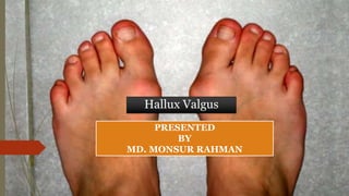
2. What is Hallux valgus?
Hallux valgus is a progressive foot deformity in which the first metatarsophalangeal (MTP)
joint is affected and is often accompanied by significant functional disability and foot pain
This joint is gradually subluxed (lateral deviation of the MTP joint) resulting in an abduction
of the first metatarsal while the phalanges adduct
This often leads to development of soft tissue and bony prominence on the medial side of what
is called a bunion
At a late stage, these changes lead to pain and functional deficit: i.e. impaired gait (lateral and
posterior weight shift, late heel rise, decreased single-limb balance, pronation deformity)
3. Clinically Relevant Anatomy
The Hallux or first toe, is the medio-distal part of the foot. It is formed by the first metatarsal
(articulates with two sesamoid bones), the first proximal phalanx and the first distal phalanx.
So the hallux is formed by three bones instead of four, unlike the other toes who have an extra
bone called the intermediate phalanx.
4. Epidemiology
The exact etiology is not well established. However, certain factors have been
considered to play a role in the development of hallux valgus;
• Gender (10x more frequent in women)
• Congenital deformity or predisposition
• Chronic Achilles tightness
• Severe flatfoot
• Hypermobility of the first metatarsocunieform joint
• Systemic disease
• Wearing tight shoes and/or heeled shoes between 20 and 39 years of age can be crucial in the
development of hallux valgus in later years.
• It is also possible that abnormal muscle insertions are partly responsible for hallux valgus.
• Hallux valgus is also associated with hip and knee OA and is inversely associated with a
higher BMI
5. Clinical Presentation
• Gait deviations in the midstance and the propulsion phase .As the body weight moves forward on a
foot on the ground, the patient will tend to keep his weight on the lateral border of the foot. This leads
to a lateral and posterior weight shift
• Patient has also a pronation deformity
• Patient is unable to supinate his / her foot and will tend to keep his body weight on the lateral border
of the foot which results in a late heel rise
• The period of single-limb support will be diminished
When a physical examination is executed, the following indications could be present:
• Lateral deviation of the MTP joint
• Swelling of first MTP joint
• Shortening of flexor hallucis brevis muscle
• Tenderness of hallux
• Weakness of hallux abductor muscles
• Pain (primary symptom)
6. Hallux valgus angle 𝝰 between the axis of
the first metatarsal and the first
proximal phalanx
The severity of the hallux deformity is
measured by (A) hallux valgus angle
and (B) intermetatarsal 1-2 angle
7. Differential Diagnosis
In the early stages, the redness and pain can be confused with an inflammation, infection or gout
of the first MTP joint.
Other forms of arthritis and their consequences for example hallux rigidus can be confused with
hallux valgus as well as being a result of the deformity. For example, a joint affected by septic
arthritis is also red and swollen.
Pain and swelling due to turf toe can be confused with hallux valgus.
Surgical or traumatic arthropathy
8. Diagnostic Procedures
Radiographs are used to determine the presence of hallux valgus by looking at the
angle formed between the longitudinal bisections of the first metatarsal and the
proximal phalanx.
If the angle is greater than 15°, hallux valgus is diagnosed. An angle of 45-50° is
considered serious
9. Management / Interventions
Non-operative treatment
• Adjustment of footwear to help in eliminating friction at the level of the medial eminence (bunion)
e.g., patients should be provided of a shoe with a wider and deeper toe box
• The condition of pes planus may be helped by an orthosis. Severe pes planus can lead to a
recurrence of hallux valgus following surgery.
• Achilles tendon contracture may require stretching or even lengthening (level of evidence: 4)
10. Operative treatment
There are several surgical procedures that we can apply depending on the severity of the
injury:
• Austin/Chevron Procedure
This procedure is frequently used for mild deformities. The osteotomy is in a "V" shape, a sagittal saw is used in a
medial to lateral direction inside the first metatarsal head. The loose fragment is then placed differently to correct the
first metatarsal angle. The fragment is fixated with pins or screws.
• Reverdin Procedure
A wedge is removed from the head of the first metatarsal head in order to get a better organization of the articular
cartilage. The wedge is situated on the dorsal side and medially based. In some cases, the surgeon decides that is
necessary to rotate the articular cartilage. A screw or K-wire is used for fixation.
• Scarf Procedure
When the deformity is moderate to sever, the scarf procedure is a frequently used option. The osteotomy is
longitudinal, in a medial to lateral direction, inside the shaft. The capital piece is moved more to the lateral side and
stabilized with two screws.
• Closing Base Wedge Procedure
Base procedures are mostly used for severe deformities, so is this one. A wedge is made in the proximal metatarsal, on
lateral side. When the wedge is removed, the distal part is translated to the lateral side causing the gap to close and
the first metatarsal to align with the second. The minimum fixation is a bicortical screw.
11. Cont…
• Lapidus Arthrodesis
This is another option when a severe deformity is observed. By removing a piece of the
articular cartilage of the medial cuneiform and the base of the first metatarsal, a fusion
between the two is created. The fixation can be external, by the use of a plate or screws.
• Akin Procedure
An extra correction can be constructed when a wedge is made on the medial side of the first
proximal phalanx. The gap is closed by pushing the distal part to the medial side and fixating it.
The similarities with the closing base wedge procedure stand out.
• First Metatarsal-Phalangeal Joint Arthrodesis
The permanent elimination of motion is the reason that this procedure is one of the last
options to treat a significant deformity. Most of the time, there is another component that
motivates the choice of this joint destructive procedure: degenerative arthrose or an unstable
joint. The proximal phalanx is correctly positioned towards the first metatarsal and is then
fixated through the joint with screws, plates or crossing K-wires.
12. Post-operative Management
For all surgical procedures, the patient is allowed to ambulate in a post-operative shoe
immediately after surgery. Patients need to wear a post-op shoe and compressive dressings for
8 weeks. Long-term follow-up has shown equally positive outcomes after Chevron osteotomy for
both patients both younger and older than 50 years of age.
13. Physical Therapy Management
Adjusted footwear with wider and deeper tip
Increase extension of MTP joint
Relieve weight-bearing stresses (orthosis)
Sesamoid Mobilization: *The physical therapist performs grade III joint mobilizations
on the medial and lateral sesamoid of the affected first MPJ. One thumb is placed on
the proximal aspect of the sesamoid and is used to apply a force from proximal to
distal that causes the sesamoid to reach the end range of motion (distal glides). These
are performed with large-amplitude rhythmic oscillations. No greater than 20° of
movement of the MPJ should be allowed during the technique.( Level of evidence 2B)
Strengthening of peroneus longus (Level of evidence 2A)
Physiotherapists should contain an expanded program, including ultrasound,
ice, electrical stimulation, MTJ mobilizations and exercises. This is more effective than
physical therapy alone. The combination will result in a increase in ROM of the MTP
joint, strength and function, and also a decrease in pain .(Level of evidence 2B)
14. Cont…
Gait Training: (Level of evidence 2B)
• Stance phase: could be trained by performing a heel-strike in its physiological
position at the lateral aspect of the heel.
• Stance phase could be followed by weight bearing of the first metatarsal during
midstance and terminal stance, with training of active push-off by the hallux
flexors, the flexor digitorum longus and brevis muscles and the lumbrical
muscles (Level of evidence 1: 2B, Level of evidence 2: 2C)
15. PHASE I - Pain Relief. Minimise Swelling & Injury Protection (Level of evidence 5)
Pain is the main reason that patients seek treatment for a bunion. Inflammation is best eased
using ice therapy, techniques (e.g. soft tissue massage, acupuncture, unloading taping techniques)
or exercises that unload the inflamed structures. Anti-inflammatory medications may help.
Orthotics can also be used to offload the bunion.
PHASE II - Restoring Normal ROM & Posture (Level of evidence 5)
As pain and inflammation settles, the focus of treatment turns to restoring normal toe and foot
joint range of motion and muscle length.
Treatment may include;
• Joint mobilization (abduction and flexion) and alignment techniques (between the first and the
second metatarsal)
• Muscle and joint stretches
• Taping
• Bunion splint or orthotic
• Bunion stretch and soft tissue release.
16. PHASE III - Restore Normal Muscle Control & Strength( Level of evidence 5)
• A foot posture correction Program to assist you to regain your normal foot posture.
Dorsiflexion Strengthening with Elastic Resistance Band
The patient is positioned in long-sitting. The centre of the resistance band is placed on the top of the
forefoot with the toes slightly pointed. The ends of the band are either held by an assistant or secured
against an immovable object. The patient then dorsiflexes the ankle, pulling "towards their nose,"
working against the resistance of the band.
Towel curls The patient spreads out a small towel on the floor, curling his/her toes around it and
pulling the towel towards them.
17. PHASE IV - Restoring Full Function
o The goal of this stage of rehabilitation is to return the patient to his/her desired activities.
o Everyone has different demands for their feet that will determine what specific treatment goals
need to be achieved.
PHASE V - Preventing a Recurrence
o Bunions will deform further with no attention and bunion-associated pain has a tendency to
return. The main reason is biomechanical.
o In addition to muscle control, the physiotherapist should assess foot biomechanics and may
recommend either a temporary off-the shelf orthotic or refer for a custom-made orthotic.
o High heeled shoes and shoes with tight or angular toe boxes should be avoided