1. Introduction The Enterobacteriaceae are a large, heterogeneous group of gram-negative rods whose natural habitat is the intestinal tract of humans and animals. The family includes many genera ( Escherichia, Shigella, Salmonella, Enterobacter, Klebsiella, Serratia, Proteus, and others). Some enteric organisms, eg, Escherichia coli, are part of the normal flora and incidentally cause disease, while others, the salmonellae and shigellae, are regularly pathogenic for humans. The Enterobacteriaceae are facultative anaerobes or aerobes, ferment a wide range of carbohydrates, possess a complex antigenic structure, and produce a variety of toxins and other virulence factors. Enterobacteriaceae, enteric gram-negative rods, and enteric bacteria, may also be called coliforms.
2. . The taxonomy of the Enterobacteriaceae is complex and rapidly changing since the introduction of techniques that measure evolutionary distance, such as nucleic acid hybridization and sequencing. More than 25 genera and 110 species or groups have been defined; however, the clinically significant Enterobacteriaceae comprise 20–25 species, and other species are encountered infrequently. The family Enterobacteriaceae have the following characteristics: They are gram-negative rods, either motile with peritrichous flagella or nonmotile; they grow on peptone or meat extract media without the addition of sodium chloride or other supplements; grow well on MacConkey's agar; grow aerobically and anaerobically (are facultative anaerobes); ferment glucose, often with gas production; are catalase-positive, oxidase-negative, and reduce nitrate to nitrite; have a 39–59% G + C DNA content.
3. Morphology & Identification Typical Organisms The Enterobacteriaceae are short gram-negative rods. Typical morphology is seen in growth on solid media in vitro, but morphology is highly variable in clinical specimens. Capsules are large and regular in klebsiella, less in enterobacter, and uncommon in the other species. Culture E coli and most of the other enteric bacteria form circular, convex, smooth colonies with distinct edges. Enterobacter colonies are similar but somewhat more mucoid. Klebsiella colonies are large and very mucoid and tend to coalesce with prolonged incubation. The salmonellae and shigellae produce colonies similar to E coli but do not ferment lactose. Some strains of E coli produce hemolysis on blood agar.
4. Growth Characteristics Carbohydrate fermentation patterns and the activity of amino acid decarboxylases and other enzymes are used in biochemical differentiation of some tests, eg, the production of indole from tryptophan, are commonly used in rapid identification systems, while others, eg, the Voges-Proskauer reaction (production of acetylmethylcarbinol from dextrose), are used less often. Culture on "differential" media that contain special dyes and carbohydrates (eg, eosin-methylene blue [EMB], MacConkey's, or deoxycholate medium) distinguishes lactose-fermenting (colored) from non-lactose-fermenting colonies (nonpigmented) and may allow rapid presumptive identification of enteric bacteria
5.
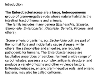
6. Enterobacter aerogenes (left) – E. coli (bright red) + Reagent: Methyl red indicator identifies pH change due to mixed acid fermentation Methyl Red (MR) (IMViC tests)
7. Enterobacter aerogenes +(left) E. coli – (right) Barritt’s reagent Tests for acetoin, precursor to 2,3 butanediol fermentation Voges – Proskauer (VP) (IMViC tests)
8.
9. . Rapid, Presumptive Identification of Gram-Negative Enteric Bacteria. Lactose Fermented Rapidly Escherichia coli: metallic sheen on differential media; motile; flat, nonviscous colonies Enterobacter aerogenes: raised colonies, no metallic sheen; often motile; more viscous growth Klebsiella pneumoniae: very viscous, mucoid growth; nonmotile Lactose Fermented Slowly Edwardsiella, Serratia, Citrobacter, Arizona, Providencia, Erwinia Lactose Not Fermented Shigella species: nonmotile; no gas from dextrose Salmonella species: motile; acid and usually gas from dextrose Proteus species: "swarming" on agar; urea rapidly hydrolyzed (smell of ammonia) Pseudomonas species soluble pigments, blue-green and fluorescing; sweetish smell
10. Many complex media have been devised to help in identification of the enteric bacteria. One such medium is triple sugar iron (TSI) agar, which is often used to help differentiate salmonellae and shigellae from other enteric gram-negative rods in stool cultures. The medium contains 0.1% glucose, 1% sucrose, 1% lactose, ferrous sulfate (for detection of H2S production), tissue extracts (protein growth substrate), and a pH indicator (phenol red). It is poured into a test tube to produce a slant with a deep butt and is inoculated by stabbing bacterial growth into the butt. If only glucose is fermented, the slant and the butt initially turn yellow from the small amount of acid produced; as the fermentation products are subsequently oxidized to CO2 and H2O and released from the slant and as oxidative decarboxylation of proteins continues with formation of amines, the slant turns alkaline (red). If lactose or sucrose is fermented, so much acid is produced that the slant and butt remain yellow (acid). Salmonellae and shigellae typically yield an alkaline slant and an acid butt.
11. Escherichia group E coli typically produces positive tests for indole, lysine decarboxylase, and mannitol fermentation and produces gas from glucose. An isolate from urine can be quickly identified as E coli by its hemolysis on blood agar, typical colonial morphology with an iridescent "sheen" on differential media such as EMB agar, and a positive spot indole test. Klebsiella-Enterobacter-Serratia Group Klebsiella species exhibit mucoid growth, large polysaccharide capsules, and lack of motility, and they usually give positive tests for lysine decarboxylase and citrate. Most Enterobacter species give positive tests for motility, citrate, and ornithine decarboxylase and produce gas from glucose. Enterobacter aerogenes has small capsules. Serratia produces DNase, lipase, and gelatinase. Klebsiella, enterobacter, and serratia usually give positive Voges-Proskauer reactions.
12. Proteus-Morganella-Providencia Group The members of this group deaminate phenylalanine, are motile, grow on potassium cyanide medium (KCN), and ferment xylose. Proteus species move very actively by means of peritrichous flagella, resulting in "swarming" on solid media unless the swarming is inhibited by chemicals, eg, phenylethyl alcohol or CLED (cystine-lactose-electrolyte-deficient) medium. Proteus species and Morganella morganii are urease-positive, while Providencia species usually are urease-negative. The proteus-providencia group ferments lactose very slowly or not at all. These bacteria typically are citrate-positive and differ from the salmonellae in that they do not decarboxylate lysine.
13. Shigella Shigellae are nonmotile and usually do not ferment lactose but do ferment other carbohydrates, producing acid but not gas. They do not produce H2S. The four Shigella species are closely related to E coli. Many share common antigens with one another and with other enteric bacteria (eg, Hafnia alvei and Plesiomonas shigelloides ). Salmonella Salmonellae are motile rods that characteristically ferment glucose and mannose without producing gas but do not ferment lactose or sucrose. Most salmonellae produce H2S. They are often pathogenic for humans or animals when ingested. Arizona is included in the salmonella group. Other Enterobacteriaceae Yersinia ,occasionally found in human infections include Edwardsiella and Ewingella, Hafnia, Cedecea, and Kluyvera .
14. Antigenic Structure Enterobacteriaceae have a complex antigenic structure. They are classified by more than 150 different heat-stable somatic O (lipopolysaccharide) antigens, more than 100 heat-labile K (capsular) antigens, and more than 50 H (Flagellar) antigens in Salmonella typhi, the capsular antigens are called Vi antigens.
15. Antigenic structures antigens are the most external part of the cell wall lipopolysaccharide and consist of repeating units of polysaccharide. Some O-specific polysaccharides contain unique sugars. O antigens are resistant to heat and alcohol and usually are detected by bacterial agglutination. Antibodies to O antigens are predominantly IgM. While each genus of Enterobacteriaceae is associated with specific O groups, a single organism may carry several O antigens. Thus, most shigellae share one or more O antigens with E coli. Occasionally, O antigens may be associated with specific human diseases, eg, specific O types of E coli are found in diarrhea and in urinary tract infections.
16. Antigenic structures contd. K antigens are external to O antigens on some but not all Enterobacteriaceae. Some are polysaccharides, including the K antigens of E coli; others are proteins. K antigens may be associated with virulence (eg, E coli strains producing K1 antigen are prominent in neonatal meningitis, and K antigens of E coli cause attachment of the bacteria to epithelial cells prior to gastrointestinal or urinary tract invasion). polysaccharides of Haemophilus influenzae or Neisseria meningitidis.
17. Colicins (Bacteriocins)Many gram-negative organisms produce bacteriocins. These virus-like bactericidal substances are produced by certain strains of bacteria active against some other strains of the same or closely related species. Their production is controlled by plasmids. Colicins are produced by E coli, marcescens by serratia, and pyocins by pseudomonas. Bacteriocin-producing strains are resistant to their own bacteriocin; Most gram-negative bacteria possess complex lipopolysaccharides in their cell walls. These substances, endotoxins, have a variety of pathophysiologic effects .Many gram-negative enteric bacteria also produce exotoxins of clinical importance.
18. Klebsiellae form large capsules consisting of polysaccharides (K antigens) covering the somatic (O or H) antigens and can be identified by capsular swelling tests with specific antisera. Human infections of the respiratory tract are caused particularly by capsular types 1 and 2; those of the urinary tract, by types 8, 9, 10, and 24. H antigens are located on flagella and are denatured or removed by heat or alcohol. . The determinants in H antigens are a function of the amino acid sequence in flagellar protein (flagellin).
19. Diseases Caused by Enterobacteriaceae Causative Organisms E coli is a member of the normal intestinal flora . Other enteric bacteria ( Proteus, Enterobacter, Klebsiella, Morganella, Providencia, Citrobacter, and Serratia species) are also found as members of the normal intestinal flora but are considerably less common than E coli . The enteric bacteria are sometimes found in small numbers as part of the normal flora of the upper respiratory and genital tracts. The enteric bacteria generally do not cause disease, and in the intestine they may even contribute to normal function and nutrition. When clinically important infections occur, they are usually caused by E coli, but the other enteric bacteria are causes of hospital-acquired infections and occasionally cause community-acquired infections. The bacteria become pathogenic only when they reach tissues outside of their normal intestinal. The most frequent sites of clinically important infection are the urinary tract, biliary tract, and other sites in the abdominal cavity, but any anatomic site (eg, prostate gland, lung, bone, meninges) can be the site of disease.
20. Some of the enteric bacteria (eg, Serratia marcescens, Enterobacter aerogenes ) are opportunistic pathogens. When normal host defenses are inadequate—particularly in infancy or old age, in the terminal stages of other diseases, after immunosuppression, or with indwelling venous or urethral catheters—localized clinically important infections can result, and the bacteria may reach the blood stream and cause sepsis .
21. Urinary Tract Infection E coli is the most common cause of urinary tract infection and accounts for approximately 90% of first urinary tract infections in young women . The symptoms and signs include urinary frequency, dysuria, hematuria, and pyuria. Flank pain is associated with upper tract infection. None of these symptoms or signs is specific for E coli infection. Urinary tract infection can result in bacteremia with clinical signs of sepsis. E coli -Associated Diarrheal Diseases E coli that cause diarrhea are extremely common worldwide. These E coli are classified by the characteristics of their virulence properties, and each group causes disease by a different mechanism.
22. Enteropathogenic E coli (EPEC) is an important cause of diarrhea in infants , especially in developing countries. EPEC previously was associated with outbreaks of diarrhea in nurseries in developed countries. EPEC adhere to the mucosal cells of the small bowel. Genetically mediated factors promote tight adherence. There is loss of microvilli (effacement), formation of filamentous actin pedestals or cup-like structures, and, occasionally, entry of the EPEC into the mucosal cells. Characteristic lesions can be seen on electron micrographs of small bowel biopsy lesions. The result of EPEC infection is watery diarrhea, which is usually self-limited but can be chronic. EPEC diarrhea has been associated with multiple specific serotypes of E coli; strains are identified by O antigen and occasionally by H antigen typing. . The duration of the EPEC diarrhea can be shortened and the chronic diarrhea cured by antibiotic treatment .
23. Enterotoxigenic E coli (ETEC) is a common cause of "traveler's diarrhea" and a very important cause of diarrhea in infants in developing countries. ETEC colonization factors specific for humans promote adherence of ETEC to epithelial cells of the small bowel. Some strains of ETEC produce a heat-labile exotoxin (LT) (MW 80,000) that is under the genetic control of a plasmid. Its subunit B attaches to the GM1 ganglioside at the brush border of epithelial cells of the small intestine and facilitates the entry of subunit A (MW 26,000) into the cell, where the latter activates adenylyl cyclase. This markedly increases the local concentration of cyclic adenosine monophosphate (cAMP), which results in intense and prolonged hypersecretion of water and chlorides and inhibits the reabsorption of sodium. The gut lumen is distended with fluid, and hypermotility and diarrhea ensue, lasting for several days. LT is antigenic and cross-reacts with the enterotoxin of Vibrio cholerae.
24. Enterohemorrhagic E coli (EHEC) produces verotoxin, named for its cytotoxic effect on Vero cells, a line of African green monkey kidney cells. There are at least two antigenic forms of the toxin. EHEC has been associated with hemorrhagic colitis, a severe form of diarrhea, and with hemolytic uremic syndrome, a disease resulting in acute renal failure, microangiopathic hemolytic anemia, and thrombocytopenia. Verotoxin has many properties that are similar to the Shiga toxin produced by some strains of Shigella dysenteriae type 1; however, the two toxins are antigenically and genetically distinct.
25. Enteroinvasive E coli (EIEC) produces a disease very similar to shigellosis. The disease occurs most commonly in children in developing countries and in travelers to these countries. Like shigella, EIEC strains are nonlactose or late lactose fermenters and are nonmotile. EIEC produce disease by invading intestinal mucosal epithelial cells. Enteroaggregative E coli (EAEC) causes acute and chronic diarrhea (> 14 days in duration) in persons in developing countries. These organisms also are the cause of food-borne illnesses in industrialized countries. They are characterized by their characteristic pattern of adherence to human cells. EAEC produce ST-like toxin (see above) and a hemolysin. Sepsis When normal host defenses are inadequate, E coli may reach the bloodstream and cause sepsis. Newborns may be highly susceptible to E coli sepsis because they lack IgM antibodies. Sepsis may occur secondary to urinary tract infection. Meningitis
26. Meningitis E coli and group B streptococci are the leading causes of meningitis in infants. Approximately 75% of E coli from meningitis cases have the K1 antigen. This antigen cross-reacts with the group B capsular polysaccharide of N meningitidis. The mechanism of virulence associated with the K1 antigen is not understood. Klebsiella-Enterobacter-Serratia; Proteus-Morganella-Providencia; and Citrobacter The pathogenesis of disease caused by these groups of enteric gram-negative rods is similar to that of the nonspecific factors in disease caused by E coli . Klebsiella K pneumoniae is present in the respiratory tract and feces of about 5% of normal individuals. It causes a small proportion (about 1%) of bacterial pneumonias. K pneumoniae can produce extensive hemorrhagic necrotizing consolidation of the lung. It occasionally produces urinary tract infection and bacteremia with focal lesions in debilitated patients. Other enterics also may produce pneumonia. K pneumoniae and Klebsiella oxytoca cause hospital-acquired infections. Two other klebsiellae are associated with inflammatory conditions of the upper respiratory tract: Klebsiella ozaenae has been isolated from the nasal mucosa in ozena, a fetid, progressive atrophy of mucous membranes; and Klebsiella rhinoscleromatis from rhinoscleroma, a destructive granuloma of the nose and pharynx. Enterobacter aerogenes This organism has small capsules, may be found free-living as well as in the intestinal tract, and causes urinary tract infections and sepsis.
27. Serratia S marcescens is a common opportunistic pathogen in hospitalized patients. Serratia (usually nonpigmented) causes pneumonia, bacteremia, and endocarditis—especially in narcotics addicts and hospitalized patients. Only about 10% of isolates form the red pigment (prodigiosin) that has long characterized Serratia marcescens. S marcescens is often multiply resistant to aminoglycosides and penicillins; infections can be treated with third-generation cephalosporins. Proteus Proteus species produce infections in humans only when the bacteria leave the intestinal tract. They are found in urinary tract infections and produce bacteremia, pneumonia, and focal lesions in debilitated patients or those receiving intravenous infusions. P mirabilis causes urinary tract infections and occasionally other infections. Proteus vulgaris and Morganella morganii are important nosocomial pathogens. Proteus species produce urease, resulting in rapid hydrolysis of urea with liberation of ammonia. Thus, in urinary tract infections with proteus, the urine becomes alkaline, promoting stone formation and making acidification virtually impossible. The rapid motility of proteus may contribute to its invasion of the urinary tract. Strains of proteus vary greatly in antibiotic sensitivity. P mirabilis is often inhibited by penicillins; the most active antibiotics for other members of the group are aminoglycosides and cephalosporins.
28. Providencia Providencia species ( Providencia rettgeri, Providencia alcalifaciens, and Providencia stuartii ) are members of the normal intestinal flora. All cause urinary tract infections and occasionally other infections and are often resistant to antimicrobial therapy. Citrobacter the disease process Citrobacter can cause urinary tract infections and sepsis. Diagnostic Laboratory Tests Specimens Specimens included urine, blood, pus, spinal fluid, sputum, or other material, as indicated by the localization of.
29. The ShigellaeThe natural habitat of shigellae is limited to the intestinal tracts of humans and other primates, where they produce bacillary dysentery. Morphology & Identification Typical OrganismsShigellae are slender gram-negative rods; coccobacillary forms occur in young cultures. Culture Shigellae are facultative anaerobes but grow best aerobically. Convex, circular, transparent colonies with intact edges reach a diameter of about 2 mm in 24 hours. Growth Characteristics All shigellae ferment glucose. With the exception of Shigella sonnei, they do not ferment lactose. The inability to ferment lactose distinguishes shigellae on differential media. Shigellae form acid from carbohydrates but rarely produce gas. They may also be divided into those that ferment mannitol and those that do not ++
30. Antigenic Structure Shigellae have a complex antigenic pattern. There is great overlapping in the serologic behavior of different species, and most of them share O antigens with other enteric bacilli. The somatic O antigens of shigellae are lipopolysaccharides. Their serologic specificity depends on the polysaccharide. There are more than 40 serotypes. Pathogenesis & Pathology Shigella infections are almost always limited to the gastrointestinal tract; bloodstream invasion is quite rare. Shigellae are highly communicable; the infective dose is on the order of 103 organisms (whereas it usually is 105–108 for salmonellae and vibrios). The essential pathologic process is invasion of the mucosal epithelial cells (eg, M cells) by induced phagocytosis, escape from the phagocytic vacuole, multiplication and spread within the epithelial cell cytoplasm, and passage to adjacent cells. Microabscesses in the wall of the large intestine and terminal ileum lead to necrosis of the mucous membrane, superficial ulceration, bleeding, and formation of a "pseudomembrane" on the ulcerated area. This consists of fibrin, leukocytes, cell debris, a necrotic mucous membrane, and bacteria. As the process subsides, granulation tissue fills the ulcers and scar tissue forms.
31. Toxins Endotoxin Upon autolysis, all shigellae release their toxic lipopolysaccharide. This endotoxin probably contributes to the irritation of the bowel wall. Shigella dysenteriae Exotoxin S dysenteriae type 1 (Shiga bacillus) produces a heat-labile exotoxin that affects both the gut and the central nervous system. The exotoxin is a protein that is antigenic (stimulating production of antitoxin) and lethal for experimental animals. Acting as an enterotoxin, it produces diarrhea as does the E coli verotoxin, perhaps by the same mechanism. In humans, the exotoxin also inhibits sugar and amino acid absorption in the small intestine. Acting as a "neurotoxin," this material may contribute to the extreme severity and fatal nature of S dysenteriae infections and to the central nervous system reactions observed in them (ie, meningismus, coma). Patients with Shigella flexneri or Shigella sonnei infections develop antitoxin that neutralizes S dysenteriae exotoxin in vitro. The toxic activity is distinct from the invasive property of shigellae in dysentery. The two may act in sequence, the toxin producing an early nonbloody, voluminous diarrhea and the invasion of the large intestine resulting in later dysentery with blood and pus in stools.
32. Clinical Findings After a short incubation period (1–2 days), there is a sudden onset of abdominal pain, fever, and watery diarrhea. The diarrhea has been attributed to an exotoxin acting in the small intestine (see above). A day or so later, as the infection involves the ileum and colon, the number of stools increases; they are less liquid but often contain mucus and blood. Each bowel movement is accompanied by straining and tenesmus (rectal spasms), with resulting lower abdominal pain. In more than half of adult cases, fever and diarrhea subside spontaneously in 2–5 days. However, in children and the elderly, loss of water and electrolytes may lead to dehydration, acidosis, and even death. The illness due to S dysenteriae may be particularly severe. On recovery, most persons shed dysentery bacilli for only a short period, but a few remain chronic intestinal carriers and may have recurrent bouts of the disease. Upon recovery from the infection, most persons develop circulating antibodies to shigellae, but these do not protect against reinfection. Diagnostic Laboratory Tests Specimens Specimens include fresh stool, mucus flecks, and rectal swabs for culture. Large numbers of fecal leukocytes and some red blood cells often are seen microscopically. Serum specimens, if desired, must be taken 10 days apart to demonstrate a rise in titer of agglutinating antibodies.
33. Culture The materials are streaked on differential media (eg, MacConkey's or EMB agar) and on selective media (Hektoen enteric agar or salmonella-shigella agar), which suppress other Enterobacteriaceae and gram-positive organisms. Colorless (lactose-negative) colonies are inoculated into triple sugar iron agar. Organisms that fail to produce H2S, that produce acid but not gas in the butt and an alkaline slant in triple sugar iron agar medium, and that are nonmotile should be subjected to slide agglutination by specific shigella antisera. Serology Normal persons often have agglutinins against several Shigella species. However, serial determinations of antibody titers may show a rise in specific antibody. Serology is not used to diagnose shigella infections. Immunity Infection is followed by a type-specific antibody response. Injection of killed shigellae stimulates production of antibodies in serum but fails to protect humans against infection. IgA antibodies in the gut may be important in limiting reinfection; these may be stimulated by live attenuated strains given orally as experimental vaccines. Serum antibodies to somatic shigella antigens are IgM.