
Recommandé
Contenu connexe
Similaire à ANATOMY OF RETINA.pptx
Similaire à ANATOMY OF RETINA.pptx (20)
Plus de MalavikaAG
Plus de MalavikaAG (15)
Dernier
Dernier (20)
ANATOMY OF RETINA.pptx
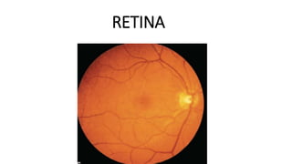
- 1. RETINA
- 2. • Inner nervous coat of eyeball • FUNDUS • Thin, transparent membrane • purplish red in colour • Formation of optical image • Thickness- 0.56mm near optic disc to 0.1mm at ora serrata • Thinnest at centre of fovea
- 3. EXTENSION • Outer surface- Bruchs membrane of choroid • Inner surface- vitreous • Posteriorly- optic nerve • Anteriorly- epithelium of iris and • ciliary body(ora serrata) • Ant- extends more on nasal side • Firmly attached- margins of optic disc & ora serrata
- 5. Optic disc Macula lutea Peripheral retina OPTIC DISC • 1.5mm in diameter & 3mm nasal to macula • Pale pinK • Depression in centre- physiological cup • Central retinal vessels • Optic nerve- lamina cribrosa • Blind spot
- 6. • MACULA – • oval,yellowish,at centre of posterior part • 5mm in diameter & 3mm temporal to optic disc • carotenoid pigment-xanthophyll(present from outer nuclear layer onward) • Central depression – fovea centralis • Sides of depression – Clivus • Floor of depression – foveola • 1,47,000 per sq.mm
- 7. • Umbo- foveolar reflex • Central bouquet of cones • Parafovea-0.5mm surrounding fovea • Perifovea-1.5mmsurrounding parafovea
- 8. • Near periphery – 1.5mm ring peripheral to temporal vascular Arcades • Equatorial retina- retina around the equator • Peripheral retina – region anterior to it
- 9. • OUTER PIGMENTED LAYER • Retinal pigment epithelium (RPE) • INNER NEUROSENSORY LAYER • Photoreceptors • External limiting membrane • Outer nuclear layer • Outer plexiform layer • Inner nuclear layer • Inner plexiform layer • Ganglion cell layer • Nerve fibre layer • Internal limiting membrane
- 10. • LAYERS OF RETINA
- 11. • EMRYOLOGICAL DEVELOPMENT • Outer pigmented layer- outer layer of optic cup • Neurosensory layer- inner layer of optic cup • anterior one fifth- columnar • Posterior four fifth • outer nuclear zone & inner marginal zone • Inner neuroblastic layer –ganglion cells,amacrine cells,Muller cells • Outer neuroblastic layer –rods,cones,bipolar cells • Macular area- superimposed nuclei of ganglion cell layer, lateral to optic disc
- 12. • DEVELOPMENT
- 13. • RETINAL PIGMENTED EPITHELIUM • Single layer of hexagonal cells • Narrow & tall Posteriorly ,flattened near ora serrata • Apical end- microvilli – project between outer segment of rods & cone • Embedded in glycosaminoglycans • Cell nucei- basal part • Melanin granules • Basal region- zona adherens,apical region- zona occludens
- 14. • FUNCTION • Absorption of light • Turn over of outer segment of rods& cones • Formation of rhodopsin& iodopsin( vitamin A) • SUB RETINAL SPACE • - Retinal detachment • Ocular albinism • -transillumination of iris
- 15. NEURAL RETINA • PHOTO RECEPTORS • BIPOLAR CELLS • GANGLION CELLS
- 16. • PHOTORECEPTORS • RODS • 110 to 125 million • Rhodopsin- scotopic vision • 40to 60micrometre long • STRUCTURE • Outer segment – cylindrical & contains lamellar disc • Eccentric cilium- 9 doublet microtubules with no Central pair • Inner segment – ellipsoid& myoid • Ellipsoid (outer segment) – mitochondria • Myoid- (inner segment- ER,Golgi apparatus,ribosomes
- 17. • Outer rod fibre • Rod granule( cell body with nucleus) • Inner rod fibre • Rod spherule
- 18. • CONES • 6.3 to 6.8 million • Iodopsin, scotopic vision • 65to 75micrometre long • Outer segment – conical,wider base & • rounded tip • Continuous with extra cellular space • Cilium • Inner segment – ellipsoid ( very plump)& myoid • Cell body with nucleus • Inner cone fibre • Cone pedicle
- 19. BIPOLAR CELLS • First order neurons (neurons in posterior root ganglion) • Rod bipolar cells several rod cells to 1to 4 ganglion cells • Flat or diffuse bipolar cells –many cone cells with many ganglion cells • Midget bipolar cell- single cone cell with single midget ganglion cell
- 20. • GANGLION CELLS • Second order neuron-nervous ganglion) • Increases from periphery to macula(10 layers) • Fovea- absent • HORIZONTAL CELLS • Associated with cones- 7 • Associateed with rods – 10 to 12 AMACRINE CELLS No axons,large cell body,abundant cytoplasm,lobulated nuclei Synapse with dendrites of ganglion cells& axons of bipolar cells
- 21. • Supporting cells- MULLER CELL • Fill in most of the space of neural retina,not occupied by neurons • On outer surface – row of zona adherents between photoreceptor cell& radial process of Muller cells- OUTER LIMITING MEMBRANE • On inner surface –muller cells have expanded termination covered by basement membrane – INNER LIMITING MEMBRANE •
- 22. • BLOOD SUPPLY • Outer 4 layers- chorio capillaries • Inner 6 layers- • Central retinal artery & vein • Course of Central retinal artery • circle of zinn or Haller
- 23. • Arterial branches run in nerve fibre layer • Foveal avascular zone- 500micronmetre in diameter • BLOOD RETINAL BARRIER • Tight junctions of RPE • Nonfenestrated endothelium of capillaries • CENTRAL RETINAL VEIN • Accompany CRA& leaves through lamina cribrosa • Lie on lateral side of artery in optic nerve • Longer course in subarachnoid space • Drains into cavernous sinus or superior ophthalmic vein
- 24. • ARRANGEMENT OF NERVE FIBRES IN RETINA
- 25. • THICKNESS OF NERVE FIBRE LAYER AT DISC • Most lateral quadrant(thinnest) • Upper and lower temporal quadrant • Most medial quadrant • Upper and lower nasal quadrant( thickest) • CLINICAL SIGNIFANCE • Papilledema- first in thickest quadrant • Glaucomatous damage- arcuate fibres(more sensitive) . Macular fibres(resistant)
- 26. CONGENITAL ANAMOLIES • Medullated nerve fibres
- 27. • COLOBOMA of optic disc- • Failure of closure of embryonic fissure • Minor defect or fully developed coloboma • HYPOPLASIA of optic disc • Small disc surrounded by yellowish ring- double ring sign • PERSISTENT HYALOID ARTERY • Bregmesters papillae • Mittendorf dot
- 29. • DRUSEN of optic disc • Intracapillary refractile bodies • Children- present as pseudopapilledema • Adults- waxy pea like irregular refractilebodies