SlideShare une entreprise Scribd logo
1  sur  257
Télécharger pour lire hors ligne
INFECTIOUS DISORDERS
Figure 6-3 Urinary Tract Infection
An infection (UTI) cause by bacteria, virus, fungus, that occurs in the urinary tract.
Risk for UTI increases when a patient has indwelling catheter, urinary retention,
urinary and fecal incontinence and poor perineal hygiene practices. Other risk factors
are renal scarring from previous infection, decrease ureteral peristalsis, presence of
urinary tract obstruction, being a female, sexual activity. The common causative
microorganisms are escherichia coli , staphylococcus, streptococcus,
enterobacter, klebsiella and aerobacter, pseudoaeriginosa.
Pathophysiology
The epithelilium of the kidneys, ureter and bladder are sterile in healthy
individuals, infection begins when bacteria enters , usually starting at the opening of
the urethra travelling up to the bladder. If the flushing or urinating cannot stop the
bacteria it can move up further to the ureters and kidney. Lower urinary tract
infection( UTI) rarely cause complications but upper UTI if untreated can spread into
the blood stream potential for chronic illness and death .
1. URETHRITIS – is inflammation of the urethra.
Causes
Microorganisms ( Escherichia coli, Chlamydia Trichomonas, Neisseria gonorrhoeae,
and herpes simplex virus type 2),trauma, or hyper-sensitivity to chemicals in products
such as vaginal deodorants, spermicidal jellies, or bubble bath detergents.
Signs and Symptoms
dysuria, frequency, urgency, and bladder spasms. A urethral discharge may
be noticed.
Medical Diagnosis
Based on patient signs and symptoms, urinalysis, and urethral smear
Medical Treatment
Antimicrobials when it is caused by microorganisms. If the patient is sexually
active, the patient and the sexual partner may be treated with antimicrobials to
prevent reinfection.
Nursing Intervention
Sitz baths are soothing and may reduce the pain . Instruct female patients to
wipe from front to back after toileting, and to void before and after sexual intercourse .
Discourage bubble baths and vaginal deodorant sprays. Instruct uncircumcised male
patients to clean the penis under the foreskin regularly. Advise patients to void after
swimming.
2. CYSTITIS - is inflammation of the urinary bladder. The most common
Causes
Bacterial contamination, prolonged immobility, renal calculi, urinary diversion,
indwelling catheters , radiation therapy, and treatment with some types of
chemotherapy.
Signs and Symptoms
urgency, frequency, dysuria, hematuria, nocturia, bladder spasms,
incontinence, and low-grade fever. Urine may be dark, tea colored, or cloudy.
Fever, fatigue, and pelvic or abdominal discomfort and bladder spasms
experienced as pain behind the symphysis pubis.
Medical Diagnosis
Urinalysis, culture, and sensitivity. The presence of bacteria does not mean that
the patient has an infection unless the patient also has white blood cells (WBCs) in
the urine.
Medical Treatment
Antibiotic , mild analgesic such as acetaminophen is useful for relieving
discomfort. Phenazopyridine (pyridium) and Oxybutynin chloride (Ditropan may be
ordered for 2 to 3 days to decrease discomfort and bladder spasms.
Nursing care
advise patient to complete the entire course of antibiotics and take analgesics as
ordered.If phenozopyradine is given, advise patient that the drug causes red-orange
urine Warm sitz bath , oral fluid intake 30m/kg of fluid per day.
To reduce risk of future infection, teach patient to, wear cotton undergraments, avoid
tight-fitting clothing in the perineal area, take shower instead of tub bath, avoid
caffeine drinks, apple, grapefruit, orange these irritates the bladder, maintain high
fluid intake and void often, Wiping from front to back after voiding for female and drink
a glass of water after swimming ,before and after intercourse to flush the bacteria.
3. PYELONEPHRITIS – inflammation of the renal pelvis. It may affect one or both
kidneys.
Cause
Acute pyelonephritis is most often caused by an ascending bacterial infection, but it
may be bloodborne. Chronic pyelonephritis most often is the result of reflux of urine
from inadequate closure of the ureterovesical junction during voiding. It is also
usually caused by long standing UTIs with relapses and reinfections, may even lead
to chronic renal failure .
Signs and Symptoms
High fever, chills, nausea, vomiting, and dysuria. Severe pain or a constant dull ache
in the flank area. The patient with chronic pyelonephritis experience fatigue,
hypertension, increase BUN and creatinine and a slight aching over one or both
kidneys.
Medical Diagnosis
Urinalysis, urine culture and sensitivity, CBC, IVP, cystoscopy
Medical Treatment
Antibiotics, urinary tract anti-septics, analgesics, and antispasmodics . Additional
medications may be needed to treat hypertension. Adults are advised to dink at least
eight 8-oz glasses of fluids daily. Intravenous fluids may be ordered if the patient has
nausea and vomiting. Dietary salt and protein restriction may be imposed on the
patient with chronic disease. follow-up cultures to determine whether the infection
has been resolved.
Nursing Interventions
Record the presence of signs and symptoms, history of previous urinary disorders,
fluid intake at least 8 oz of glasses a day, advise patient to complete the entire course
of antibiotics and take analgesics as ordered. Limit physical activity and exercise ,
protein and salt dietary restrictions if advised by the physician.
KIDNEY TRANSPLANT
- Is the surgical implantation of a human kidney from a compatible donor to a
recipient.
DONOR:
LIVING
1. ABO Screening
2. Tissue specific antigen & human leukocyte antigen histocompatibility
3. Excellent health
4. Full functioning kidneys
5. Emotionally ready
6. Full understanding of the process
CADAVER:
1. Below 60 yrs of age
2. Brain death
3. Normal renal function
4. No metastatic disease, HIV, Hep. B
S/S OF REJECTION
oliguria
fever
pain
tenderness over transplant site
hypertension
creatinine increase
COMPLICATION POST TRANSPLANT
rejection
infection
malignancy (basal and squamous cell carcinoma.-skin,lips, vulva, lungs
cardiovascular – hypertension
IMMUNOSUPPRESSIVE DRUGS
Cyclosporine (Neoral)- blocks interleukin
Azathioprine (imuran) – blocks DNA preventing
lymphocyte proliferation
Corticosteroids (prednisone)- block cytokines
Cyclophosphamide (cytoxan)
Tacrolimus (prograft) – blocks calcineurin and T cell
Mycophenolate (cellcept)- inhibit B & T lymphocyte
OKT 3 - antilymphocyte globulin
NURSING MANAGEMENT FOR RENAL TRANSPLANT
• Maintain fluid balance by monitoring I & O
• Monitor laboratory studies for graft function and electrolyte balance
• Monitor for s/s of graft rejection
• Monitor wound drainage
• Adhere to immunosuppression schedule
• Monitor blood glucose levels related to high glucocorticoids
• Arrange supply of medications prior to discharge to prevent disruption of
immunosupressions
• Arrange post discharge follow - up appointments
NURSING DIAGNOSES POST KIDNEY TRANSPLANT
• Altered nutrition : more than body requirements related to side effects of
immunosuppressant agents
Less than body requirement: related to increased caloric needs after
transplant
• Altered protection and risk for infection related to immunosuppression required after
transplantation
• Effective management of Therapeutic Regimen related to post-transplantation
regimen
• Pain related to transplantation surgery
• Risk for Ineffective Individual Coping after transplantation related to increased stress,
anxiety, fear, and lifestyle changes
• Risk for injury: rejection of transplanted organ related to impaired
immunocompetence
INTERNAL JUGULAR CATHETER
- a central venous catheter inserted through the internal jugular vein.
SUBCLAVIAN CATHETER
- Central venous access via the subclavian vein has several advantages over other
possible locations.
ARTERIOVENOUS GRAFT – artificial graft made of gore-tex or a bovine carotid
artery is used
Risks & Complications in Hemodialysis
• Hypotension
• Blood loss from technical problem
• Muscle cramps
• Disequilibrium syndrome
• Air embolism
• Hypoglycemia
• Cardiac arrythmia
Nursing Care Before and During dialysis
• Check the dialysis order { BFR, duration, bath, UFR, heparin, K+ } * SLED
• Chart client’s weight { DRY WEIGHT}
• Assess vital signs
• Check patency of vascular access
• Withhold antihypertensives
• Ensure bed rest with frequent position change
• Monitor closely for complications
• Monitor blood line connections, arterial and venous and transmembrane pressure
in the dialysis machine
Nursing Care Post Hemodialysis
• Check VS Nursing
• Record post dialysis weight
• Resume all medications
• Apply pressure dressing on decannulated sites
• Protect IJ/subclavian catheter with sterile dressing
REVIEW OF ANATOMY AND PHYSIOLOGY OF THE RENAL SYSTEM
Figure 6-1 Urinary System
The renal system is responsible for maintaining homeostasis in the body by carefully
regulating fluid and electrolytes, acid – base balance, removing wastes, and providing
hormones responsible for red blood cell production, hypertension and bone
metabolism. The renal system is composed of the upper and lower urinary tract.
Lower Urinary System
A. Bladder
The bladder is an extra peritoneal organ that lies behind the symphysis pubis .
Its main function is for storage of urine. As volume of urine increases, starting from
300-500 ml, awareness of the need to void develops. Voluntary voiding is
accomplished by stimulation of the parasympathetic nerve fibers causing coordinated
contraction of the detrusor muscle and the bladder body.
B. Urethra
The urethra drains urine from the bladder to an exterior opening of the body, the
external urethral orifice. In females, the urethra is about 3 to 4 cm. (1.5 in.). In males,
the urethra is about 15 to 20 cm (6 to 8 in.) Micturition, or urination, is the process of
releasing urine from the bladder into the urethra.
Upper Urinary System
Figure 6-2 Structure of the Kidneys
Kidneys
The two kidneys lie on the posterior wall of the abdomen outside the peritoneal cavity.
Each kidney of the adult human that weighs about 150g is about the size of an
indented region called the hilum through which passes the renal artery and vein,
lymphatic, nerve supply. The outer part is the cortex and inner region called medulla.
The medulla is divided into multiple cone shaped masses called renal pyramids. The
base of each pyramid terminates in the papilla, which projects into the space of renal
pelvis, a funnel shaped continuation of the upper end of the ureter. The outer border
of the pelvic is divided into minor calyces, the walls up the calyces, pelvis that contain
contractile elements that propel the urine toward the bladder, where urine is stored
until it is emptied by micturition.
The functional unit of the kidney is the nephron. Millions of nephrons are present
in each human kidney which aid in the urine production and process of removing
metabolic waste products from the blood. These significant structures extend between
the cortex and the medulla. At one end of the nephron is closed, expanded and folded
into a double-walled cuplike structure called the Bowman’s capsule. This capsule
encloses glomerulus, the nephron’s primary structure in filtering function.
Functions of Kidney
Kidney performs different functions in order to maintain homeostasis in the body
by excreting metabolic waste products and reabsorbing necessary elements for the
body. The following are the functions of the kidney:
1. Formation of urine
The formation of urine happens in three phases which are filtration, reabsorption
and secretion. Each of these processes happens in the body in order to create
homeostasis by removing those metabolic waste products and reabsorbing helpful
substances.
a. Filtration. The filtration process is nonselective, passive process which forms
essential blood plasma without blood protein but both of it is normally too large to
pass through the filtration membrane. If any of the two appeared in the urine, it would
mean that there is a problem in the glomerular filters. The water and solute are
smaller than proteins that are forced through the capillary walls and pores of
Bowman’s capsule into the renal tubule.
b. Reabsorption. Tubular reabsorption is achieved by active and passive transfer
mechanism Sodium, potassium, calcium, phosphate, and uric acid are actively
reabsorbed. Urea, water, chloride, some bicarbonates and some phosphate are
passively reabsorbed. Most reabsorption occurs in the proximal tubule which
conserves needed substances but does not reabsorb metabolic waste products.
c. Secretion. Some of substances such as hydrogen and potassium ion, creatinine,
and ammonia, move from the peritubular capillary blood and secreted by the tubule
cells into the filtration. Excreting nitrogenous waste products , unnecessary and
excess substances .
2. Body’s Water Volume Regulator
Regulation of water in the body contained in the blood is greatly influenced by
antidiuretic hormone (ADH), also called vasopressin. Vasopressin is produced in the
hypothalamus and stored in nearby pituitary gland. Receptors in the brain monitor the
blood’s water concentration causing the release of ADH in the bloodstream if the
amount of salt and other substances is too high. ADH in the bloodstream causes
more water to be reabsorbed into the bloodstream. In the absence of ADH, the
collecting ducts become impermeable to solute and water, making it less
concentrated than plasma and the urine is diluted.
3. Excretion of Metabolic Waste Products
The kidney functions as the body’s main excretory organ, eliminating the body’s
metabolic waste products and serves as the primary mechanism for excreting drug
metabolites. 25g to 30g of urea is produced as the end product of protein breakdown
and excreted daily making it the major waste product of protein metabolism. Other
waste products are creatinine, phosphates and sulfates. Uric acid, formed as a waste
product of purine metabolism, is also eliminated in the urine.
4. Blood Pressure Regulator
Regulating blood pressure is linked to the kidneys' ability to excrete enough
sodium chloride (salt) to preserve normal sodium balance, extracellular fluid volume
and blood volume.
5. Regulation of Acid-Base Balance
The kidney also adjusts the body’s acid-base balance to prevent such blood
disorders as acidosis and alkalosis. It helps maintain normal pH by retaining or
excreting hydrogen ions and regenerating lost buffer. The kidneys excrete acids that
the lungs cannot excrete and they can excrete hydrogen ions or reabsorb bicarbonate
to correct acidosis. They can reverse this process to correct alkalosis. Only renal
mechanisms can remove metabolic acids and excess bases from the body.
6. Regulation of Red Blood Cell Production
Decreasing amount of oxygen in the renal blood flow activates the release of
erythropoietin. Erythropoietin stimulates the bone marrow to produce red blood cells,
thereby increasing the amount of hemoglobin available to carry oxygen.
7. Vitamin D Synthesis
The kidneys are also responsible for the final conversion of inactive vitamin D to its
active form, 1,25-dihydroxychole-calciferol. Vitamin D is necessary for maintaining
normal calcium balance in the body.
8. Secretion of Prostaglandins
The kidneys also produce prostaglandin E and prostacyclin, which have a
vasodilatory effect and are important in maintaining renal blood flow.
DIALYSIS
• A technique in which a substance move from the blood through a semi permeable
membrane into a dialysis solution.
3 PHYSICAL PRINCIPLES (osmosis. Diffusion, ultrafiltration)
Indications of dialysis
1. GFR less than 5 to 1O ml/min
2. Manifestations of uremic syndrome
TWO TYPES
1. Hemodialysis – removal of waste and water by circulating the blood into a
dialyzer through a dialysis machine.
2. Peritoneal dialysis – repeated cycles of instilling dialysate into the peritoneal
cavity.
4 BASIC GOALS OF DIALYSIS
1. Remove waste products from protein metabolism.
2 .Maintain safe concentration of serum electrolytes
3. Correction of acidosis and replenish blood’s bicarbonate system.
4. Removal of excess water
Figure 6-8 Hemodialysis
Components of Hemodialysis
• Dialyzer – Artificial filter containing fine fibers acting as artificial kidney
• Dialysate - Dialysis solution or bath containing sodium, potassium, calcium,
chloride, magnesium, bicarbonate, ph, glucose use to remove toxins , nitrogenous
waste products and excess water from the blood
• Vascular access - direct method of circulating the blood into the dialyzer
• Dialysis machine
Figure 6-9 AVF
External shunt – silastic cannula in the forearm or leg inserted into the artery and the
vein to form an external blood path.
Arteriovenous fistula (AVF) – anastomosis of the artery and the vein
Arteriovenous graft – artificial graft made of gore-tex or a bovine carotid artery is
used
Internal Jugular Catheter – A dialysis catheter containing arterial and venous lumens
inserted in the jugular vein
Complications in Hemodialysis
• Hypotension – Decrease blood pressure as a consequence of reduce cardiac output
secondary to excessive water and sodium removal
114
• Blood loss from technical problem – Blood loss related to blood clotting in the circuit
or blood loss due to loose connections .
• Muscle cramps – Involuntary muscle contraction due to excessive water and sodium
removal
• Disequilibrium syndrome – A neurologic deterioration during dialysis manifested as
headache,nausea,vomiting due to marked decrease of blood urea nitrogen in the
cerebrospinal fluid and brain tissue.
• Air embolism – Obstruction of the circulation by air related to air entry in the blood
circuit
• Hypoglycemia – Low blood sugar concentration related to glucose diffusing out of
the blood
• Cardiac arrythmia- Disturbance in the electrical impulse formation or conduction
related to electrolyte and ph changes during dialysis or underlying heart disease
Nursing Care Before and During dialysis
• Check the dialysis order { BFR, duration, bath, UFR, heparin, K+ } * SLED
• Chart client’s weight { DRY WEIGHT}
• Assess vital signs
• Check patency of vascular access
• Withhold antihypertensives
• Ensure bed rest with frequent position change
• Monitor closely for complications
• Monitor blood line connections, arterial and venous and transmembrane pressure
in the dialysis machine
Nursing Care Post Hemodialysis
• Check VS Nursing
• Record post dialysis weight
• Resume all medications
• Apply pressure dressing on decannulated sites
• Protect IJ/subclavian catheter with sterile dressing
NURSING CARE FOR AVF/GRAFT
• Assess for bruit or thrill
• Assess for bleeding, pain
• May squeeze rubber ball to help in the development of the vessel
• Avoid venipuncture, blood pressure taking on the arm with the vascular access
• Avoid sleeping and wearing of tight fitting clothing on access extremity
• Instruct client not to lift heavy objects using the extremity with the vascular access.
• keep dressing dry
• Keep catheter taped to the skin and prevent pulling of the catheter to prevent
accidental removal
the patient to clamp remaining tail of the catheter and apply direct pressure at
insertion site with occlusive dressing if accidental removal of catheter happens
PERITONEAL DIALYSIS
- repeated cycles of instilling dialysate into the peritoneal cavity and
the peritoneum is the dialyzing membrane.
CYCLES OF PD
1.inflow ( infusion) – the solution is introduced into the peritoneum through a
catheter by gravity
115
• tenckhoff catheter, a siliconized rubber catheter, inserted 3-5 cm below
umbilicus, stabilized by dacron cuffs where fibroblast and blood vessel grow, fixing the
catheter in place, occurring 1-2 weeks after insertion
2. dwell (equilibrium) – solution is retained in the peritoneal cavity for a prescribed
period to allow better exchange and clearance.
3. Drain ( outflow) – process of removing the previously retained solution in the
peritoneal cavity by gravity.
Peritoneal Dialysis Complications
• drainage obstruction – Presence of fibrin or clots in the tubings
• Difficulty of breathing – Dialysis solution in the peritoneum during dwell time may
push the diaphragm
• Bleeding – Bloody dialysate or in the PD catheter insertion site related to the
procedure, manipulation or trauma
• Peritonitis – Microorganism gaining entry into the peritoneum related to poor aseptic
technique during the dialysis procedure
Nursing Responsibilities
•VS and complication monitoring
•Pre warm the solution
•Documentation of the three phases of PD (Solution use, time infused, retain, drain
including the amount, color, appearance )
Table 6-7 Sample Documentation OF Peritoneal Dialysis
Date Vital
Signs
Meds
Added
to
Solution
IN
Solution
OUT
Difference
+ / -
Cumulative
Difference
Remarks
No of
Cycles
BP PR Time
Started
Time
Finished
Vol Time
Started
Time
Finished
Vol
1 Perisol
2.5%
8:30 8:45 1L 10:45 11:00 1200 -200 Slightly
bloody
2 11:00 11:15 1L 1:00 1:25 1300 -300 -500
3 1:25 1:40 1L 340 4:00 800 +200 -300
4 4:15 6:30 11:00 1100 -100 Clear
GLOMERULAR DISORDER
Nephrotic Syndrome - is an alteration of kidney function caused by increased
glomerular basement membrane permeability to plasma protein (albumin). Altered
glomerular permeability result in characteristic symptoms of gross proteinuria,
generalized edema (anasarca), hypoalbuminemia, oliguria, and increased serum lipid
level (hyperlipidemia). It maybe primary, wherein the pathology is the kidney itself or
secondary of which nephrotic syndrome is the renal manifestation of a systemic
disease .Nephrotic syndrome , usually occurs in children between ages 2-6 but may
affect adults , both sexes and any race.
Pathophysiology
Normally large protein cannot pass through the glomerulus. Proteinuria occurs
because of changes to capillary endothelial cells of the glomerulus.The mechanism of
damage to these structures is unknown in primary and secondary glomerular
diseases, but evidence suggests that T cells may upregulate a circulating permeability
factor or downregulate an inhibitor of permeability factor in response to unidentified
immunogens and cytokines. Other possible factors include hereditary defects in
proteins that are integral to the slit diaphragms of the glomeruli, activation of
complement leading to damage of the glomerular epithelial cells and loss of the
negatively charged groups attached to proteins of the GBM.
Figure 6-5 Pathophysiology of Nephrotic Syndrome
Signs and Symptoms
Massive proteinuria , hypoalbuminemia, generalized edema, hyperlipidemia
lipiduria , hypercoagulability and hypertension.
Medical Diagnosis
Urinalysis, serum albumin, renal ultrasound and biopsy. Serologic studies for
infection and immune abnormalities e.g. antinuclear antibody.
Medical/Pharmacologic Management
• Blood-pressure medications , ACE inhibitors and ARBs, which curb the pressure in
the glomeruli and lower the amount of protein in the urine
• Diuretics to reduce swelling
• Cholesterol-lowering drugs
• Blood thinners, or anticoagulants
• Corticosteroids
• Limit salt to reduce swelling and low in saturated fats and cholesterol diet.
• Dialysis if conservative management is not effective .
Nursing Management
1. limit oral fluid intake, low sodium, protein and saturated food sources diet
2. I & O
3. Skin care
4. Reverse isolation, vit c rich diet
5. Monitor lymphocytes & wbc
6. Monitor complications ( pulmonary edema, hypertension, CHF, renal
failure,stroke, )
7. Report fever, malaise & adverse effects of meds
Nursing Diagnoses
a. Fluid Volume
b. Imbalanced Nutrition: Less Than Body Requirements
c. Fatigue
d. Deficient Knowledge
e. Risk For Infection
OBSTRUCTIVE DISORDERS
Figure 6- 4 Renal Calculi ( Urolithiasis )
Renal calculi is the formation of stones in the urinary tract . These are crystalline
structures that form from the components of urine.
Pathophysiology
Most calculi are precipitations of calcium salts (phosphate and oxalate),uric acid,
magnesium ammonium phosphate (struvite) or cystine. These substances are
normally found in the urine.
Factors Fostering Calculi Formation
Concentrated urine
Excessive intake of vitamin D, animal protein, oxalates, sodium, sucrose,
vitamin C, calcium based antacids
Familial history
Immobility, urine stasis, sedentary lifestyle
Altered urine pH
Lack of kidney substance that inhibits calculi formation
Signs and Symptoms
Pain depends on the location of the stone. Nausea, vomiting ,hematuria and
signs and symptoms of UTI
Medical Diagnosis
Urinalysis, urine culture and sensitivity ,IVP, ultrasound
Computed tomography (CT) scan
Medical ,Surgical and Pharmacologic Management
Phases of Stone Management
1. Acute phase
- narcotics, antispasmodic, anti -emetic, warm bath to relieve flank pain
2. Elimination of stone
- Waiting to be passed out
- Mechanical intervention
- Surgical intervention
3. Long term prevention of recurrence
- OFI (3-4l/day), diet, medication
Pharmacologic Management
1.potassium citrate therapy - It attaches to calcium in the urine, preventing the
formation of mineral crystals ; prevents the urine from becoming too acidic .
2.Thiazides – increases urinary Ca excretion
3. Allopurinol – prevents formation of uric acid nidus
FOR SMALL STONES
4. Alpha blockers or α-adrenergic-antagonist - It relax muscle tension in the ureter and
facilitate passage
E.g. tamsulosin
5. Sodium bicarbonate
6. Pain relievers
Surgical and Non-surgical Management of Stone
Often calculi pass spontaneously, if it does not pass and symptoms continues , other
options may be used to destroy or remove the calculus.
Surgical management are indicated if there is progressive renal damage,
obstruction of urine flow , presence of infection and severe pain. The surgical
procedures are Nephrolithotomy (removal of kidney stones) and ureterolithotomy
(removal of stones in the ureters) .
For non-surgical approach, options include ureteral stent placement, lithotripsy,
cystoscopic stone removal, and percutaneous nephrolithotomy.
Ureteral Stent- are small tubes inserted into the ureter to treat or prevent a blockage
that prevents the flow of urine from the kidney to the bladder.
Lithotripsy is a process of eliminating a calculus in the renal pelvis, ureter , bladder
by crushing the stone. It can be accomplished by Extracorporeal shock wave
lithotripsy (ESWL) which utilizing sound, laser, or shockwave energy with a use of a
lithotripter. It is guided by an ultrasound probe, the energy is directed to the stone
through a water-filled cushion.
Table 6-1 Complications of ESWL
Relate d to Shock Fragments Related to Shock waves
colic Bruising /hematoma
Incomplete fragment urosepsis
Blocking of ureters by the multiple stone
fragments
Cystoscopy/Ureteroscopy - use of lighted scope or a tube inserted into the urethra
into the bladder and ureters to remove stone and uses a laser fiber to crush
the stone in the case of ureteroscopy.
Percutaneous nephrolithotomy – The surgeon creates a tunnel directly through the
skin into the kidney and uses ultrasound or electrohydrolysis to break the stone into
pieces. this approach is usually used when stones are large and cannot be broken
with lithotripsy.
Dietary Management
Prevention of stone recurrence is important .Apart from high fluid intake to keep urine
diluted dietary restrictions are also important and the dietary restrictions depend
on the type of stone
Table 6- 2 Recommended Diet for Stone Management
Acid Ash Diet ( To acidify the urine) Alkaline Ash Diet ( To alkalinize the
urine)
Calcium Stones
cranberry, prune juice,meat, egg,
poultry,fish,grapes, whole grains
limit milk and other dairy products
Oxalate Stones
milk, vegetables, fruits except
prunes, cranberry, plums
Avoid tea, chocolate, rhubarb,
spinach
Uric Acid Stones -
Reduce foods high in purine like
liver, brain, kidneys, venison,
shellfish, meat soup, gravies,
legumes
Nursing Management
1. administer prescribed analgesics
2. reassure client that most stones smaller than 4mm can pass out spontaneously
3. provide education to prevent future and recurrence of stone
4. encourage OFI
5. instruct client to avoid foods that contribute to the diagnosed type of stone
Nursing Diagnoses
Acute pain related to renal calculi
Ineffective coping related to anxiety , low activity level and inability to perform
ADL
Impaired urinary elimination related to renal calculi
Risk for infection
Nutrition imbalance, less than body requirements related to nausea
NEUROGENIC BLADDER
- - urinary bladder malfunction due to neurologic dysfunction emanating from internal
or external trauma, disease or injury.
- Interference of the bladder normal mechanism cause by the disruption of the central
and peripheral nervous system
Review of The Neuro Anatomy
- The normal function of the urinary bladder is to store and expel urine in a
coordinated, controlled fashion. This coordinated activity is regulated by the central
and peripheral nervous systems. Normal voiding is essentially a spinal reflex ,
modulated by the central nervous system (brain and spinal cord), which coordinates
function of the bladder and urethra.
The brain receives input via afferent pathways that ascend from the bladder
and provide feedback on how full the bladder is. Higher brain centers then determine
whether it is socially acceptable to void and trigger downstream structures to permit or
suppress the voiding reflex. The pons is a major relay center between the brain and
the bladder. The mechanical process of urination is coordinated by the pons in the
area known as the pontine micturition center (PMC).
The PMC coordinates the urethral sphincter relaxation and detrusor contraction to
facilitate urination. Emotions, experienced in higher brain centers, may exert
downstream effects on the PMC, which is why some people can experience
incontinence with excitement or fear. The spinal cord functions as a long
communication pathway between the brainstem and the sacral spinal cord ( terminal
portion of the spinal cord, situated at the lower back in the lumbar area responsible for
bladder contractions) When the sacral cord receives the sensory information from the
bladder, this signal travels up the spinal cord to the pons and then ultimately to higher
brain centers. a spinal cord injury can lead to urinary frequency, urgency, and urge
incontinence, which may be complicated by difficulty emptying the bladder. This
occurs because the urinary bladder and the sphincter are no longer coordinated
Pathophysiology
Normal bladder function relies on information travelling through neural pathways from
the cerebral cortex, through the spinal cord, and on to the bladder to coordinate
normal micturition and urinary continence. When this pathway is damaged, it can
result in loss of bladder sensation and also the loss of the coordination between
urethral sphincter and its muscles, these muscles may not contract even when the
bladder fills or the person has the urge to void, leading to bladder dysfunction such as
urinary incontinence and retention (Mauk 2012).
TYPES OF NEUROGENIC BLADDER
A. Spastic- Sensory and voluntary control of urination is disrupted partially or totally.
The stimuli generated by bladder filling cause frequent spontaneous detrusor muscle
contraction and involuntary emptying caused by disruption of CNS transmission
above the sacral spinal cord segment.
Causes:
1) Spinal cord injury- most common cause
2) Stroke
3) MS- immune mediated inflammatory disease attacking the myelin and axons
4) CNS lesions
B. Flaccid- damage to the sacral spinal cord at the level of the reflex arc, cauda
equina, sacral nerve roots leading to loss of detrusor muscle tone resulting to
overdistention, weak and ineffective detrusor muscle contraction.
Causes:
1) Spinal shock phase (6-12 weeks)in SCI
2) Myelomeningocele or meningocele – type of neural tube birth defect wherein the
backbone and spinal canal don’t close before the baby is born; a type of spinal bifida
3) Peripheral neuropathies- DM most common cause ,metabolic derangement of the
Schwann cell results in segmental demyelination and impaired nerve conduction.
4) MS
5) Chronic alcoholism
6) Prolonged overdistention of the bladder
Symptoms of Neurogenic Bladder
Overactive bladder, frequent urination, stress incontinence.urge incontinence ,urinary
retention, underactive bladder ( bladder cannot send signal when full)
Diagnostic Tests
Table 6- 3 Neurogenic Bladder Diagnostics
DIAGNOSTIC TEST PURPOSE PROCEDURE NURSING
RESPONSIBILITY
1) Urine culture,
Urianalysis
Detect UTI Urine sample Midstream clean
catch
2) Creatinine, BUN
* ascending infection,
vesicoureteral reflux in
neurogenic bladder can
damage kidneys
Determine
kidney
damage
Blood extraction Avoid meat
3) Postvoid bladder
scan
*>50mL means
ineffective detrusor
muscle contraction Less
than 50mL PVR is
adequate bladder
emptying
Over 200mL PVR
indicates inadequate
emptying
Children-
More than 20mL PVR is
considered abnormal
Elderly-
50-100 ml PVR is
considered normal
Measures
residual urine
in the bladder
shortly void;
this can be
accomplished
through
ultrasound,
bladder scan, or
by directly
measuring the
urine volume
drained by a
urinary catheter.
Urinary
catheterization is
the gold standard
for measuring the
post-void
residual.
Explain the
procedure
4) Cystometography To evaluate
bladder filling
and detrusor
muscle
function
Shows the
Double lumen
catheter is
inserted into the
bladder, 50 ml is
introduce in one
lumen and
Explain the
procedure
relationship of
intravesical
pressure with
the changing
volume of
urine in the
bladder
pressure is
recorded through
the other lumen
10cms
water for
50-400ml
of urine
5) CYSTOSCOPY To determine
presence of
bladder
lesions
Flexible
cyctoscope
inserted into the
bladder , using
also video system
and irrigating
medium
Explain procedure,
local or GA maybe
use,
Supine or lithotomy
position
MEDICAL MANAGEMENT OF NEUROGENIC BLADDER
GOAL: Maintain continence and avoid complication associated with overfilling or
incomplete emptying of bladder through self-care. Thus, teaching is the primary
intervention.
I. MEDICATIONS
GOAL:
- increase or decrease contractility of detrusor muscle.
- increase/decrease internal sphincter tone.
- relax external urethral sphincter.
Table 6-4 Medications For Neurogenic Bladder
ANTICHOLINERGIC DRUG TO
PREVENT SPASTIC BLADDER
CHOLINERGIC DRUGS TO
STIMULATE MICTURITION
1. Darifenacim (Enablex)
2. Oxybutynin (Ditropan )
3. Solifenacin succinate (vesicare)
4. Trospium (santural)
5. Tolterodine (destrol)
6. Propantheline bromide
(pro-bannthine)
7. Flavoxate hcl (urispas)
*inhibits response to acetylcholine
relaxing the detrusor muscle and
increase sphincter tone
*acetylcholine is a neurotransmitter
causing muscle to contract,activate
pain responses,regulates
endocrine and REM sleep
functions
1. bethanechol chloride (Urecholine)
*stimulates parasympathetic NS
increasing detrusor muscle tone
producing contraction strong enough to
produce micturition
II. NUTRITION
-Moderate to increase OFI to decrease UTI and stone formation
- Diet to acidify urine (cranberry)
III.BLADDER RETRAINING
Measures to stimulate reflex voiding for patients with spastic neurogenic bladder
• Stroke or pinch abdomen, inner thigh, glans penis (trigger points to stimulate
urination)
• Pulling pubic hair
• Tapping suprapubic region
• Inserting gloved finger to rectum and gently stretch anal sphincter
• Crede’s method- applying suprapubic pressure with finger of one or both hands
ALERT: may stimulate SNS causing sudden increase of blood pressure for patients
with SCI (autonomic dysreflexia -medical emergency )
• Valsalva maneuver- bearing down while holding one’s breath
CATHETERIZATION- intermittent for patients with SCI
SURGICAL MANAGEMENT
Maybe required when urination can’t be effectively managed using conservative
measures.
1) Rhizotomy- destruction of the nerve supply to the detrusor muscle or the external
sphincter for patients with hyperreflexia or spasticity.
2) Urinary diversion-
3) Implantation of artificial sphincter
Nursing Management
1. Promote urinary drainage and continence
2. Prevent complication
3. Teach patient and family self-care techniques
Nursing Diagnoses
1) Impaired urinary elimination related to impaired bladder innervation
2) Toileting self-care deficit related to neurologic injury
3) Risk for impaired skin integrity related to urinary incontinence
4) Risk for infection related to impaired urinary reflex
RENAL EXCRETORY STUDY
URINALYSIS - assessed the nature of the urine produce and evaluates the
color,ph and specific gravity, determines the presence of glucose (glycosuria,
protein, blood(hematuria), ketones ( ketonuria) and analyzes sediment for
cells (WBC, called pyuria, casts bacteria, crystals)
Nursing Responsibilities:
Cleanse perineal area, spread labia and cleanse meatus from front to back using
antiseptic sponge, for males, retract foreskin for uncircumcised penis and cleanse
glans antiseptic sponge, must be analyze 1 hour after of collection, For 24 Hour urine
collection ( creatinine clearance) discard the first voided urine, collect all subsequent
urine in a sterile container for 24 hours.
Kidney, ureter ,bladder X-ray (KUB ) - plain film abdominal flat plate x-ray
identifying the number and size of kidneys with tumors, malformations and
calculi ; no special preparations needed
IVP – fluoroscopic visualization of the irinary tract after injection with a
radiopaque dye
Nursing Responsibilities - Test for iodine sensitivity, enema before the procedure, 8
hours, NPO, push fluids after.
ULTRASOUND – non- invasive visualization of the kidney, ureter, bladder
through the use of sound waves
Nursing Responsibilities- Supine position, NPO not required, cleanse conducting
gel from skin after the procedure
CYSTOSCOPY – use of lighted scope to inspect the bladder (CYSTOSCOPE);
maybe use to remove tumors, stones, or other foreign materials or use to
implant radium, place catheters in the ureters.
Nursing Responsibilities Before the Procedure
Explain the procedure will be done under local or general anesthesia, secure consent,
administer sedative as ordered, NPO for general anesthesia, or NPO after light
breakfast for local anesthesia, enema as ordered ,let client assume a lithotomy
position
Post Procedure
Mild analgesic or warm sitz bath to relieve pain, I &O and temperature monitoring,
explain that
hematuria expected post 24-48 hours, assess for clots, burning sensation upon
urination maybe felt, and
force fluids.
RENAL BIOPSY – removal of kidney tissue for microscopic study
Open method promotes better visualization but high risk for infection, close method
none by aspiration with a fine needle and has less risk for infection
Nursing Responsibilities before and during the procedure
Secure consent,NPO after midnight,assess hemoglobin and coagulation
studies ,assist client to assume
Prone position with a pillow below the abdomen, apply pressure for 20 minutes on the
aspirated area after the procedure
Post Procedure
Flat bedrest for 24 hours, monitor for hemorrhage, hypotension, dizziness,
tachycardia ,pallor , back, flank and shoulder pain, avoid strenuous activities,
coughing, sneezing and straining, encourage fluid to avoid urinary retention and clots,
monitor hemoglobin, assess for hematuria and if present should cease 24 hours
after the procedure. Administer analgesics
Blood chemistry and Hemoglobin tests - these tests or panels are groups of
tests that measure many chemical substances in the blood that are released
from body tissues or are produced .For kidney function, creatinine and blood
urea nitrogen .
Nursing Responsibilities – NPO is not required for BUN , hemoglobin and
creatinine , instruct the client not to eat red meat a day prior to creatinine
test ,intake of red meat can affect the result.
RENAL FAILURE
- A condition wherein the kidneys cannot remove the body’s metabolic waste
products or when it cannot perform its regulatory functions. Different causes may lead
to renal failure. There are two types of renal failure, acute renal failure( ARF) or acute
kidney injury (AKI) and chronic renal failure (CRF).
ACUTE RENAL FAILURE
Acute Renal Failure occurs with reversible clinical syndrome wherein there is a
sudden and almost complete loss of kidney function. This type of Renal Failure may
last for days, weeks or even months. Some of the patients with ARF may die and for
some may progress to chronic renal failure.
Categories of AKI
Prerenal - occurs in 60% to 70% of cases which result from impaired blood flow or
if the kidneys do not receive sufficient blood that leads to hypoperfusion of the kidney
and decreases GFR.
intrarenal - This results from parenchymal damage to the glomeruli or kidney
tubules, resulting from prolonged renal ischemia resulting from myoglobinuria
Postrenal ARF is usually the result of an obstruction somewhere distal to the kidney.
Pressure increases in the kidney tubules and eventually the GFR decreases.
Blockage causes urine to back up and harm the kidneys. Common causes are urinary
tract obstruction like calculi, tumors, benign prostatic hyperplasia or an enlarged
prostate, kinked ureter, and blood clots. Increase in blood urea nitrogen and
creatinine are noted in postrenal ARF.
Table 6-5 Causes of AKI
Stages of Acute Renal Failure
1. Initiation period
At this phase the kidney begins with the initial injury and ends when oliguria
develops. Its manifestation is the reduced blood flow to the nephrons. This causes
decreased reabsorption of water, electrolytes, and excretion of protein wastes and
excess metabolic substances. This phase can last from hours to days and is
characterized by urine output at 30 cc per hour or less.
2. Oliguria period
At this period, this is caused by reduction in the glomerular filtration rate. There is
an increase in the serum concentration of creatinine, urea, uric acid, and the
intracellular cations like potassium and magnesium. In this phase the uremic
symptoms first appear and may develop life-threatening condition. Other
manifestations would include hyperkalemia, hypernatremia, hyperphosphatemia,
hypocalcemia, hypermagnesemia and metabolic acidosis. Decreasing renal function
happens with increasing nitrogen retention with urine output of less than 400 ml /24
hours and may last for 1 to 2 weeks.
3. Diuresis period
At this period, there is a gradual increase in urine output, which signals that
glomerulus filtration has started to recover. The laboratory results show an increased
BUN and creatinine result. The urine output ranges from 3 to 5 liters per day due to
partial regenerated tubule’s inability to concentrate urine which lasts for 2 to 3 weeks.
4. Recovery period
This period signals the improvement of renal function and may take 3 to 12
months. Laboratory values return to the patient’s normal value.
Figure 6-6 Pathophysiology of AKI
Pre Intra Post
↓ BLD supply
renal ischemia
tubular damage
↓ GFR
↓ urine output
↑ retention of waste
Precipitation occurs
(CHON/HGB liberated)
Interferes with urine
excretion
kidney swells
necrosis
obstruction
↑intrarenal pressure
↓ GFR
↓ output /retention of
waste
Signs and symptoms
The symptomatology of acute kidney injury depends on the cause.
Figure 6-7 Common Symptoms of AKI
Diagnostic Management
1. History taking , physical exam
2. Identify precipitating cause
3. Creatinine, BUN, Serum electrolytes
4. Urinalysis
Medical Management
1. Treatment of precipitating cause
2. Fluid restriction
3. Dietary management
4. Dialysis
5. Total Parenteral Nutrition (TPN) if indicated
6. Measures to decrease potassiumK (most life-threatening disturbance)
a. administer cation – exchange resin
1. sodium polysterene sulfonate (kayexalate)
2. Calcium Polysterene Sulfonate (Kalimate)
Major site of potassium exchange is the colon if enema is administered and
Intestinal tract for oral administration . Sorbitrol is often administered with kayexalate
to induce diarrhea type effect.
b. IV glucose and insulin – This will cause the potassium to move into the
cell, decreasing the serum potassium serum level .
c. Ca gluconate - Protects the heart from the effects of hyperkalemia
d. Bicarb IV - Corrects acidosis
e . Dialysis - A procedure to remove waste products and excess fluid from
the blood when the kidneys stop functioning properly.
f. Dietary restriction – restrict fruits high in potassium like citrus fruits, banana,
grapes, watermelon ,for protein intake 1 gm /kg during oliguric phase and
sodium 2 g / day.
Nursing Management
Nursing goal of treating patients with acute renal failure is to correct or eliminate any
reversible causes of kidney failure. Provide support by taking accurate measurements
of intake and output, including all body fluids, monitor vital signs and maintain proper
electrolyte balance.
1. Precise I/O monitoring
2. Vital signs
3. Dietary management
4. Fluid management base on the clinical manifestation
5. daily weight
6. Assess signs and symptoms of electrolyte imbalance such as mental status,
neuromuscular status and cardiac rate and rhythm.
Nursing Diagnoses
1. Fluid volume excess related to impaired kidney function
2. Decrease cardiac output related to high output renal failure (diuretic phase)
3. Fluid volume deficit related to high output renal failure (diuretic phase)
4. Potential for complications of immobility related to therapeutic restrictions
5. Deficient Knowledge related to condition and treatment
6. Risk for Imbalanced Nutrition: Less Than Body Requirements
CHRONIC RENAL FAILURE
Chronic renal failure (CRF), or End Stage Renal Disease (ESRD), is a progressive,
irreversible deterioration in renal function in which the body’s ability to maintain
metabolic and fluid and electrolyte balance fails, resulting in uremia or azotemia.
The development of uremia happens and adversely affects every system in the body.
The greater the buildup, the greater are the symptoms and are considered fatal.
The pathology of CRF Occurs in 4 stages and the conditions that may contribute to
ESRD include systemic diseases such as diabetes mellitus, hypertension, chronic
glomerulonephritis, pyelonephritis, obstruction of the urinary tract. Hereditary lesions
include polycystic kidney disease, vascular disorders, autoimmune disorders,
infections, medications which are nephrotoxic.
Stage 1. Decreased Renal Reserve
In this stage, the residual renal function is 40-75% than the normal and patient
is asymptomatic with normal BUN and plasma creatinine. Excretory and regulatory
renal functions are undamaged.
Stage 2. Renal Insufficiency
The renal function is 20-40% marked decrease in glomerular filtration rate, solute
clearances, ability to concentrate urine and hormone secretion. The signs and
symptoms include rising BUN, plasma creatinine, mild azotemia, polyuria, nocturia
and anemia.
Stage 3. Renal Failure
Renal function is approximately 5% of normal. Serum urea & creatinine levels rise
rapidly and Urine output is less than 500ml/day .Symptoms of uremia develop
Stage 4 End Stage Renal Disease
End Stage Renal Disease (ESRD) with residual renal function less than 15%
or normal. Its excretory, regulatory and hormonal renal functions are severely
impaired.
111
Unable to maintain homeostasis like fluids and electrolytes imbalance may occur as
well as pH imbalances. Noticeable are elevated BUN and plasma creatinine, anemia,
hyperphosphatemia, hypocalcemia, hyperkalemia, fluid overload, usually oliguria .
Uremic syndrome may occur and all body.
Table 6- 6 Clinical Manifestations of CRF
System Involved Clinical Manifestation
Integumentary Skin
Nails
Hair
Yellow,gray,bronze
pigmentations,pallor,dry
Brittle and thin
dry and coarse
Cardiovascular Hypertension, myocardial dysfunction,
CHF,
Pericarditis
Metabolic Carbohydrate intolerance
Hyperlipidemia
Protein wasting
Skeletal Hypocalcemia
Hyperphosphatemia
Bone demineralization
Pulmonary volume overload, decreased pulmonary
macrophage activity and infections
Gastrointestinal Nausea,vomiting, to gastritis, gastric and
duodenal ulcers
Hematopoietic Anemia, bleeding
Reproductive Diminish fertility
Changes in menstruation
Medical Management
1. Serum studies (nitrogenous wastes, electrolytes)
2. Complete blood count
3. Urinalysis / Culture
4. 24 hour creatinine clearance (to determine GFR
Medical Treatment
Goals:
To prevent further damage to the kidneys
To promote recovery of renal function
To prevent complications
1. Pharmacologic (anti-hypertensive, erythropoletin, iron supplement, phosphate
binding, calcium supplement, anti seizure, ketoanalogues, Sodium Bicarbonate,
Potassium resin exchange (Kayexalate)
2. Nutritional - reduce protein potassium food sources , fluids
3. Blood transfusion to correct anemia
4. Correction of fluid and electrolyte imbalance
5. Dialysis
6. Kidney transplant
SODIUM
(135 to 145 mEq/L)
Sodium is one of the most important elements in the body. It accounts for 90% of extracellular
fluid cations (positively charged ions) and is the most abundant solute in extracellular fluid. The
normal serum sodium levels range from 135 to 145 mEq/L.
The body needs sodium to maintain proper extracellular fluid osmolality (concentration). Sodium
attracts fluids and helps preserve the extracellular fluid volume and fluid distribution in the body.
It also helps transmits impulses in nerve and muscle fibers (Lippincott, 2016).
Sodium imbalances affect the osmolality of ECF and water distribution between the fluid
compartments. When sodium levels are low (hyponatremia), water is drawn into the cells of the
body, causing them to swell. In contrast, high levels of sodium in ECF (hypernatremia) draw
water out of body cells, causing them to shrink (Lemone, 2011).
The kidney is the primary regulator of sodium balance in the body. The kidney excretes or
conserves sodium in response to changes in vascular volume. A fall in blood volume prompts
several mechanisms that lead to sodium and water retention:
HYPONATREMIA
Hyponatremia, a common electrolyte imbalance, refers to sodium
deficiency in relation to the amount of water in the body. It usually results from a
loss of sodium from the body, but it may also be caused by water gains that
dilute extracellular fluid (ECF).
Classifications:
(1) Hypovolemic both sodium and water levels decrease in the extracellular area, but sodium
loss is greater than water loss.
Causes: vomiting, diarrhea, fistulas, gastric suctioning, excessive sweating, cystic fibrosis,
burns, wound drainage, osmotic diuresis,adrenal insudfficiency, diuretic use
(2) Hypervolemic, both water and sodium levels increase in the extracellular area, but the
water gain is more impressive. Serum sodium levels are diluted and edema also occurs.
Causes: heart failure, liver failure, nephrotic syndrome, excessive administration of hypotonic IV
fluids, hyperaldosteronism
(3) Isovolemic sodium levels may appear low because too much fluid is in the body. Patients
may not exhibit signs of fluid volume excess, and total body sodium remains stable.
Causes: glucocorticoid deficiency, hypothyroidism, renal failure
Pathophysiology
Normally, the body gets rid of excess water by secreting less Antidiuretic Hormone
(ADH); less ADH causes diuresis. For that to happen, the nephrons must be functioning
normally, receiving and excreting excess water and reabsorbing sodium.
Hyponatremia develops when this regulatory function goes haywire. Serum sodium level
s decrease, and fluid shifts occur. When the blood vessels contain more water and less sodium,
fluid moves by osmosis from the extracellular area into the more concentrated intracellular area.
With more fluid in the cells and less in the blood vessels, cerebral edema and hypovolemia (fluid
volume deficit) can occur.
Manifestations
The manifestations of hyponatremia depend on the rapidity of onset, the severity, and the cause
of the imbalance. If the condition develops slowly, manifestations are usually not experienced
until the serum sodium levels reach 125 mEq/L. In addition, the manifestations of hyponatremia
vary, depending on extracellular fluid volume.
a. Poor skin turgor
b. Dry mucosa
c. Decrease saliva production
d. Orthostatic hypotension
e. Nausea, abdominal cramping
f. Neurologic changes such as: lethargy, confusion, signs of increase ICP, muscle twitch,
seizure
Laboratory Findings
Serum osmolality less than 280 mOsm/kg (dilute blood)
Serum sodium level less than 135 mEq/L (low sodium level in blood)
Urine specific gravity less than 1.010
Elevated hematocrit and plasma protein levels
Medical Management
• Generally, treatment varies with the cause and severity of hyponatremia.
Hypervolemia/isovolemia
a. Fluid restrictions
b. Oral sodium supplements
Hyponatremia
a. Give isotonic IV fluids (e.g. normal saline)
b. Offer High sodium foods
In SEVERE cases:
a. Infusion of hypertonic saline solution (3% or 5% saline)
Nursing Management
Watch patients at risk for hyponatremia, including those with heart failure, cancer, or GI
disorders with fluid losses. Review patient’s medications, noting those that are associated with
hyponatremia. For patients who develop hyponatremia, take the following actions:
• Monitor and record vital signs, especially blood pressure and pulse, and watch for orthostatic
hypotension and tachycardia
• Monitor neurologic status frequently Report any deterioration in level of consciousness.
• Accurately measure and record intake and output.
• Assess skin turgor at least every 8 hours for signs of dehydration.
• Restrict fluid intake as ordered (fluid restriction is the primary treatment for dilutional
hyponatremia).
• Administer oral sodium supplements as ordered.
• Provide a safe environment for patients with altered thought processes
• Seizure precautions
HYPERNATREMIA
Hypernatremia, a less common problem than hyponatremia, refers to
excess of sodium relative to the amount of water in the body.
Pathophysiology
The cells play a role in maintaining sodium balance. When serum osmolality increases
because of hypernatremia, fluid moves by osmosis from inside the cell to outside the cell. As
fluid leaves the cells, they become dehydrated and shrink.
Manifestations
Thirst is the first manifestation of hypernatremia. If thirst is not relieved, the primary
manifestations relate to altered neurologic function. Initial lethargy, weakness, and irritability can
progress to seizures, coma, and death in severe hypernatremia. Both the severity of the sodium
excess and the rapidity of its onset affect the manifestations of hypernatremia. You may also
observe the following:
a. Low grade fever
b. Flushed skin
c. Dry mucous membranes
d. Oliguria
e. Orthostatic hypotension
Laboratory Findings
The following laboratory and diagnostic tests may be ordered with the following findings:
Serum sodium levels are greater than 145 mEq/L
Serum osmolality is greater than 295 mOsm/kg
Medical Management
Treatment for hypernatremia varies with the cause. The underlying disorder must be
corrected, and serum sodium levels and related diagnostic tests must be monitored.
Note that the fluids should be given gradually over 48 hours to avoid shifting water into brain
cells.
Nursing Management
•Intravenous fluid replacement (dextrose 5% in water) to return serum sodium levels to
normal
• Sodium intake restrictions
•
Nursing Management
If patient develop hypernatremia, take the following measures:
• Monitor and record vital signs, especially blood pressure and pulse
• Check neurologic status frequently.
• Report any deterioration in the level of consciousness
• Carefully measure and record intake and output.
• Assess skin and mucous membranes for signs of breakdown and infection
• Monitor serum sodium level and report any increase.
• Assist with oral hygiene. Lubricate the patient’s lips frequently.
• Provide a safe environment for confused or agitated patients
PHOSPHOROUS
(1.2 to 3.0 meq/L)
The primary anion, or negatively charged ion, found in the intracellular fluid. It’s
contained in the body as phosphate.
Functions:
a. Integral part of acid base buffer system
b. Regulate ATP use for muscle contraction, nerve transmission, electrolyte transport
c. Regulates 2,3 DPG (diphosphoglycerate) a compound in red blood cells that facilitates
oxygen delivery from the red blood cells to the tissues.
d. Regulates 2,3 DPG (diphosphoglycerate) a substance in RBC affecting oxygen affinity
HYPOPHOSPHATEMIA
Hypophosphatemia occurs when the serum phosphorous level
falls below 1.2 mEq/L. although this condition generally indicates a
deficiency of phosphorous, it can occur under various circumstances
when total body phosphorous stores are normal
Causes:
a. Respiratory alkalosis one of the most common cause of hypophosphatemia, can stem
from a number of conditions that produce hyperventilation
b. Malabsorption syndromes
c. Diuretic use, (Thiazides, Loop diuretics, Acetazolamide)
Pathophysiology
Most effects of hypophosphatemia result from depletion of ATP and impaired oxygen delivery to
the cells due to a deficiency of the red blood cell enzyme 2,3-DPG. Severe hypophosphatemia
affects virtually every major organ system:
Manifestations
a. Muscle weakness, most common symptom
b. malaise, weakened hand grasp, myalgia (pain in the muscles)
c. rhabdomyolysis (skeletal muscle destruction), can occur with altered muscle cell activity
d. Osteomalacia/ Fracture, due to loss of bone density
e. Bruising and bleeding
Laboratory Findings
Diagnostic test results may indicate hypophosphatemia or a related condition:
a. serum phosphorous level less than 1.2 mEq/L
b. elevated Creatinine Kinase level if Rhabdomyolysis is present
c. X-ray studies that reveal skeletal changes typical of osteomalacia or bone fractures
d. Abnormal electrolytes (decreased magnesium levels and increased calcium levels)
Medical Management
Treatment varies with the severity and cause of the condition:
a. Treat the underlying cause
b. Oral and intravenous supplements
Nursing Interventions
Nursing care should focus on careful monitoring, safety measures and interventions to
restore normal serum phosphorous levels.
a. Monitor vital signs.
b. Assess the patient frequently for evidence of decreasing muscle strength, such as weak hand
grasps or slurred speech, and document findings regularly.
c. Assist in ambulation and activities of daily living, if needed, and keep essential objects near
the patient to prevent accidents.
HYPERPHOSPHATEMIA
Hyperphosphatemia is a serum phosphate level greater than
4.5mg/dL. As with other electrolyte imbalances, it may be the result of
impaired phosphate excretion, excess intake, or a shift of phosphate from
the intracellular space into extracellular fluids.
Causes:
a. Renal Failure
b. Hypoparathyroidism impairs less synthesis of parathyroid hormone(PTH), when less PTH
is synthesized, less phosphorous is excreted from the kidneys.
c. Respiratory acidosis
d. Increase tissue breakdown
Pathophysiology
Effects of high phosphorous level are actually due to hypocalcemia; calcium suppresses cellular
excitability.
Manifestations
a. Hyperphosphatemia causes few symptoms (tetany, tissue calcification)
b. Symptoms occurring result from decrease calcium secondary to reciprocity
Laboratory Findings:
The following diagnostic test results may indicate hyperphosphatemia or a related
condition such as hypocalcemia:
a. Serum phosphorous above normal
b. Low serum calcium level
Medical Management:
a. Treat underlying cause
b. Restrict dietary intake
c. Administer phosphate binding antacid, this may decrease absorption of phosphorous in the
gastrointestinal system
Nursing Management
a. Monitor vital signs
b. Monitor intake and output.( if urine output falls below 30 mL/hour, immediately notify the
doctor) Decreased urine output can seriously affect renal clearance of excess serum
phosphorous
c. Monitor serum phosphorous and calcium levels
d. Monitor signs of tetany, such as positive Trousseau’s and Chvostek’s sign
POTASSIUM
(3.5 to 5.0 mEq/L)
A major cation (ion with a positive charge) in intracellular fluid, potassium plays a critical
role in many metabolic cell functions. Only 2% fluid; 98% of the body’s potassium is found in
extracellular fluid. That significant difference affects nerve impulse transmission. The normal
serum (ECF) potassium level is 3.5 to 5.0 mEq/L. To maintain balance, potassium must be
replaced daily through diet. Virtually all foods contain potassium, although some foods and
fluids are richer sources of this element than others.
Aldosterone helps regulate potassium elimination by the kidneys. An increased
potassium concentration in ECF stimulates aldosterone production by the adrenal gland. The
kidneys respond to aldosterone by increasing potassium excretion. Changes in aldosterone
secretion can profoundly affect the serum potassium level.
Potassium directly affects how well the body cells, nerves, and muscles function by:
• Maintaining cells’ electrical neutrality and osmolality
• Aiding neuromuscular transmission of nerve impulses
• Assisting skeletal and cardiac muscle contraction and electrical conductivity
• Affecting acid-base balance in relationship to the hydrogen ion.
HYPOKALEMIA
Hypokalemia is an abnormally low serum potassium level . It
usually results from excess potassium loss, although hospitalized patients
may be at risk for hypokalemia because of inadequate potassium intake.
Pathophysiology
Inadequate intake and excessive output of potassium can cause a moderate drop in its
level, upsetting the balance and causing a potassium deficiency. Potassium shifts from the
extracellular space to the intracellular space and hides in the cells. Because the cells contain
more potassium than usual, less can be measured in the blood.
Causes:
a. Inadequate potassium intake
b. Severe GI losses, e.g. suction, lavage, prolonged vomiting can deplete the body’s potassium
supply as a result potassium levels drop
c. Drug associated, e.g. diuretics,(esp. thiazides and furosemides), corticosteroids, insulin
d. Decrease bowel motility
Manifestations
The signs and symptoms of a low potassium level reflect how important the electrolyte is to
normal body functions.
a. Skeletal muscle weakness, especially in the legs a sign of a moderate loss of potassium.
This also includes paresthesia and leg cramps
b. Decreased bowel sounds, constipation, paralytic ileus
c. Weak and irregular pulse
d. Orthostatic hypotension and palpitations
e. ECG changes, flattened or inverted T wave, prominent U wave
Figure 3-1 (Left) Normal ECG wave ; (Right) Inverted T wave
Laboratory Findings
The following test results may develop to confirm the diagnosis of hypokalemia:
a. Serum potassium level less than 3.5 mEQ/L
b. Increased 24-hour urine level
c. Characteristic ECG changes
Medical Management
a. Identify and treat the cause treatment for hypokalemia focuses on restoring a normal
potassium balance, preventing serious complications, and removing or treating the underlying
causes.
57
b. Oral and IV replacements (40-80 meq/day – Kalium durule or K IV), it can be safely given at
the same time
Nursing Management
Careful monitoring and skilled interventions can help prevent hypokalemia and spare
your patient from its associated complications. The following actions are considered.
a. Monitor vital signs, especially blood pressure hypokalemia is commonly associated with
hypovolemia, which can cause orthostatic hypotension
b. Check heart rate and rhythm and ECG tracings in patients with serum potassium level less
than 3 meQ/L hypovolemia causes tachyarrthymias
c. Assess respiratory rate, depth and pattern hypokalemia may weaken or paralyze
respiratory muscles. Notify the doctor immediately if respirations become shallow and rapid.
d. Monitor serum potassium levels changes in serum potassium level can lead to serious
cardiac complications.
e. Administer potassium infusions cautiously.
HYPERKALEMIA
Hyperkalemia can result from inadequate excretion of potassium,
excessively high intake of potassium, or a shift of potassium from the ICF
to the ECF. Hyperkalemia affects neuromuscular and cardiac function.
Causes:
a. Increased dietary intake
b. Blood transfusion (stored blood) serum potassium level increases longer the blood is
stored.
c. Rapid IV potassium administration
d. Renal insufficiency
e. Metabolic acidosis, potassium shift to ECF in exchange of H ions
Pathophysiology
Potassium move from the extracellular to the intracellular compartment and increases
cell excitability, so that cells respond to stimuli of less intensity and may actually discharge
independently without a stimulus.
Manifestations
Signs and symptoms of hyperkalemia reflect its effects on neuromuscular and cardiac
functioning in the body.
a. Nausea, abdominal cramping and diarrhea the early signs of hyperkalemia due to smooth-
muscle hyperactivity
b. Muscle weakness that in turn lead to flaccid paralysis
c. Decreased heart rate, irregular pulse, decreased cardiac output, hypotension
d. ECG changes, elevated T wave
Figure 3-2 Elevated T wave
Laboratory Findings
The following test results help confirm the diagnosis and determine the severity of
hyperkalemia:
a. Serum potassium level greater than 5 mEq/L
b. Decreased arterial pH, indicating acidosis
c. ECG abnormalities
Medical Management
Treatment for hyperkalemia is aimed at lowering the potassium level, treating its cause,
stabilizing the myocardium, and promoting renal and gastrointestinal excretion of potassium.
The severity of hyperkalemia dictates how it should be treated.
a. Diet restrictions
b. May administer loop diuretics for mild hyperkalemia to increase potassium loss from the
body or to resolve any acidosis present
c. Sodium polystyrene sulfonate (Kayexalate) a cation-exchange resin (common treatment
for hyperkalemia)
d. Hemodialysis, if patient has renal failure
Nursing Interventions
Patients at risk for hyperkalemia require frequent monitoring of serum potassium and
other electrolyte levels. Take these actions.
a. Assess vital signs.
b. Monitor the patient’s intake and output Report an output of less than 30 mL/hour.
c. Prepare to administer a slow calcium chloride or gluconate IV infusion to counteract the
myocardial depressant effects of hyperkalemia
d. Keep in mind when giving Kayexalate that serum sodium levels may rise. Watch for signs of
heart failure.
e. Monitor ECG changes
FLUIDS AND
ELECTROLYTES
By Amie Perez De Jesus
- NCM 212 CONCEPT LECTURE -
OBJECTIVES OF THE COURSE
After the 45-hour learning provision the learner is expected to:
Identify the different terminologies related to fluid and electrolyte imbalance;
Describe the anatomy and physiology and functions of the affected systems;
Recognize the different causes of disorders that affects fluid and electrolyte imbalance;
Describe the normal variations in assessment findings;
Explain the pathophysiology of the common disease condition affecting fluid and electrolyte
imbalances;
Identify abnormal findings that may indicate alterations in the given disorders;
Illustrate the manifestations of different disorders related to imbalances;
Delineate normal and abnormal findings of specific diagnostic tests related to imbalances;
Discuss specific medical management for clients with problems in fluids and electrolytes;
Appreciate importance of proper holistic nursing care for client’s experiencing such
imbalances;
Recognize the need for self-evaluation and progress completion on this self- learning
module; and
Appreciate the importance of self-learning.
FOUNDATIONAL
CONCEPTS AND
ASSESSMENTS
KEY ELEMENTS UNDERLYING FLUID
AND ELECTROLYTE IMBALANCE
01
THE CELL MEMBRANE
CELL
● smallest autonomous functional unit of the
body
● in its fetal form it is undifferentiated , but as
growth continues the cell differentiates into
specific tissue types, forming organs and
systems.
CELL WALL
● a semipermeable membrane that separates
the intracellular from the extracellular
components, allowing for an exchange in an
effort for the cell to obtain energy, synthesize
complex molecules, participate in electrical
events, and replicate.
COMPOSITION
PHOSPHOLIPIDS
● arranged in one end hydrophilic and the other
end hydrophobic
Hydrophylic head (loving water)
● faces the outside of the membrane, retaining
water and adhering to the neighboring cell
Hydrophobic tail (hating water)
● associates with other fatty groups to exclude
the hydrophilic groups
COMPOSITION
PROTEINS
● second major component of the cell
membrane where most of the functions of
the cellular membrane occur
● They transport lipid-insoluble particles
acting as carriers to pass these
compounds directly through the
membrane. Some proteins form ion
channels for the exchange of electrolytes.
The type of protein involved depends on
the cell’s function.
COMPOSITION
CELL COAT
● long chains of complex carbohydrates make up glycoproteins, glycolipids and lectins that
form the outside surface of the cell. This intricate coat helps in cell-to-cell recognition
and adhesion.
The Cell Membrane
BODY FLUID
COMPOSITION
02
BODY FLUID COMPOSITION
WATER
● primary component of body fluids
● provides a medium for the transport and exchange of nutrients and
other substances such as oxygen, carbon dioxide, and metabolic
wastes to and from cells;
● provides a medium for metabolic reactions within cells
● assists in regulating body temperature through the evaporation of
perspiration.
● constitutes about 60% of the total body weight, but this amount
varies with age, gender, and the amount of body fat
● Total body water decreases from 45% to 50% of total body weight
with obesity and with aging
BODY FLUID COMPOSITION
WATER
● In the person who is obese, the proportion of water
to total body weight is less than in the person of
average weight
● In a person who is very thin, the proportion of water
to total body weight is greater than in the person of
average weight.
● Adult females have a greater ratio of fat to lean
tissue mass than adult males; therefore, they have a
lower percentage of total body water.
WATER
Functions:
1. Temperature regulation
2. Transport of materials to/from the cells
3. Aqueous medium for cellular
metabolism (provides a medium for
metabolic reaction)
4. Assist in food digestion (hydrolysis)
5. Acts as solvent in which solute are
available for cell function
6. Maintain blood volume
7. Medium of waste excretion
8. Cushion body parts from injury
WATER
Factors affecting body water:
1) AGE
Infant 70-80%
Adult 50-60%
Elderly 45-50%
2) SEX
Male - 60%
Female - 50%
3) BODY FATS
-Fat cells contains little H2O
*To maintain normal fluid balance, body water intake and
output should be approximatelyequal. The average fluid
intake and output is about 2500ml over a 24-hour period.
WATER
INSENSIBLE WATER LOSS
● occurs through the skin, lungs and feces
● can increase significantlyduring
a) Exercise
b) High environmental temperatures
c) During illnessesthat respiratory rate, perspiration or GI losses
Table 1-1 Approximate Values Of 24hour Fluid Gain And Loss Of An Adult
ELECTROLYTES
● Body fluids contain both water molecules and chemical
compounds
● Either remain intact in solution or dissociate into
discrete particles
● Substances that dissociate in solution to form charged
particle called ions.
● Cations are positively charged electrolytes; anions are
negatively charged electrolytes
● Electrically charged particles and is expressed in terms
of milliequivalent per liter (meq/L)
*IONS – dissociated electrolyte particles which carry
either (+) (-) charge
(+) charge = cations
(-) charge = anions
BODY FLUID COMPOSITION
4 MAJOR FUNCTIONS OF
ELECTROLYTES:
1. Assisting with regulation of water
balance
2. Regulating and maintaining acid-base
balance
3. Contributing to enzyme reactions
4. Essential for neuromuscular activity
ELECTROLYTES
ELECTROLYTES
ELECTROLYTES
COMMON ELECTROLYTES
ELECTROLYTE
ION
DISTRIBUTION IN BODY
FLUID
BASIC FUNCTIONS DIETARY
SOURCES
ECF(mEq/L) ICF (mEq/L)
Sodium (Na+) 135-154 15-20 -regulates fluid volume
within ECF compartment
-regulates vascular osmotic
pressure
-controls water distributioj
between ECF and ICF
compartments
-participates in conduction
of nerve impulses
-maintains neuromuscular
excitability
-table salt
-cheese, milk,
processed meat,
poulty, shellfish,
fish, eggs and
foods preserved
with salt (eg ham
and bacon)
COMMON ELECTROLYTES
ELECTROLYTE
ION
DISTRIBUTION IN BODY
FLUID
BASIC FUNCTIONS DIETARY
SOURCES
ECF(mEq/L) ICF (mEq/L)
Potassium 3.5-5 150-155 -regulates osmolality of ICF
-participates in transmission
of nerve impulses
-promotes contraction of
skeletal and smooth
muscles
-regulates acid-base
balance by cellular
exchange of hydrogen ions
-Fruits,
especially
bananas,
oranges, and
dried fruits
-vegetables
-meats
-nuts
COMMON ELECTROLYTES
ELECTROLYTE
ION
DISTRIBUTION IN BODY
FLUID
BASIC FUNCTIONS DIETARY
SOURCES
ECF(mEq/L) ICF (mEq/L)
Calcium 4.5-5.5 1-2 -provides strength and
durability to bones and teeth
-establishes thickness and
strength of cell membranes
-promotes transmission of
nerve impulses
-maintains neuromuscular
excitability
-essential for blood
coagulation
-activates enzyme reactions
and hormone secretions
-dairy products
(milk, cheese,
and yogurt
-sardines, whole
grains and green
leafy vegetables
COMMON ELECTROLYTES
ELECTROLYTE
ION
DISTRIBUTION IN BODY
FLUID
BASIC FUNCTIONS DIETARY
SOURCES
ECF(mEq/L) ICF (mEq/L)
Magnesium 4.5-5.5 27-29 -activates enzyme systems,
mainly those associated
with vit. B metabolism and
the use of potassium
calcium and protein.
-promotes regulation of
serum calcium, phosphorus
and potassium levels
-promotes neuromuscular
activity
-green leafy
vegetables,
whole grains,
fish and nuts
SERUM COMPONENT
SERUM COMPONENT VALUES
CONVENTIONAL SI
Sodium 135-145 mEq/L 135-145 mmol/L
Chloride 98-106 mEq/L 98-106 mmol/L
Bicarbonate 22-26 mEq/L 22-26 mmol/L
Calcium 8.5- 10.0 mEq/L 2.1-2.6 mmol/L
Potassium 3.5-5.0 mEq/L 3.5-5.0 mmol/L
Phosphate/inorganic
phosphorus
1.7-2.6 mEq/L (2.5-4.5 mg/dl) 0.8-1.5 mmol/L
Magnesium 1.6-2.6 mg/dl (1.3-2.1 mEq/L) 0.8-1.3 mmol/L
Serum osmolality 275-295 mOsm/kg 275-295 mmol/kg
BODY FLUID COMPARTMENT
(DISTRIBUTION)
03
Body Fluid is classified by its location
inside or outside the cells. Capillary and
cell membranes separate total body
fluids into two main compartments: the
intracellular and extracellular.
BODY FLUID COMPARTMENT (DISTRIBUTION)
Total body water is the
equivalent of the fluids that exist
in all the fluid compartments.
This approximately 60% of the
body weight of an average adult.
Expressed in kilograms, 1L of
fluid is equivalent of 2.2 lbs (1kg).
*TOTAL BODY WATER*
Intracellular fluid (ICF) is found within
cells. ICF is essential for normal cell
function, providing a medium for
metabolic processes.
• Make’s up 2/3 of the body’s water or
40% of Body weight
• Larger of the two compartments
• Rich in electrolytes, potassium,
magnesium, inorganic and organic
phosphates and proteins
INTRACELLULAR FLUID (ICF) (40%)
Extracellular fluid (ECF) is located
outside of cells
• contains all the fluid outside the cells
• accounts for 20% of Body weight
• rich in electrolytes: sodium, chloride
and bicarbonate
EXTRACELLULAR FLUID (ECF) (20%)
a) INSTERSTITIAL FLUID (15%)
• located in the spaces between most cells of the body
• accounts for approximately 15% of Body weight
b) INTRAVASCULAR FLUID (5%)
• blood vessel compartments
• called “plasma”, is contained within the arteries, veins, and
capillaries
c) TRANSCELLULAR FLUID (1%)
• includes urine; digestive secretions; perspiration; and
cerebrospinal, pleural, synovial, intraocular,gonadal, and pericardial
fluids
• the transcellular space contributes approximately 1% of the body
fluid, and significant gains and losses do not occur on a daily basis
ECF is further classified by location:
MECHANISM OF FLUID
TRANSPORT
04
Four chemical and physiologic processes control the movement of fluid,
electrolytes, and other molecules across membranes between the
intracellular and interstitial space and the interstitial space and plasma.
These processes are osmosis, diffusion, filtration, and active transport.
MECHANISM OF FLUID TRANSPORT
Active transport allows molecules to move across
cell membranes and epithelial membranes against
a concentration gradient. This movement requires
energy (adenosine triphosphate [ATP]) and a
carrier mechanism to maintain a higher
concentration of a substance on one side of the
membrane than on the other.
Complex sugar, ions, large cells, proteins, and
other particles are transported in this process.
ACTIVE TRANSPORT
Types of Active Transport:
• Primary Active Transport-uses the initial
source of energy to carry the substance
• Secondary Active Transport (cotransport)-
harnesses the energy obtained from the
primary active transport and uses it as a
cotransporter of a secondary substance
ACTIVE TRANSPORT
SODIUM-POTASSIUM PUMP
The sodium-potassium pump moves sodium ions out of and potassium ions into the cell. This
pump is powered by ATP. For each ATP that is broken down, 3 sodium ions move out and 2
potassium ions move in.
EXAMPLES OF PRIMARY ACTIVE TRANSPORT
ENDOCYTOSIS
plasma membrane surrounds the substance being transported and
transport the substance into the cell
EXAMPLES OF PRIMARY ACTIVE TRANSPORT
• Phagocytosis or cellular eating
Occurs when the dissolved materials enter the
cell. The plasma membrane engulfs the solid
material, forming a phagocytic vesicle.
• Pinocytosis or cellular drinking
Occurs when the plasma membrane folds inward
to form a channel allowing dissolved substances
to enter the cell. When the channel is closed, the
liquid is encircled within a pinocytic vesicle.
• Receptor-mediated endocytosis (RME)
also called clathrin-mediated endocytosis, is a
process by which cells absorb metabolites,
hormones, proteins – and in some cases viruses
– by the inward budding of the plasma
membrane.
MAIN KINDS OF ENDOCYTOSIS
EXOCYTOSIS 11
manufactured substances are pack in the secretory vesicles that fuse
with the plasma and are released outside the cell
EXAMPLES OF PRIMARY ACTIVE TRANSPORT
Exocytosis Vs Endocytosis
https://www.youtube.com/watch?v=2-icEADP0J4&t=3s
• In these biological process, there is
no need for any energy for
transporting the molecules, as the
biochemicals move from higher to
the lower concentration.
• This process is carried out to
maintain the balance and the
equilibrium level in a cell.
• All the wastes molecules including,
water and carbon dioxide is
separated and moved out of the cell
using passive transport.
PASSIVE TRANSPORT
OSMOSIS
Osmosis is the process by which water moves across a selectively permeable membrane from
an area of lower solute concentration to an area of higher solute concentration. A selectively
permeable membrane allows water molecules to cross but is relatively impermeable to
dissolved substances (solutes). Osmosis continues until the solute concentration on both sides
of the membrane is equal. (Lemone, 2017)
TYPES OF PASSIVE TRANSPORT
OSMOSIS
• the distribution of water from a lesser
area of solute concentration to a higher
area of solute concentration
• the direction is determined by the
concentration of particles on either
side of the cell membrane
• osmosis stops when the concentration
of solutes on both sides of the
membrane becomes equalized
• osmosis the primary process that
controls body fluid movement between
the ICF and ECF compartments
TYPES OF PASSIVE TRANSPORT
OSMOSIS
OSMOLALITY
• Osmolality, or concentration of a solution, refers to the number of solutes
per kilogram of water (by weight); it is reported in milliosmoles per
kilogram (mOsm/kg). The osmolality of the ECF depends chiefly on sodium
concentration. Serum osmolality may be estimated by doubling the serum
sodium concentration (approximately 142 mEq/L).Glucose and urea
contribute to the osmolality of ECF, although to a lesser extent than
sodium.
• normal serum osmolality is 280-300 mOsm/kg
OSMOLARITY
• The number of solutes per Liter of Fluid
• Expressed as mOsm/L of total solution
• Normal range:
For adults, the normal result range is between 285 and 295.
For children, it’s between 275 and 290.
OSMOLALITY
OSMOLARITY
OSMOTIC PRESSURE
• the power of a solution to draw water
across a membrane
ONCOTIC PRESSURE
• also called colloid osmotic pressure (COP)
• osmotic pressure exerted by plasma
proteins in the vessels (e.g., albumin)
• proteins in the bloodstream exert oncotic
pressure to pull fluid out of the interstitial
space into the intravascular space to
maintain fluid balance and osmolality
• average COP is 28mmHg, a pressure that
remains constant across the capillary
OSMOSIS
refers to the effect a solution’s osmotic pressure has on water movement across the cell
membrane of cells within that solution
TONICITY
ISOTONIC SOLUTION
• have the same concentration of solutes as plasma
• cells placed in an isotonic solution will neither shrink
nor swell because there is no net gain or loss of water
within the cell, and no change in cell volume
HYPERTONIC SOLUTION
• have a greater concentration of solutes than plasma
• in their presence, water is drawn out of a cell, causing
it to shrink
HYPOTONIC SOLUTION
• have a lower solute concentration than plasma
• when red blood cells are placed in a hypotonic
solution, water moves into the cells, causing them to
swell; rupture (hemolysis) of cells may occur with
extremely hypotonic solutions
TONICITY
DIFFUSION
Diffusion is the process by which solute molecules move from an area of
high solute concentration to an area of low solute concentration to
become evenly distributed.
TYPES OF PASSIVE TRANSPORT
SIMPLE DIFFUSION
Simple diffusion
occurs by the random
movement of particles
through a solution.
Water, carbon dioxide,
oxygen, and solutes
move between plasma
and the interstitial
space by simple
diffusion through the
capillary membrane.
Water and solutes
move into the cell by
passing through
protein channels or by
dissolving in the lipid
cell membrane.
TYPES OF DIFFUSION
FACILITATED DIFFUSION
Facilitated diffusion, also
called carrier-mediated
diffusion, allows large
water-soluble molecules,
such as glucose and amino
acids, to diffuse across cell
membranes.
Proteins embedded in the
cell membrane function as
carriers, helping large
molecules cross the
membrane.
The rate of diffusion is
influenced by a number of
factors, such as the
concentration of solute and
the availability of carrier
proteins in the cell
membrane.
TYPES OF DIFFUSION
SIMPLE DIFFUSION VS FACILITATED DIFFUSION
https://www.youtube.com/watch?v=MS5igwkWKe8
FILTRATION
Filtration is the process
by which water and
dissolved substances
(solutes) move from an
area of high hydrostatic
pressure to an area of low
hydrostatic pressure. This
usually occurs across
capillary membranes.
TYPES OF PASSIVE TRANSPORT
HYDROSTATIC
PRESSURE
• created by the
pumping action of
the heart and gravity
against the capillary
wall
• filtration occurs in
the glomerulus of the
kidneys, as well as at
the arterial end of
capillaries
FILTRATION
https://www.youtube.com/watch?v=yJEsQcc_Bec
FLUID REGULATION
05
Homeostasis requires
several regulatory
mechanisms and processes
to maintain the balance
between fluid intake and
excretion. These include
thirst, the kidneys, the
renin–angiotensin–
aldosterone mechanism,
antidiuretic hormone, and
atrial natriuretic peptide.
These mechanisms affect
the volume, distribution,
and composition of body
fluids.
FLUID REGULATION
Thirst is the primary regulator of water intake.
Thirst plays an important role in maintaining fluid
balance and preventing dehydration.
The thirst center, located in the hypothalamus, is
stimulated when the blood volume drops
because of water losses or when serum
osmolality increases.
The thirst mechanism is highly effective in
regulating extracellular sodium levels.
Increased sodium in ECF increases serum
osmolality, stimulating the thirst center.
Fluid intake in turn reduces the sodium
concentration of ECF and lowers serum
osmolality.
Conversely, a drop in serum sodium and low
serum osmolality inhibit the thirst center.
(Lemone,2017)
THIRST
NOTE:
The thirst mechanism declines with
aging, making older adults more
vulnerable to dehydration and
hyperosmolality.
Patients with an altered level of
consciousness or who are unable to
respond to thirst, such as intubated
patients and artificially fed patients,
are also at risk.
THIRST
https://www.youtube.com/watch?v=9fzcAqmiK4A&t=68
s
The kidneys are primarily responsible
for regulating fluid volume and
electrolyte balance in the body.
They regulate the volume and
osmolality of body fluids by controlling
the excretion of water and
electrolytes.
In adults, about 170 L of plasma are
filtered through the glomeruli every
day.
By selectively reabsorbing water and
electrolytes, the kidneys maintain the
volume and osmolality of body fluids.
About 99% of the glomerular filtrate is
reabsorbed, and only about 1500 mL of
urine is produced over a 24-hour
period. (Lemone, 2017)
KIDNEYS
• Besides regulating fluid
balance, the kidneys also
contribute to regulation of
electrolyte levels.
• Selectively secreting or
reabsorbing electrolytes
achieves body balance.
• Two important electrolytes
regulated by the kidneys are
hydrogen and bicarbonate.
• Regulation of these two ions
contributes significantly to
the body’s overall acid-base
balance. (Daniels, 2007)
KIDNEYS
• Regulate ECF volume and osmolality
by selective retention and excretion
of body fluids
• Regulation of electrolyte levels in the
ECF by selective retention of needed
substances and excretion of
unneeded substances
• Regulate ph of ECF by retention of
hydrogen ions
• Excretion of metabolic wastes and
toxic substances
Major Functions of Kidneys in maintaining normal
fluid balance
The lungs participate in the maintenance
of fluid balance by excreting moisture
during exhalation.
Under normal conditions, this contributes
about 300ml per 24hours to the fluid
output of the body.
However, in cases of increased respiratory
rate, as may occur with fever or
respiratory problems, such as pneumonia,
the amount of fluid lost during respiration
can increase significantly, potentially
leading to fluid deficits.
The lungs also play a role in acid-base
balance by regulating excretion of carbon
dioxide (CO2). (Daniels, 2007)
RESPIRATORY SYSTEM
RENIN-ANGIOTENSIN-ALDOSTERONE SYSTEM
https://www.youtube.com/watch?v=PDE2qdS2ZvY&t=1
85s
Antidiuretic hormone (ADH), released by the posterior
pituitary gland, regulates water excretion from the
kidneys.
Osmoreceptors in the hypothalamus respond to
increases in serum osmolality and decreases in blood
volume, stimulating ADH production and release.
ADH acts on the distal tubules of the kidney, making them
more permeable to water and thus increasing water
reabsorption.
With increased water reabsorption, urine output falls,
blood volume is restored, and serum osmolality drops as
the water dilutes body fluids.
Increased amounts of ADH are also released in response
to stress situations such as nausea, pain, surgery and
anesthesia, narcotics, and nicotine.
Its release is inhibited by alcohol and medications such
as phenytoin, as well as by increased blood volume and
decreased serum osmolality.
ANTI DIURETIC HORMONE(vasopressin)
• Atrial natriuretic peptide (ANP) is a
hormone released by atrial muscle
cells in response to distention from
fluid overload.
• ANP affects several body systems,
including the cardiovascular, renal,
neural, GI, and endocrine systems,
but it primarily opposes the renin–
angiotensin–aldosterone system by
inhibiting renin secretion and
blocking the secretion and sodium-
retaining effects of aldosterone.
• As a result, ANP promotes sodium
wasting and increased urine output.
ATRIAL NATRIURETIC PEPTIDE
ATRIAL NATRIURETIC PEPTIDE MECHANISM
https://www.youtube.com/watch?v=bWZqN1my5sQ
Cortisol increases glomerular
filtration rate, and renal plasma flow
from the kidneys thus increasing
phosphate excretion, as well as
increasing sodium and water
retention and potassium excretion in
high amounts acting as aldosterone (in
high amounts cortisol is converted to
cortisone which acts on mineral
corticoid receptor mimicking the
effect of aldosterone).
It also increases sodium and water
absorption and potassium excretion in
the intestines.[27]
CORTISOL
Parathyroid hormone is the
most important endocrine
regulator of calcium and
phosphorus concentration in
extracellular fluid.
This hormone is secreted
from cells of the parathyroid
glands and finds its major
target cells in bone and
kidney.
PARATHYROID
HORMONE
FLUID SHIFTS
06
OSMOTIC FORCES are the principal determinant f water
distribution in the body. Water accounts for the osmotic
pressure in the tissues and cells of the body. Osmotic
pressure is actually determined by the movement or draw
of water through a selectively permeable membrane
toward an area of greater solute concentration. To
determine the osmotic pressure of a solute, the
osmolality of the solution must be determine.
Osmolality- number of osmoles (Osm) of a substance
contained within a kilogram of water
Osmole- number of molecules in 1g molecular weight of
undissociated solute
A solution with 1 Osm in each kg of water has an
osmolality of 1 Osm/kg, and a solution with an osmolality
of 1 milliosmole (mOsm) per kilogram contains 0.001
Osm/kg solute.
Normal serum osmolality is 285-295 mOsm/kg.
OSMOTIC FORCES IN WATER DISTRIBUTION
Capillaries are formed of endothelium
MOVEMENT OF FLUIDS AT THE CAPILLARY MEMBRANE
Substances move through the gaps or spaces of the endothelium
The process occurs by diffusion, so that equilibrium exist at the capillary
line
Starling’s law – dynamic equilibrium- achieved between the hydrostatic
pressure of the blood and the colloid osmotic pressure within the
capillaries
Blood enters the capillary in a hydrostatic pressure that is generated by the heart.
As fluid filters out of the capillary into the tissue spaces, the hydrostatic pressure
decreases.
The high pressure exerted at the arteriolar end of the capillary is a simple outward
force, or pushing force, which moves fluid from the vessel to the interstitial spaces
The hydrostatic pressure exerted at the arteriolar end of the capillary averages 30-
40 mm Hg, but drops to approximately 10-15 mm Hg at the venous end
The hydrostatic pressure provides an outward force, enhanced by a negative
interstitial pressure and the interstitial fluid colloid osmotic pressure (ISCOP)
MOVEMENT OF FLUIDS AT THE CAPILLARY MEMBRANE
The pressure exerted by the plasma proteins in the vessels, the colloid osmotic or oncotic
pressure (COP), is an osmotic pulling or inward force that draws water toward it.
Plasma proteins primarily exert their colloidal effect by drawing water back into the vessel
The average COP is 28 mm Hg, a pressure that remains constant across the capillary.
The small amounts of protein that escape into the tissue spaces during the process of fluid
movement cannot be reabsorbed by the blood vessels.
These excesses of fluid and protein are absorbed by the lymphatic system and returned through
lymphatic channels to the blood.
The lymphatic system carries away proteins and large matter from the tissue spaces directly into
the blood capillaries.
MOVEMENT OF FLUIDS AT THE CAPILLARY MEMBRANE
https://www.youtube.com/watch?v=ZJVUTkgYhhg&t=99s
EDEMA
• palpable swelling produced by
expansion of the interstitial fluid
volume may be localized or
generalized, pitting or nonpitting,
depending on its cause
• The mechanism causing skin edema
also can cause fluid shifts in other
vulnerable areas of the body. These
fluid shifts are sometimes termed
third-space shifts
PLASMA TO INSTERTITIAL
https://www.youtube.com/watch?v=6ecmOuCIoNc
• Third spacing is a shift of fluid from the
vascular space into an area where it is not
available to support normal physiologic
processes.
• Fluid may be sequestered in the abdomen or
bowel, or in other actual or potential body
spaces as the pleural or peritoneal space.
• Fluid may also become trapped within soft
tissues following trauma or burns.
• The trapped fluid represents a volume loss
and is unavailable for normal physiologic
processes.
• In many cases, fluid is sequestered in
interstitial tissues and is unavailable to
support cardiovascular function.
PLASMA TO INSTERTITIAL
Decreased colloid osmotic pressure in the
capillary
e.g. Burns, Liver failure
Increased capillary hydrostatic pressure
e.g. Congestive Heart Failure
Increased capillary permeability
e.g. Burns, Allergic Reactions
Lymphatic obstruction or increased
interstitial colloid osmotic pressure
e.g. Surgical removal of lymph structures
MAJOR CAUSES OF EDEMATOUS STATE
Diuretic therapy
Elevating the
affected extremity
Elastic support
stockings in the
morning
Albumin IV
MANAGEMENT OF EDEMA
INSTERTITIAL TO PLASMA
Movement back
of edema to
circulatory
volume
e.g. excessive
administration
of hypertonic
solution
THANK YOU FOR
LISTENING
NCM 212 Concept Lecture on Fluids and Electrolyte
FLUID VOLUME
IMPAIRMENT
BY AMIE PEREZ DE JESUS
OBJECTIVES
At the end of the lecture, you will be able to:
• Differentiate fluid volume deficit and fluid
volume excess in terms of the following:
• Etiology
• Pathophysiology
• Manifestation
• Diagnostics
• Medical management
• Nursing management
• Nursing diagnosis
• Understand and demonstrate IV therapy.
There are primarily two types of fluid imbalances
(a) Fluid volume deficit and (b) Fluid volume excess.
Both type of imbalances can be life-threatening and are
often seen in acute care settings.
Patients with many underlying pathologies develop one of
these fluid imbalances and without careful management,
serious and critical conditions may develop
FLUID VOLUME IMPAIRMENT
01
FLUID
VOLUME
DEFICIT
Fluid volume deficit (FVD) is a decrease in intravascular,
interstitial, and/or intracellular fluid in the body.
Fluid volume deficits may be the result of excessive fluid
losses, insufficient fluid intake, or failure of regulatory
mechanisms and fluid shifts within the body.
FVD is a relatively common problem that may exist alone or
in combination with other electrolyte or acid–base
imbalances. (Lemone)
FLUID VOLUME DEFICIT
ISOOSMOLAR FLUID VOLUME DEFICIT
Occurs when sodium and water are lost in equal amounts.
HYPEROSMOLAR FLUID VOLUME DEFICIT
Occurs when more fluid is lost than sodium, resulting in
higher serum osmolality than normal ( > 295 mOsm/kg)
HYPOOSMOLAR FLUID VOLUME DEFICIT
Occurs when electrolyte loss is greater than fluid (rare).
FLUID VOLUME DEFICIT
excessive loss of GI fluids from vomiting, diarrhea,
GI suctioning, intestinal fistulas, and intestinal
drainage.
diuretics, renal disorders, endocrine disorders,
excessive exercise, hot environment, hemorrhage,
and chronic abuse of laxatives and/or enemas.
inability to access fluids, inability to request or to
swallow fluids, oral trauma, or altered thirst
mechanisms.
Older adults are at particular risk for fluid volume
deficit.
FLUID VOLUME DEFICIT
ETIOLOGY
FLUID VOLUME DEFICIT
PATHOPHYSIOLOGY
Fluid volume deficit can develop slowly or rapidly, depending on the type of fluid loss.
Loss of extracellular fluid volume can lead to hypovolemia, decreased circulating blood
volume.
Electrolytes often are lost along with fluid, resulting in an isotonic fluid volume deficit.
FLUID VOLUME DEFICIT
PATHOPHYSIOLOGY
When both water and electrolytes are lost, the serum sodium level remains normal,
although levels of other electrolytes such as potassium may fall.
Fluid is drawn into the vascular compartment from the interstitial spaces as the body
attempts to maintain tissue perfusion.
This eventually depletes fluid in the intracellular compartment as well.
FLUID VOLUME DEFICIT
PATHOPHYSIOLOGY
Hypovolemia stimulates regulatory mechanisms to maintain circulation.
The sympathetic nervous system is stimulated, as is the thirst mechanism.
ADH and aldosterone are released, prompting sodium and water retention by the kidneys.
Severe fluid loss, as in hemorrhage, can lead to shock and cardiovascular collapse.
FLUID VOLUME DEFICIT
MANIFESTATION
● Rapid weight loss is a good indicator of fluid volume deficit.
○ Each liter of body fluid weighs about 1 kg (2.2 lb).
○ The severity of the fluid volume deficit can be estimated by the
percentage of rapid weight loss:
○ A loss of 2% of body weight represents a mild FVD; 5%, moderate FVD;
and 8% or greater, severe FVD (Metheny, 2000).
● Loss of interstitial fluid causes skin turgor to diminish. When pinched, the skin
of a patient with FVD remains elevated.
● Postural or orthostatic hypotension is a sign of hypovolemia. A drop of more
than 15 mmHg in systolic blood pressure when changing from a lying to
standing position often indicates loss of intravascular volume.
FLUID VOLUME DEFICIT
MANIFESTATION
● Venous pressure falls as well, causing flat neck
veins, even when the patient is recumbent.
● Compensatory mechanisms to conserve water and
sodium and maintain circulation account for many of
the manifestations of fluid volume deficit, such as
tachycardia; pale, cool skin (vasoconstriction); and
decreased urine output.
● The specific gravity of urine increases as water is
reabsorbed in the tubules.
Fluids and Electrolytes Compiled Notes.pdf

Contenu connexe

Similaire à Fluids and Electrolytes Compiled Notes.pdf

Urinary tract disorder medical surgical nursing.ppt
Urinary tract disorder  medical surgical nursing.pptUrinary tract disorder  medical surgical nursing.ppt
Urinary tract disorder medical surgical nursing.pptssuser47b89a
 
URINARY SYSTEM DISORDERS of ROBB, FILAMER
URINARY SYSTEM DISORDERS of ROBB, FILAMERURINARY SYSTEM DISORDERS of ROBB, FILAMER
URINARY SYSTEM DISORDERS of ROBB, FILAMERshenell delfin
 
urinary tract infection for B.sC STUDENTS
urinary tract infection for B.sC STUDENTSurinary tract infection for B.sC STUDENTS
urinary tract infection for B.sC STUDENTSvenbarani
 
Urinary Tract Infection in Children
Urinary Tract Infection in ChildrenUrinary Tract Infection in Children
Urinary Tract Infection in ChildrenKadeen Stewart
 
URINARY TRACT I-WPS Office.pptx
URINARY TRACT I-WPS Office.pptxURINARY TRACT I-WPS Office.pptx
URINARY TRACT I-WPS Office.pptxPrasannaKasina
 
Presentation on small intestine disorder
Presentation on small intestine disorder Presentation on small intestine disorder
Presentation on small intestine disorder RakhiYadav53
 
PowerPoint on Urinary Tract Infections(UTI)
PowerPoint on Urinary Tract Infections(UTI)PowerPoint on Urinary Tract Infections(UTI)
PowerPoint on Urinary Tract Infections(UTI)ManjotSingh536760
 
Disorders of micturation
Disorders of micturationDisorders of micturation
Disorders of micturationNatangwe Tangi
 
URINARY TRACT INFECTION IN CHILDREN
URINARY TRACT INFECTION IN CHILDRENURINARY TRACT INFECTION IN CHILDREN
URINARY TRACT INFECTION IN CHILDRENMandeepSingh1214
 
UTI slides urinary tract infection slides
UTI slides urinary tract infection slidesUTI slides urinary tract infection slides
UTI slides urinary tract infection slidesArshdeepBhullar3
 
medicine.Kidney3.(dr.alaa)
medicine.Kidney3.(dr.alaa)medicine.Kidney3.(dr.alaa)
medicine.Kidney3.(dr.alaa)student
 
urinary tract disorders.pptx
urinary tract disorders.pptxurinary tract disorders.pptx
urinary tract disorders.pptxMohammedAbdela7
 
Urinary tract infections (UTI) & Renal vascular diseases
Urinary tract infections (UTI) & Renal vascular diseasesUrinary tract infections (UTI) & Renal vascular diseases
Urinary tract infections (UTI) & Renal vascular diseasesyuyuricci
 
Urinary track infection
Urinary track infectionUrinary track infection
Urinary track infectionasifimon99
 
therputics 2 chapter4 urinary tract infections noor batarseh.ppt
therputics 2 chapter4 urinary tract infections noor batarseh.ppttherputics 2 chapter4 urinary tract infections noor batarseh.ppt
therputics 2 chapter4 urinary tract infections noor batarseh.pptDuaaMichael
 

Similaire à Fluids and Electrolytes Compiled Notes.pdf (20)

Urinary tract disorder medical surgical nursing.ppt
Urinary tract disorder  medical surgical nursing.pptUrinary tract disorder  medical surgical nursing.ppt
Urinary tract disorder medical surgical nursing.ppt
 
Group 2
Group 2Group 2
Group 2
 
URINARY SYSTEM DISORDERS of ROBB, FILAMER
URINARY SYSTEM DISORDERS of ROBB, FILAMERURINARY SYSTEM DISORDERS of ROBB, FILAMER
URINARY SYSTEM DISORDERS of ROBB, FILAMER
 
urinary tract infection for B.sC STUDENTS
urinary tract infection for B.sC STUDENTSurinary tract infection for B.sC STUDENTS
urinary tract infection for B.sC STUDENTS
 
Urinary Tract Infection in Children
Urinary Tract Infection in ChildrenUrinary Tract Infection in Children
Urinary Tract Infection in Children
 
Urology
UrologyUrology
Urology
 
URINARY TRACT I-WPS Office.pptx
URINARY TRACT I-WPS Office.pptxURINARY TRACT I-WPS Office.pptx
URINARY TRACT I-WPS Office.pptx
 
Presentation on small intestine disorder
Presentation on small intestine disorder Presentation on small intestine disorder
Presentation on small intestine disorder
 
PowerPoint on Urinary Tract Infections(UTI)
PowerPoint on Urinary Tract Infections(UTI)PowerPoint on Urinary Tract Infections(UTI)
PowerPoint on Urinary Tract Infections(UTI)
 
Disorders of micturation
Disorders of micturationDisorders of micturation
Disorders of micturation
 
URINARY TRACT INFECTION IN CHILDREN
URINARY TRACT INFECTION IN CHILDRENURINARY TRACT INFECTION IN CHILDREN
URINARY TRACT INFECTION IN CHILDREN
 
14. UTIs.ppt
14. UTIs.ppt14. UTIs.ppt
14. UTIs.ppt
 
Uti
UtiUti
Uti
 
UTI slides urinary tract infection slides
UTI slides urinary tract infection slidesUTI slides urinary tract infection slides
UTI slides urinary tract infection slides
 
medicine.Kidney3.(dr.alaa)
medicine.Kidney3.(dr.alaa)medicine.Kidney3.(dr.alaa)
medicine.Kidney3.(dr.alaa)
 
urinary tract disorders.pptx
urinary tract disorders.pptxurinary tract disorders.pptx
urinary tract disorders.pptx
 
Urinary tract infections (UTI) & Renal vascular diseases
Urinary tract infections (UTI) & Renal vascular diseasesUrinary tract infections (UTI) & Renal vascular diseases
Urinary tract infections (UTI) & Renal vascular diseases
 
Urinary track infection
Urinary track infectionUrinary track infection
Urinary track infection
 
therputics 2 chapter4 urinary tract infections noor batarseh.ppt
therputics 2 chapter4 urinary tract infections noor batarseh.ppttherputics 2 chapter4 urinary tract infections noor batarseh.ppt
therputics 2 chapter4 urinary tract infections noor batarseh.ppt
 
PUERPERAL SEPSIS & UTI.ppt
PUERPERAL SEPSIS & UTI.pptPUERPERAL SEPSIS & UTI.ppt
PUERPERAL SEPSIS & UTI.ppt
 

Plus de MargaretValdehueza

Acidosis and Alkalosis Mnemonics.pdf
Acidosis and Alkalosis Mnemonics.pdfAcidosis and Alkalosis Mnemonics.pdf
Acidosis and Alkalosis Mnemonics.pdfMargaretValdehueza
 
Basic Concepts of Fluid and Electrolyte Theraphy.pdf
Basic Concepts of Fluid and Electrolyte Theraphy.pdfBasic Concepts of Fluid and Electrolyte Theraphy.pdf
Basic Concepts of Fluid and Electrolyte Theraphy.pdfMargaretValdehueza
 
Fluids and Electrolytes Compilation.pdf
Fluids and Electrolytes Compilation.pdfFluids and Electrolytes Compilation.pdf
Fluids and Electrolytes Compilation.pdfMargaretValdehueza
 
Fluids and Electrolytes Complete Notes.pdf
Fluids and Electrolytes Complete Notes.pdfFluids and Electrolytes Complete Notes.pdf
Fluids and Electrolytes Complete Notes.pdfMargaretValdehueza
 
Fluids and Electrolytes Notes.pdf
Fluids and Electrolytes Notes.pdfFluids and Electrolytes Notes.pdf
Fluids and Electrolytes Notes.pdfMargaretValdehueza
 

Plus de MargaretValdehueza (7)

ACID-BASE-BALANCE (1).pdf
ACID-BASE-BALANCE (1).pdfACID-BASE-BALANCE (1).pdf
ACID-BASE-BALANCE (1).pdf
 
Acidosis and Alkalosis Mnemonics.pdf
Acidosis and Alkalosis Mnemonics.pdfAcidosis and Alkalosis Mnemonics.pdf
Acidosis and Alkalosis Mnemonics.pdf
 
Basic Concepts of Fluid and Electrolyte Theraphy.pdf
Basic Concepts of Fluid and Electrolyte Theraphy.pdfBasic Concepts of Fluid and Electrolyte Theraphy.pdf
Basic Concepts of Fluid and Electrolyte Theraphy.pdf
 
Fluids & Electrolytes.pdf
Fluids & Electrolytes.pdfFluids & Electrolytes.pdf
Fluids & Electrolytes.pdf
 
Fluids and Electrolytes Compilation.pdf
Fluids and Electrolytes Compilation.pdfFluids and Electrolytes Compilation.pdf
Fluids and Electrolytes Compilation.pdf
 
Fluids and Electrolytes Complete Notes.pdf
Fluids and Electrolytes Complete Notes.pdfFluids and Electrolytes Complete Notes.pdf
Fluids and Electrolytes Complete Notes.pdf
 
Fluids and Electrolytes Notes.pdf
Fluids and Electrolytes Notes.pdfFluids and Electrolytes Notes.pdf
Fluids and Electrolytes Notes.pdf
 

Dernier

Top Rated Pune Call Girls (DIPAL) ⟟ 8250077686 ⟟ Call Me For Genuine Sex Serv...
Top Rated Pune Call Girls (DIPAL) ⟟ 8250077686 ⟟ Call Me For Genuine Sex Serv...Top Rated Pune Call Girls (DIPAL) ⟟ 8250077686 ⟟ Call Me For Genuine Sex Serv...
Top Rated Pune Call Girls (DIPAL) ⟟ 8250077686 ⟟ Call Me For Genuine Sex Serv...Dipal Arora
 
Premium Bangalore Call Girls Jigani Dail 6378878445 Escort Service For Hot Ma...
Premium Bangalore Call Girls Jigani Dail 6378878445 Escort Service For Hot Ma...Premium Bangalore Call Girls Jigani Dail 6378878445 Escort Service For Hot Ma...
Premium Bangalore Call Girls Jigani Dail 6378878445 Escort Service For Hot Ma...tanya dube
 
Dehradun Call Girls Service {8854095900} ❤️VVIP ROCKY Call Girl in Dehradun U...
Dehradun Call Girls Service {8854095900} ❤️VVIP ROCKY Call Girl in Dehradun U...Dehradun Call Girls Service {8854095900} ❤️VVIP ROCKY Call Girl in Dehradun U...
Dehradun Call Girls Service {8854095900} ❤️VVIP ROCKY Call Girl in Dehradun U...Sheetaleventcompany
 
Call Girls Mysore Just Call 8250077686 Top Class Call Girl Service Available
Call Girls Mysore Just Call 8250077686 Top Class Call Girl Service AvailableCall Girls Mysore Just Call 8250077686 Top Class Call Girl Service Available
Call Girls Mysore Just Call 8250077686 Top Class Call Girl Service AvailableDipal Arora
 
Most Beautiful Call Girl in Bangalore Contact on Whatsapp
Most Beautiful Call Girl in Bangalore Contact on WhatsappMost Beautiful Call Girl in Bangalore Contact on Whatsapp
Most Beautiful Call Girl in Bangalore Contact on WhatsappInaaya Sharma
 
Call Girls Coimbatore Just Call 8250077686 Top Class Call Girl Service Available
Call Girls Coimbatore Just Call 8250077686 Top Class Call Girl Service AvailableCall Girls Coimbatore Just Call 8250077686 Top Class Call Girl Service Available
Call Girls Coimbatore Just Call 8250077686 Top Class Call Girl Service AvailableDipal Arora
 
💚Call Girls In Amritsar 💯Anvi 📲🔝8725944379🔝Amritsar Call Girl No💰Advance Cash...
💚Call Girls In Amritsar 💯Anvi 📲🔝8725944379🔝Amritsar Call Girl No💰Advance Cash...💚Call Girls In Amritsar 💯Anvi 📲🔝8725944379🔝Amritsar Call Girl No💰Advance Cash...
💚Call Girls In Amritsar 💯Anvi 📲🔝8725944379🔝Amritsar Call Girl No💰Advance Cash...Sheetaleventcompany
 
Models Call Girls In Hyderabad 9630942363 Hyderabad Call Girl & Hyderabad Esc...
Models Call Girls In Hyderabad 9630942363 Hyderabad Call Girl & Hyderabad Esc...Models Call Girls In Hyderabad 9630942363 Hyderabad Call Girl & Hyderabad Esc...
Models Call Girls In Hyderabad 9630942363 Hyderabad Call Girl & Hyderabad Esc...GENUINE ESCORT AGENCY
 
Call Girls Mumbai Just Call 8250077686 Top Class Call Girl Service Available
Call Girls Mumbai Just Call 8250077686 Top Class Call Girl Service AvailableCall Girls Mumbai Just Call 8250077686 Top Class Call Girl Service Available
Call Girls Mumbai Just Call 8250077686 Top Class Call Girl Service AvailableDipal Arora
 
Call Girls Rishikesh Just Call 9667172968 Top Class Call Girl Service Available
Call Girls Rishikesh Just Call 9667172968 Top Class Call Girl Service AvailableCall Girls Rishikesh Just Call 9667172968 Top Class Call Girl Service Available
Call Girls Rishikesh Just Call 9667172968 Top Class Call Girl Service Availableperfect solution
 
(Low Rate RASHMI ) Rate Of Call Girls Jaipur ❣ 8445551418 ❣ Elite Models & Ce...
(Low Rate RASHMI ) Rate Of Call Girls Jaipur ❣ 8445551418 ❣ Elite Models & Ce...(Low Rate RASHMI ) Rate Of Call Girls Jaipur ❣ 8445551418 ❣ Elite Models & Ce...
(Low Rate RASHMI ) Rate Of Call Girls Jaipur ❣ 8445551418 ❣ Elite Models & Ce...parulsinha
 
Call Girls Hyderabad Just Call 8250077686 Top Class Call Girl Service Available
Call Girls Hyderabad Just Call 8250077686 Top Class Call Girl Service AvailableCall Girls Hyderabad Just Call 8250077686 Top Class Call Girl Service Available
Call Girls Hyderabad Just Call 8250077686 Top Class Call Girl Service AvailableDipal Arora
 
Russian Call Girls Lucknow Just Call 👉👉7877925207 Top Class Call Girl Service...
Russian Call Girls Lucknow Just Call 👉👉7877925207 Top Class Call Girl Service...Russian Call Girls Lucknow Just Call 👉👉7877925207 Top Class Call Girl Service...
Russian Call Girls Lucknow Just Call 👉👉7877925207 Top Class Call Girl Service...adilkhan87451
 
Russian Call Girls Service Jaipur {8445551418} ❤️PALLAVI VIP Jaipur Call Gir...
Russian Call Girls Service  Jaipur {8445551418} ❤️PALLAVI VIP Jaipur Call Gir...Russian Call Girls Service  Jaipur {8445551418} ❤️PALLAVI VIP Jaipur Call Gir...
Russian Call Girls Service Jaipur {8445551418} ❤️PALLAVI VIP Jaipur Call Gir...parulsinha
 
Coimbatore Call Girls in Thudiyalur : 7427069034 High Profile Model Escorts |...
Coimbatore Call Girls in Thudiyalur : 7427069034 High Profile Model Escorts |...Coimbatore Call Girls in Thudiyalur : 7427069034 High Profile Model Escorts |...
Coimbatore Call Girls in Thudiyalur : 7427069034 High Profile Model Escorts |...chennailover
 
Call Girls Service Jaipur {9521753030 } ❤️VVIP BHAWNA Call Girl in Jaipur Raj...
Call Girls Service Jaipur {9521753030 } ❤️VVIP BHAWNA Call Girl in Jaipur Raj...Call Girls Service Jaipur {9521753030 } ❤️VVIP BHAWNA Call Girl in Jaipur Raj...
Call Girls Service Jaipur {9521753030 } ❤️VVIP BHAWNA Call Girl in Jaipur Raj...khalifaescort01
 
Call Girls Amritsar Just Call 8250077686 Top Class Call Girl Service Available
Call Girls Amritsar Just Call 8250077686 Top Class Call Girl Service AvailableCall Girls Amritsar Just Call 8250077686 Top Class Call Girl Service Available
Call Girls Amritsar Just Call 8250077686 Top Class Call Girl Service AvailableDipal Arora
 
Call Girls in Lucknow Just Call 👉👉7877925207 Top Class Call Girl Service Avai...
Call Girls in Lucknow Just Call 👉👉7877925207 Top Class Call Girl Service Avai...Call Girls in Lucknow Just Call 👉👉7877925207 Top Class Call Girl Service Avai...
Call Girls in Lucknow Just Call 👉👉7877925207 Top Class Call Girl Service Avai...adilkhan87451
 
Independent Call Girls Service Mohali Sector 116 | 6367187148 | Call Girl Ser...
Independent Call Girls Service Mohali Sector 116 | 6367187148 | Call Girl Ser...Independent Call Girls Service Mohali Sector 116 | 6367187148 | Call Girl Ser...
Independent Call Girls Service Mohali Sector 116 | 6367187148 | Call Girl Ser...karishmasinghjnh
 

Dernier (20)

Top Rated Pune Call Girls (DIPAL) ⟟ 8250077686 ⟟ Call Me For Genuine Sex Serv...
Top Rated Pune Call Girls (DIPAL) ⟟ 8250077686 ⟟ Call Me For Genuine Sex Serv...Top Rated Pune Call Girls (DIPAL) ⟟ 8250077686 ⟟ Call Me For Genuine Sex Serv...
Top Rated Pune Call Girls (DIPAL) ⟟ 8250077686 ⟟ Call Me For Genuine Sex Serv...
 
Premium Bangalore Call Girls Jigani Dail 6378878445 Escort Service For Hot Ma...
Premium Bangalore Call Girls Jigani Dail 6378878445 Escort Service For Hot Ma...Premium Bangalore Call Girls Jigani Dail 6378878445 Escort Service For Hot Ma...
Premium Bangalore Call Girls Jigani Dail 6378878445 Escort Service For Hot Ma...
 
Dehradun Call Girls Service {8854095900} ❤️VVIP ROCKY Call Girl in Dehradun U...
Dehradun Call Girls Service {8854095900} ❤️VVIP ROCKY Call Girl in Dehradun U...Dehradun Call Girls Service {8854095900} ❤️VVIP ROCKY Call Girl in Dehradun U...
Dehradun Call Girls Service {8854095900} ❤️VVIP ROCKY Call Girl in Dehradun U...
 
Call Girls Mysore Just Call 8250077686 Top Class Call Girl Service Available
Call Girls Mysore Just Call 8250077686 Top Class Call Girl Service AvailableCall Girls Mysore Just Call 8250077686 Top Class Call Girl Service Available
Call Girls Mysore Just Call 8250077686 Top Class Call Girl Service Available
 
Most Beautiful Call Girl in Bangalore Contact on Whatsapp
Most Beautiful Call Girl in Bangalore Contact on WhatsappMost Beautiful Call Girl in Bangalore Contact on Whatsapp
Most Beautiful Call Girl in Bangalore Contact on Whatsapp
 
Call Girls Coimbatore Just Call 8250077686 Top Class Call Girl Service Available
Call Girls Coimbatore Just Call 8250077686 Top Class Call Girl Service AvailableCall Girls Coimbatore Just Call 8250077686 Top Class Call Girl Service Available
Call Girls Coimbatore Just Call 8250077686 Top Class Call Girl Service Available
 
💚Call Girls In Amritsar 💯Anvi 📲🔝8725944379🔝Amritsar Call Girl No💰Advance Cash...
💚Call Girls In Amritsar 💯Anvi 📲🔝8725944379🔝Amritsar Call Girl No💰Advance Cash...💚Call Girls In Amritsar 💯Anvi 📲🔝8725944379🔝Amritsar Call Girl No💰Advance Cash...
💚Call Girls In Amritsar 💯Anvi 📲🔝8725944379🔝Amritsar Call Girl No💰Advance Cash...
 
Models Call Girls In Hyderabad 9630942363 Hyderabad Call Girl & Hyderabad Esc...
Models Call Girls In Hyderabad 9630942363 Hyderabad Call Girl & Hyderabad Esc...Models Call Girls In Hyderabad 9630942363 Hyderabad Call Girl & Hyderabad Esc...
Models Call Girls In Hyderabad 9630942363 Hyderabad Call Girl & Hyderabad Esc...
 
Call Girls Mumbai Just Call 8250077686 Top Class Call Girl Service Available
Call Girls Mumbai Just Call 8250077686 Top Class Call Girl Service AvailableCall Girls Mumbai Just Call 8250077686 Top Class Call Girl Service Available
Call Girls Mumbai Just Call 8250077686 Top Class Call Girl Service Available
 
Call Girls Rishikesh Just Call 9667172968 Top Class Call Girl Service Available
Call Girls Rishikesh Just Call 9667172968 Top Class Call Girl Service AvailableCall Girls Rishikesh Just Call 9667172968 Top Class Call Girl Service Available
Call Girls Rishikesh Just Call 9667172968 Top Class Call Girl Service Available
 
(Low Rate RASHMI ) Rate Of Call Girls Jaipur ❣ 8445551418 ❣ Elite Models & Ce...
(Low Rate RASHMI ) Rate Of Call Girls Jaipur ❣ 8445551418 ❣ Elite Models & Ce...(Low Rate RASHMI ) Rate Of Call Girls Jaipur ❣ 8445551418 ❣ Elite Models & Ce...
(Low Rate RASHMI ) Rate Of Call Girls Jaipur ❣ 8445551418 ❣ Elite Models & Ce...
 
🌹Attapur⬅️ Vip Call Girls Hyderabad 📱9352852248 Book Well Trand Call Girls In...
🌹Attapur⬅️ Vip Call Girls Hyderabad 📱9352852248 Book Well Trand Call Girls In...🌹Attapur⬅️ Vip Call Girls Hyderabad 📱9352852248 Book Well Trand Call Girls In...
🌹Attapur⬅️ Vip Call Girls Hyderabad 📱9352852248 Book Well Trand Call Girls In...
 
Call Girls Hyderabad Just Call 8250077686 Top Class Call Girl Service Available
Call Girls Hyderabad Just Call 8250077686 Top Class Call Girl Service AvailableCall Girls Hyderabad Just Call 8250077686 Top Class Call Girl Service Available
Call Girls Hyderabad Just Call 8250077686 Top Class Call Girl Service Available
 
Russian Call Girls Lucknow Just Call 👉👉7877925207 Top Class Call Girl Service...
Russian Call Girls Lucknow Just Call 👉👉7877925207 Top Class Call Girl Service...Russian Call Girls Lucknow Just Call 👉👉7877925207 Top Class Call Girl Service...
Russian Call Girls Lucknow Just Call 👉👉7877925207 Top Class Call Girl Service...
 
Russian Call Girls Service Jaipur {8445551418} ❤️PALLAVI VIP Jaipur Call Gir...
Russian Call Girls Service  Jaipur {8445551418} ❤️PALLAVI VIP Jaipur Call Gir...Russian Call Girls Service  Jaipur {8445551418} ❤️PALLAVI VIP Jaipur Call Gir...
Russian Call Girls Service Jaipur {8445551418} ❤️PALLAVI VIP Jaipur Call Gir...
 
Coimbatore Call Girls in Thudiyalur : 7427069034 High Profile Model Escorts |...
Coimbatore Call Girls in Thudiyalur : 7427069034 High Profile Model Escorts |...Coimbatore Call Girls in Thudiyalur : 7427069034 High Profile Model Escorts |...
Coimbatore Call Girls in Thudiyalur : 7427069034 High Profile Model Escorts |...
 
Call Girls Service Jaipur {9521753030 } ❤️VVIP BHAWNA Call Girl in Jaipur Raj...
Call Girls Service Jaipur {9521753030 } ❤️VVIP BHAWNA Call Girl in Jaipur Raj...Call Girls Service Jaipur {9521753030 } ❤️VVIP BHAWNA Call Girl in Jaipur Raj...
Call Girls Service Jaipur {9521753030 } ❤️VVIP BHAWNA Call Girl in Jaipur Raj...
 
Call Girls Amritsar Just Call 8250077686 Top Class Call Girl Service Available
Call Girls Amritsar Just Call 8250077686 Top Class Call Girl Service AvailableCall Girls Amritsar Just Call 8250077686 Top Class Call Girl Service Available
Call Girls Amritsar Just Call 8250077686 Top Class Call Girl Service Available
 
Call Girls in Lucknow Just Call 👉👉7877925207 Top Class Call Girl Service Avai...
Call Girls in Lucknow Just Call 👉👉7877925207 Top Class Call Girl Service Avai...Call Girls in Lucknow Just Call 👉👉7877925207 Top Class Call Girl Service Avai...
Call Girls in Lucknow Just Call 👉👉7877925207 Top Class Call Girl Service Avai...
 
Independent Call Girls Service Mohali Sector 116 | 6367187148 | Call Girl Ser...
Independent Call Girls Service Mohali Sector 116 | 6367187148 | Call Girl Ser...Independent Call Girls Service Mohali Sector 116 | 6367187148 | Call Girl Ser...
Independent Call Girls Service Mohali Sector 116 | 6367187148 | Call Girl Ser...
 

Fluids and Electrolytes Compiled Notes.pdf

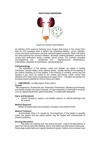
  • 1. INFECTIOUS DISORDERS Figure 6-3 Urinary Tract Infection An infection (UTI) cause by bacteria, virus, fungus, that occurs in the urinary tract. Risk for UTI increases when a patient has indwelling catheter, urinary retention, urinary and fecal incontinence and poor perineal hygiene practices. Other risk factors are renal scarring from previous infection, decrease ureteral peristalsis, presence of urinary tract obstruction, being a female, sexual activity. The common causative microorganisms are escherichia coli , staphylococcus, streptococcus, enterobacter, klebsiella and aerobacter, pseudoaeriginosa. Pathophysiology The epithelilium of the kidneys, ureter and bladder are sterile in healthy individuals, infection begins when bacteria enters , usually starting at the opening of the urethra travelling up to the bladder. If the flushing or urinating cannot stop the bacteria it can move up further to the ureters and kidney. Lower urinary tract infection( UTI) rarely cause complications but upper UTI if untreated can spread into the blood stream potential for chronic illness and death . 1. URETHRITIS – is inflammation of the urethra. Causes Microorganisms ( Escherichia coli, Chlamydia Trichomonas, Neisseria gonorrhoeae, and herpes simplex virus type 2),trauma, or hyper-sensitivity to chemicals in products such as vaginal deodorants, spermicidal jellies, or bubble bath detergents. Signs and Symptoms dysuria, frequency, urgency, and bladder spasms. A urethral discharge may be noticed. Medical Diagnosis Based on patient signs and symptoms, urinalysis, and urethral smear Medical Treatment Antimicrobials when it is caused by microorganisms. If the patient is sexually active, the patient and the sexual partner may be treated with antimicrobials to prevent reinfection. Nursing Intervention Sitz baths are soothing and may reduce the pain . Instruct female patients to wipe from front to back after toileting, and to void before and after sexual intercourse . Discourage bubble baths and vaginal deodorant sprays. Instruct uncircumcised male
  • 2. patients to clean the penis under the foreskin regularly. Advise patients to void after swimming. 2. CYSTITIS - is inflammation of the urinary bladder. The most common Causes Bacterial contamination, prolonged immobility, renal calculi, urinary diversion, indwelling catheters , radiation therapy, and treatment with some types of chemotherapy. Signs and Symptoms urgency, frequency, dysuria, hematuria, nocturia, bladder spasms, incontinence, and low-grade fever. Urine may be dark, tea colored, or cloudy. Fever, fatigue, and pelvic or abdominal discomfort and bladder spasms experienced as pain behind the symphysis pubis. Medical Diagnosis Urinalysis, culture, and sensitivity. The presence of bacteria does not mean that the patient has an infection unless the patient also has white blood cells (WBCs) in the urine. Medical Treatment Antibiotic , mild analgesic such as acetaminophen is useful for relieving discomfort. Phenazopyridine (pyridium) and Oxybutynin chloride (Ditropan may be ordered for 2 to 3 days to decrease discomfort and bladder spasms. Nursing care advise patient to complete the entire course of antibiotics and take analgesics as ordered.If phenozopyradine is given, advise patient that the drug causes red-orange urine Warm sitz bath , oral fluid intake 30m/kg of fluid per day. To reduce risk of future infection, teach patient to, wear cotton undergraments, avoid tight-fitting clothing in the perineal area, take shower instead of tub bath, avoid caffeine drinks, apple, grapefruit, orange these irritates the bladder, maintain high fluid intake and void often, Wiping from front to back after voiding for female and drink a glass of water after swimming ,before and after intercourse to flush the bacteria. 3. PYELONEPHRITIS – inflammation of the renal pelvis. It may affect one or both kidneys. Cause Acute pyelonephritis is most often caused by an ascending bacterial infection, but it may be bloodborne. Chronic pyelonephritis most often is the result of reflux of urine from inadequate closure of the ureterovesical junction during voiding. It is also usually caused by long standing UTIs with relapses and reinfections, may even lead to chronic renal failure . Signs and Symptoms High fever, chills, nausea, vomiting, and dysuria. Severe pain or a constant dull ache in the flank area. The patient with chronic pyelonephritis experience fatigue, hypertension, increase BUN and creatinine and a slight aching over one or both kidneys. Medical Diagnosis Urinalysis, urine culture and sensitivity, CBC, IVP, cystoscopy Medical Treatment
  • 3. Antibiotics, urinary tract anti-septics, analgesics, and antispasmodics . Additional medications may be needed to treat hypertension. Adults are advised to dink at least eight 8-oz glasses of fluids daily. Intravenous fluids may be ordered if the patient has nausea and vomiting. Dietary salt and protein restriction may be imposed on the patient with chronic disease. follow-up cultures to determine whether the infection has been resolved. Nursing Interventions Record the presence of signs and symptoms, history of previous urinary disorders, fluid intake at least 8 oz of glasses a day, advise patient to complete the entire course of antibiotics and take analgesics as ordered. Limit physical activity and exercise , protein and salt dietary restrictions if advised by the physician.
  • 4. KIDNEY TRANSPLANT - Is the surgical implantation of a human kidney from a compatible donor to a recipient. DONOR: LIVING 1. ABO Screening 2. Tissue specific antigen & human leukocyte antigen histocompatibility 3. Excellent health 4. Full functioning kidneys 5. Emotionally ready 6. Full understanding of the process CADAVER: 1. Below 60 yrs of age 2. Brain death 3. Normal renal function 4. No metastatic disease, HIV, Hep. B S/S OF REJECTION oliguria fever pain tenderness over transplant site hypertension creatinine increase COMPLICATION POST TRANSPLANT rejection infection malignancy (basal and squamous cell carcinoma.-skin,lips, vulva, lungs cardiovascular – hypertension IMMUNOSUPPRESSIVE DRUGS
  • 5. Cyclosporine (Neoral)- blocks interleukin Azathioprine (imuran) – blocks DNA preventing lymphocyte proliferation Corticosteroids (prednisone)- block cytokines Cyclophosphamide (cytoxan) Tacrolimus (prograft) – blocks calcineurin and T cell Mycophenolate (cellcept)- inhibit B & T lymphocyte OKT 3 - antilymphocyte globulin NURSING MANAGEMENT FOR RENAL TRANSPLANT • Maintain fluid balance by monitoring I & O • Monitor laboratory studies for graft function and electrolyte balance • Monitor for s/s of graft rejection • Monitor wound drainage • Adhere to immunosuppression schedule • Monitor blood glucose levels related to high glucocorticoids • Arrange supply of medications prior to discharge to prevent disruption of immunosupressions • Arrange post discharge follow - up appointments NURSING DIAGNOSES POST KIDNEY TRANSPLANT • Altered nutrition : more than body requirements related to side effects of immunosuppressant agents Less than body requirement: related to increased caloric needs after transplant • Altered protection and risk for infection related to immunosuppression required after transplantation • Effective management of Therapeutic Regimen related to post-transplantation regimen • Pain related to transplantation surgery • Risk for Ineffective Individual Coping after transplantation related to increased stress, anxiety, fear, and lifestyle changes • Risk for injury: rejection of transplanted organ related to impaired immunocompetence INTERNAL JUGULAR CATHETER - a central venous catheter inserted through the internal jugular vein. SUBCLAVIAN CATHETER - Central venous access via the subclavian vein has several advantages over other possible locations. ARTERIOVENOUS GRAFT – artificial graft made of gore-tex or a bovine carotid artery is used Risks & Complications in Hemodialysis • Hypotension • Blood loss from technical problem • Muscle cramps • Disequilibrium syndrome • Air embolism • Hypoglycemia
  • 6. • Cardiac arrythmia Nursing Care Before and During dialysis • Check the dialysis order { BFR, duration, bath, UFR, heparin, K+ } * SLED • Chart client’s weight { DRY WEIGHT} • Assess vital signs • Check patency of vascular access • Withhold antihypertensives • Ensure bed rest with frequent position change • Monitor closely for complications • Monitor blood line connections, arterial and venous and transmembrane pressure in the dialysis machine Nursing Care Post Hemodialysis • Check VS Nursing • Record post dialysis weight • Resume all medications • Apply pressure dressing on decannulated sites • Protect IJ/subclavian catheter with sterile dressing
  • 7. REVIEW OF ANATOMY AND PHYSIOLOGY OF THE RENAL SYSTEM Figure 6-1 Urinary System The renal system is responsible for maintaining homeostasis in the body by carefully regulating fluid and electrolytes, acid – base balance, removing wastes, and providing hormones responsible for red blood cell production, hypertension and bone metabolism. The renal system is composed of the upper and lower urinary tract. Lower Urinary System A. Bladder The bladder is an extra peritoneal organ that lies behind the symphysis pubis . Its main function is for storage of urine. As volume of urine increases, starting from 300-500 ml, awareness of the need to void develops. Voluntary voiding is accomplished by stimulation of the parasympathetic nerve fibers causing coordinated contraction of the detrusor muscle and the bladder body. B. Urethra The urethra drains urine from the bladder to an exterior opening of the body, the external urethral orifice. In females, the urethra is about 3 to 4 cm. (1.5 in.). In males, the urethra is about 15 to 20 cm (6 to 8 in.) Micturition, or urination, is the process of releasing urine from the bladder into the urethra. Upper Urinary System Figure 6-2 Structure of the Kidneys
  • 8. Kidneys The two kidneys lie on the posterior wall of the abdomen outside the peritoneal cavity. Each kidney of the adult human that weighs about 150g is about the size of an indented region called the hilum through which passes the renal artery and vein, lymphatic, nerve supply. The outer part is the cortex and inner region called medulla. The medulla is divided into multiple cone shaped masses called renal pyramids. The base of each pyramid terminates in the papilla, which projects into the space of renal pelvis, a funnel shaped continuation of the upper end of the ureter. The outer border of the pelvic is divided into minor calyces, the walls up the calyces, pelvis that contain contractile elements that propel the urine toward the bladder, where urine is stored until it is emptied by micturition. The functional unit of the kidney is the nephron. Millions of nephrons are present in each human kidney which aid in the urine production and process of removing metabolic waste products from the blood. These significant structures extend between the cortex and the medulla. At one end of the nephron is closed, expanded and folded into a double-walled cuplike structure called the Bowman’s capsule. This capsule encloses glomerulus, the nephron’s primary structure in filtering function. Functions of Kidney Kidney performs different functions in order to maintain homeostasis in the body by excreting metabolic waste products and reabsorbing necessary elements for the body. The following are the functions of the kidney: 1. Formation of urine The formation of urine happens in three phases which are filtration, reabsorption and secretion. Each of these processes happens in the body in order to create homeostasis by removing those metabolic waste products and reabsorbing helpful substances. a. Filtration. The filtration process is nonselective, passive process which forms essential blood plasma without blood protein but both of it is normally too large to pass through the filtration membrane. If any of the two appeared in the urine, it would mean that there is a problem in the glomerular filters. The water and solute are smaller than proteins that are forced through the capillary walls and pores of Bowman’s capsule into the renal tubule. b. Reabsorption. Tubular reabsorption is achieved by active and passive transfer mechanism Sodium, potassium, calcium, phosphate, and uric acid are actively reabsorbed. Urea, water, chloride, some bicarbonates and some phosphate are passively reabsorbed. Most reabsorption occurs in the proximal tubule which conserves needed substances but does not reabsorb metabolic waste products. c. Secretion. Some of substances such as hydrogen and potassium ion, creatinine, and ammonia, move from the peritubular capillary blood and secreted by the tubule cells into the filtration. Excreting nitrogenous waste products , unnecessary and excess substances . 2. Body’s Water Volume Regulator Regulation of water in the body contained in the blood is greatly influenced by antidiuretic hormone (ADH), also called vasopressin. Vasopressin is produced in the hypothalamus and stored in nearby pituitary gland. Receptors in the brain monitor the blood’s water concentration causing the release of ADH in the bloodstream if the amount of salt and other substances is too high. ADH in the bloodstream causes more water to be reabsorbed into the bloodstream. In the absence of ADH, the collecting ducts become impermeable to solute and water, making it less concentrated than plasma and the urine is diluted. 3. Excretion of Metabolic Waste Products The kidney functions as the body’s main excretory organ, eliminating the body’s metabolic waste products and serves as the primary mechanism for excreting drug
  • 9. metabolites. 25g to 30g of urea is produced as the end product of protein breakdown and excreted daily making it the major waste product of protein metabolism. Other waste products are creatinine, phosphates and sulfates. Uric acid, formed as a waste product of purine metabolism, is also eliminated in the urine. 4. Blood Pressure Regulator Regulating blood pressure is linked to the kidneys' ability to excrete enough sodium chloride (salt) to preserve normal sodium balance, extracellular fluid volume and blood volume. 5. Regulation of Acid-Base Balance The kidney also adjusts the body’s acid-base balance to prevent such blood disorders as acidosis and alkalosis. It helps maintain normal pH by retaining or excreting hydrogen ions and regenerating lost buffer. The kidneys excrete acids that the lungs cannot excrete and they can excrete hydrogen ions or reabsorb bicarbonate to correct acidosis. They can reverse this process to correct alkalosis. Only renal mechanisms can remove metabolic acids and excess bases from the body. 6. Regulation of Red Blood Cell Production Decreasing amount of oxygen in the renal blood flow activates the release of erythropoietin. Erythropoietin stimulates the bone marrow to produce red blood cells, thereby increasing the amount of hemoglobin available to carry oxygen. 7. Vitamin D Synthesis The kidneys are also responsible for the final conversion of inactive vitamin D to its active form, 1,25-dihydroxychole-calciferol. Vitamin D is necessary for maintaining normal calcium balance in the body. 8. Secretion of Prostaglandins The kidneys also produce prostaglandin E and prostacyclin, which have a vasodilatory effect and are important in maintaining renal blood flow.
  • 10. DIALYSIS • A technique in which a substance move from the blood through a semi permeable membrane into a dialysis solution. 3 PHYSICAL PRINCIPLES (osmosis. Diffusion, ultrafiltration) Indications of dialysis 1. GFR less than 5 to 1O ml/min 2. Manifestations of uremic syndrome TWO TYPES 1. Hemodialysis – removal of waste and water by circulating the blood into a dialyzer through a dialysis machine. 2. Peritoneal dialysis – repeated cycles of instilling dialysate into the peritoneal cavity. 4 BASIC GOALS OF DIALYSIS 1. Remove waste products from protein metabolism. 2 .Maintain safe concentration of serum electrolytes 3. Correction of acidosis and replenish blood’s bicarbonate system. 4. Removal of excess water Figure 6-8 Hemodialysis Components of Hemodialysis • Dialyzer – Artificial filter containing fine fibers acting as artificial kidney • Dialysate - Dialysis solution or bath containing sodium, potassium, calcium, chloride, magnesium, bicarbonate, ph, glucose use to remove toxins , nitrogenous waste products and excess water from the blood • Vascular access - direct method of circulating the blood into the dialyzer • Dialysis machine
  • 11. Figure 6-9 AVF External shunt – silastic cannula in the forearm or leg inserted into the artery and the vein to form an external blood path. Arteriovenous fistula (AVF) – anastomosis of the artery and the vein Arteriovenous graft – artificial graft made of gore-tex or a bovine carotid artery is used Internal Jugular Catheter – A dialysis catheter containing arterial and venous lumens inserted in the jugular vein Complications in Hemodialysis • Hypotension – Decrease blood pressure as a consequence of reduce cardiac output secondary to excessive water and sodium removal 114 • Blood loss from technical problem – Blood loss related to blood clotting in the circuit or blood loss due to loose connections . • Muscle cramps – Involuntary muscle contraction due to excessive water and sodium removal
  • 12. • Disequilibrium syndrome – A neurologic deterioration during dialysis manifested as headache,nausea,vomiting due to marked decrease of blood urea nitrogen in the cerebrospinal fluid and brain tissue. • Air embolism – Obstruction of the circulation by air related to air entry in the blood circuit • Hypoglycemia – Low blood sugar concentration related to glucose diffusing out of the blood • Cardiac arrythmia- Disturbance in the electrical impulse formation or conduction related to electrolyte and ph changes during dialysis or underlying heart disease Nursing Care Before and During dialysis • Check the dialysis order { BFR, duration, bath, UFR, heparin, K+ } * SLED • Chart client’s weight { DRY WEIGHT} • Assess vital signs • Check patency of vascular access • Withhold antihypertensives • Ensure bed rest with frequent position change • Monitor closely for complications • Monitor blood line connections, arterial and venous and transmembrane pressure in the dialysis machine Nursing Care Post Hemodialysis • Check VS Nursing • Record post dialysis weight • Resume all medications • Apply pressure dressing on decannulated sites • Protect IJ/subclavian catheter with sterile dressing NURSING CARE FOR AVF/GRAFT • Assess for bruit or thrill • Assess for bleeding, pain • May squeeze rubber ball to help in the development of the vessel • Avoid venipuncture, blood pressure taking on the arm with the vascular access • Avoid sleeping and wearing of tight fitting clothing on access extremity • Instruct client not to lift heavy objects using the extremity with the vascular access. • keep dressing dry • Keep catheter taped to the skin and prevent pulling of the catheter to prevent accidental removal the patient to clamp remaining tail of the catheter and apply direct pressure at insertion site with occlusive dressing if accidental removal of catheter happens PERITONEAL DIALYSIS - repeated cycles of instilling dialysate into the peritoneal cavity and the peritoneum is the dialyzing membrane. CYCLES OF PD 1.inflow ( infusion) – the solution is introduced into the peritoneum through a catheter by gravity 115 • tenckhoff catheter, a siliconized rubber catheter, inserted 3-5 cm below umbilicus, stabilized by dacron cuffs where fibroblast and blood vessel grow, fixing the catheter in place, occurring 1-2 weeks after insertion
  • 13. 2. dwell (equilibrium) – solution is retained in the peritoneal cavity for a prescribed period to allow better exchange and clearance. 3. Drain ( outflow) – process of removing the previously retained solution in the peritoneal cavity by gravity. Peritoneal Dialysis Complications • drainage obstruction – Presence of fibrin or clots in the tubings • Difficulty of breathing – Dialysis solution in the peritoneum during dwell time may push the diaphragm • Bleeding – Bloody dialysate or in the PD catheter insertion site related to the procedure, manipulation or trauma • Peritonitis – Microorganism gaining entry into the peritoneum related to poor aseptic technique during the dialysis procedure Nursing Responsibilities •VS and complication monitoring •Pre warm the solution •Documentation of the three phases of PD (Solution use, time infused, retain, drain including the amount, color, appearance ) Table 6-7 Sample Documentation OF Peritoneal Dialysis Date Vital Signs Meds Added to Solution IN Solution OUT Difference + / - Cumulative Difference Remarks No of Cycles BP PR Time Started Time Finished Vol Time Started Time Finished Vol 1 Perisol 2.5% 8:30 8:45 1L 10:45 11:00 1200 -200 Slightly bloody 2 11:00 11:15 1L 1:00 1:25 1300 -300 -500 3 1:25 1:40 1L 340 4:00 800 +200 -300 4 4:15 6:30 11:00 1100 -100 Clear
  • 14. GLOMERULAR DISORDER Nephrotic Syndrome - is an alteration of kidney function caused by increased glomerular basement membrane permeability to plasma protein (albumin). Altered glomerular permeability result in characteristic symptoms of gross proteinuria, generalized edema (anasarca), hypoalbuminemia, oliguria, and increased serum lipid level (hyperlipidemia). It maybe primary, wherein the pathology is the kidney itself or secondary of which nephrotic syndrome is the renal manifestation of a systemic disease .Nephrotic syndrome , usually occurs in children between ages 2-6 but may affect adults , both sexes and any race. Pathophysiology Normally large protein cannot pass through the glomerulus. Proteinuria occurs because of changes to capillary endothelial cells of the glomerulus.The mechanism of damage to these structures is unknown in primary and secondary glomerular diseases, but evidence suggests that T cells may upregulate a circulating permeability factor or downregulate an inhibitor of permeability factor in response to unidentified immunogens and cytokines. Other possible factors include hereditary defects in proteins that are integral to the slit diaphragms of the glomeruli, activation of complement leading to damage of the glomerular epithelial cells and loss of the negatively charged groups attached to proteins of the GBM. Figure 6-5 Pathophysiology of Nephrotic Syndrome Signs and Symptoms Massive proteinuria , hypoalbuminemia, generalized edema, hyperlipidemia lipiduria , hypercoagulability and hypertension. Medical Diagnosis Urinalysis, serum albumin, renal ultrasound and biopsy. Serologic studies for infection and immune abnormalities e.g. antinuclear antibody. Medical/Pharmacologic Management • Blood-pressure medications , ACE inhibitors and ARBs, which curb the pressure in the glomeruli and lower the amount of protein in the urine • Diuretics to reduce swelling • Cholesterol-lowering drugs • Blood thinners, or anticoagulants
  • 15. • Corticosteroids • Limit salt to reduce swelling and low in saturated fats and cholesterol diet. • Dialysis if conservative management is not effective . Nursing Management 1. limit oral fluid intake, low sodium, protein and saturated food sources diet 2. I & O 3. Skin care 4. Reverse isolation, vit c rich diet 5. Monitor lymphocytes & wbc 6. Monitor complications ( pulmonary edema, hypertension, CHF, renal failure,stroke, ) 7. Report fever, malaise & adverse effects of meds Nursing Diagnoses a. Fluid Volume b. Imbalanced Nutrition: Less Than Body Requirements c. Fatigue d. Deficient Knowledge e. Risk For Infection
  • 16. OBSTRUCTIVE DISORDERS Figure 6- 4 Renal Calculi ( Urolithiasis ) Renal calculi is the formation of stones in the urinary tract . These are crystalline structures that form from the components of urine. Pathophysiology Most calculi are precipitations of calcium salts (phosphate and oxalate),uric acid, magnesium ammonium phosphate (struvite) or cystine. These substances are normally found in the urine. Factors Fostering Calculi Formation Concentrated urine Excessive intake of vitamin D, animal protein, oxalates, sodium, sucrose, vitamin C, calcium based antacids Familial history Immobility, urine stasis, sedentary lifestyle Altered urine pH Lack of kidney substance that inhibits calculi formation Signs and Symptoms Pain depends on the location of the stone. Nausea, vomiting ,hematuria and signs and symptoms of UTI Medical Diagnosis Urinalysis, urine culture and sensitivity ,IVP, ultrasound Computed tomography (CT) scan Medical ,Surgical and Pharmacologic Management Phases of Stone Management 1. Acute phase - narcotics, antispasmodic, anti -emetic, warm bath to relieve flank pain 2. Elimination of stone - Waiting to be passed out - Mechanical intervention - Surgical intervention 3. Long term prevention of recurrence - OFI (3-4l/day), diet, medication
  • 17. Pharmacologic Management 1.potassium citrate therapy - It attaches to calcium in the urine, preventing the formation of mineral crystals ; prevents the urine from becoming too acidic . 2.Thiazides – increases urinary Ca excretion 3. Allopurinol – prevents formation of uric acid nidus FOR SMALL STONES 4. Alpha blockers or α-adrenergic-antagonist - It relax muscle tension in the ureter and facilitate passage E.g. tamsulosin 5. Sodium bicarbonate 6. Pain relievers Surgical and Non-surgical Management of Stone Often calculi pass spontaneously, if it does not pass and symptoms continues , other options may be used to destroy or remove the calculus. Surgical management are indicated if there is progressive renal damage, obstruction of urine flow , presence of infection and severe pain. The surgical procedures are Nephrolithotomy (removal of kidney stones) and ureterolithotomy (removal of stones in the ureters) . For non-surgical approach, options include ureteral stent placement, lithotripsy, cystoscopic stone removal, and percutaneous nephrolithotomy. Ureteral Stent- are small tubes inserted into the ureter to treat or prevent a blockage that prevents the flow of urine from the kidney to the bladder. Lithotripsy is a process of eliminating a calculus in the renal pelvis, ureter , bladder by crushing the stone. It can be accomplished by Extracorporeal shock wave lithotripsy (ESWL) which utilizing sound, laser, or shockwave energy with a use of a lithotripter. It is guided by an ultrasound probe, the energy is directed to the stone through a water-filled cushion. Table 6-1 Complications of ESWL Relate d to Shock Fragments Related to Shock waves colic Bruising /hematoma Incomplete fragment urosepsis Blocking of ureters by the multiple stone fragments Cystoscopy/Ureteroscopy - use of lighted scope or a tube inserted into the urethra into the bladder and ureters to remove stone and uses a laser fiber to crush the stone in the case of ureteroscopy. Percutaneous nephrolithotomy – The surgeon creates a tunnel directly through the skin into the kidney and uses ultrasound or electrohydrolysis to break the stone into pieces. this approach is usually used when stones are large and cannot be broken with lithotripsy.
  • 18. Dietary Management Prevention of stone recurrence is important .Apart from high fluid intake to keep urine diluted dietary restrictions are also important and the dietary restrictions depend on the type of stone Table 6- 2 Recommended Diet for Stone Management Acid Ash Diet ( To acidify the urine) Alkaline Ash Diet ( To alkalinize the urine) Calcium Stones cranberry, prune juice,meat, egg, poultry,fish,grapes, whole grains limit milk and other dairy products Oxalate Stones milk, vegetables, fruits except prunes, cranberry, plums Avoid tea, chocolate, rhubarb, spinach Uric Acid Stones - Reduce foods high in purine like liver, brain, kidneys, venison, shellfish, meat soup, gravies, legumes Nursing Management 1. administer prescribed analgesics 2. reassure client that most stones smaller than 4mm can pass out spontaneously 3. provide education to prevent future and recurrence of stone 4. encourage OFI 5. instruct client to avoid foods that contribute to the diagnosed type of stone Nursing Diagnoses Acute pain related to renal calculi Ineffective coping related to anxiety , low activity level and inability to perform ADL Impaired urinary elimination related to renal calculi Risk for infection Nutrition imbalance, less than body requirements related to nausea
  • 19. NEUROGENIC BLADDER - - urinary bladder malfunction due to neurologic dysfunction emanating from internal or external trauma, disease or injury. - Interference of the bladder normal mechanism cause by the disruption of the central and peripheral nervous system Review of The Neuro Anatomy - The normal function of the urinary bladder is to store and expel urine in a coordinated, controlled fashion. This coordinated activity is regulated by the central and peripheral nervous systems. Normal voiding is essentially a spinal reflex , modulated by the central nervous system (brain and spinal cord), which coordinates function of the bladder and urethra. The brain receives input via afferent pathways that ascend from the bladder and provide feedback on how full the bladder is. Higher brain centers then determine whether it is socially acceptable to void and trigger downstream structures to permit or suppress the voiding reflex. The pons is a major relay center between the brain and the bladder. The mechanical process of urination is coordinated by the pons in the area known as the pontine micturition center (PMC). The PMC coordinates the urethral sphincter relaxation and detrusor contraction to facilitate urination. Emotions, experienced in higher brain centers, may exert downstream effects on the PMC, which is why some people can experience incontinence with excitement or fear. The spinal cord functions as a long communication pathway between the brainstem and the sacral spinal cord ( terminal portion of the spinal cord, situated at the lower back in the lumbar area responsible for bladder contractions) When the sacral cord receives the sensory information from the bladder, this signal travels up the spinal cord to the pons and then ultimately to higher brain centers. a spinal cord injury can lead to urinary frequency, urgency, and urge incontinence, which may be complicated by difficulty emptying the bladder. This occurs because the urinary bladder and the sphincter are no longer coordinated Pathophysiology Normal bladder function relies on information travelling through neural pathways from the cerebral cortex, through the spinal cord, and on to the bladder to coordinate normal micturition and urinary continence. When this pathway is damaged, it can result in loss of bladder sensation and also the loss of the coordination between urethral sphincter and its muscles, these muscles may not contract even when the bladder fills or the person has the urge to void, leading to bladder dysfunction such as urinary incontinence and retention (Mauk 2012). TYPES OF NEUROGENIC BLADDER A. Spastic- Sensory and voluntary control of urination is disrupted partially or totally. The stimuli generated by bladder filling cause frequent spontaneous detrusor muscle contraction and involuntary emptying caused by disruption of CNS transmission above the sacral spinal cord segment. Causes: 1) Spinal cord injury- most common cause 2) Stroke 3) MS- immune mediated inflammatory disease attacking the myelin and axons 4) CNS lesions
  • 20. B. Flaccid- damage to the sacral spinal cord at the level of the reflex arc, cauda equina, sacral nerve roots leading to loss of detrusor muscle tone resulting to overdistention, weak and ineffective detrusor muscle contraction. Causes: 1) Spinal shock phase (6-12 weeks)in SCI 2) Myelomeningocele or meningocele – type of neural tube birth defect wherein the backbone and spinal canal don’t close before the baby is born; a type of spinal bifida 3) Peripheral neuropathies- DM most common cause ,metabolic derangement of the Schwann cell results in segmental demyelination and impaired nerve conduction. 4) MS 5) Chronic alcoholism 6) Prolonged overdistention of the bladder Symptoms of Neurogenic Bladder Overactive bladder, frequent urination, stress incontinence.urge incontinence ,urinary retention, underactive bladder ( bladder cannot send signal when full) Diagnostic Tests Table 6- 3 Neurogenic Bladder Diagnostics DIAGNOSTIC TEST PURPOSE PROCEDURE NURSING RESPONSIBILITY 1) Urine culture, Urianalysis Detect UTI Urine sample Midstream clean catch 2) Creatinine, BUN * ascending infection, vesicoureteral reflux in neurogenic bladder can damage kidneys Determine kidney damage Blood extraction Avoid meat 3) Postvoid bladder scan *>50mL means ineffective detrusor muscle contraction Less than 50mL PVR is adequate bladder emptying Over 200mL PVR indicates inadequate emptying Children- More than 20mL PVR is considered abnormal Elderly- 50-100 ml PVR is considered normal Measures residual urine in the bladder shortly void; this can be accomplished through ultrasound, bladder scan, or by directly measuring the urine volume drained by a urinary catheter. Urinary catheterization is the gold standard for measuring the post-void residual. Explain the procedure 4) Cystometography To evaluate bladder filling and detrusor muscle function Shows the Double lumen catheter is inserted into the bladder, 50 ml is introduce in one lumen and Explain the procedure
  • 21. relationship of intravesical pressure with the changing volume of urine in the bladder pressure is recorded through the other lumen 10cms water for 50-400ml of urine 5) CYSTOSCOPY To determine presence of bladder lesions Flexible cyctoscope inserted into the bladder , using also video system and irrigating medium Explain procedure, local or GA maybe use, Supine or lithotomy position MEDICAL MANAGEMENT OF NEUROGENIC BLADDER GOAL: Maintain continence and avoid complication associated with overfilling or incomplete emptying of bladder through self-care. Thus, teaching is the primary intervention. I. MEDICATIONS GOAL: - increase or decrease contractility of detrusor muscle. - increase/decrease internal sphincter tone. - relax external urethral sphincter. Table 6-4 Medications For Neurogenic Bladder ANTICHOLINERGIC DRUG TO PREVENT SPASTIC BLADDER CHOLINERGIC DRUGS TO STIMULATE MICTURITION 1. Darifenacim (Enablex) 2. Oxybutynin (Ditropan ) 3. Solifenacin succinate (vesicare) 4. Trospium (santural) 5. Tolterodine (destrol) 6. Propantheline bromide (pro-bannthine) 7. Flavoxate hcl (urispas) *inhibits response to acetylcholine relaxing the detrusor muscle and increase sphincter tone *acetylcholine is a neurotransmitter causing muscle to contract,activate pain responses,regulates endocrine and REM sleep functions 1. bethanechol chloride (Urecholine) *stimulates parasympathetic NS increasing detrusor muscle tone producing contraction strong enough to produce micturition
  • 22. II. NUTRITION -Moderate to increase OFI to decrease UTI and stone formation - Diet to acidify urine (cranberry) III.BLADDER RETRAINING Measures to stimulate reflex voiding for patients with spastic neurogenic bladder • Stroke or pinch abdomen, inner thigh, glans penis (trigger points to stimulate urination) • Pulling pubic hair • Tapping suprapubic region • Inserting gloved finger to rectum and gently stretch anal sphincter • Crede’s method- applying suprapubic pressure with finger of one or both hands ALERT: may stimulate SNS causing sudden increase of blood pressure for patients with SCI (autonomic dysreflexia -medical emergency ) • Valsalva maneuver- bearing down while holding one’s breath CATHETERIZATION- intermittent for patients with SCI SURGICAL MANAGEMENT Maybe required when urination can’t be effectively managed using conservative measures. 1) Rhizotomy- destruction of the nerve supply to the detrusor muscle or the external sphincter for patients with hyperreflexia or spasticity. 2) Urinary diversion- 3) Implantation of artificial sphincter Nursing Management 1. Promote urinary drainage and continence 2. Prevent complication 3. Teach patient and family self-care techniques Nursing Diagnoses 1) Impaired urinary elimination related to impaired bladder innervation 2) Toileting self-care deficit related to neurologic injury 3) Risk for impaired skin integrity related to urinary incontinence 4) Risk for infection related to impaired urinary reflex
  • 23. RENAL EXCRETORY STUDY URINALYSIS - assessed the nature of the urine produce and evaluates the color,ph and specific gravity, determines the presence of glucose (glycosuria, protein, blood(hematuria), ketones ( ketonuria) and analyzes sediment for cells (WBC, called pyuria, casts bacteria, crystals) Nursing Responsibilities: Cleanse perineal area, spread labia and cleanse meatus from front to back using antiseptic sponge, for males, retract foreskin for uncircumcised penis and cleanse glans antiseptic sponge, must be analyze 1 hour after of collection, For 24 Hour urine collection ( creatinine clearance) discard the first voided urine, collect all subsequent urine in a sterile container for 24 hours. Kidney, ureter ,bladder X-ray (KUB ) - plain film abdominal flat plate x-ray identifying the number and size of kidneys with tumors, malformations and calculi ; no special preparations needed IVP – fluoroscopic visualization of the irinary tract after injection with a radiopaque dye Nursing Responsibilities - Test for iodine sensitivity, enema before the procedure, 8 hours, NPO, push fluids after. ULTRASOUND – non- invasive visualization of the kidney, ureter, bladder through the use of sound waves Nursing Responsibilities- Supine position, NPO not required, cleanse conducting gel from skin after the procedure CYSTOSCOPY – use of lighted scope to inspect the bladder (CYSTOSCOPE); maybe use to remove tumors, stones, or other foreign materials or use to implant radium, place catheters in the ureters. Nursing Responsibilities Before the Procedure Explain the procedure will be done under local or general anesthesia, secure consent, administer sedative as ordered, NPO for general anesthesia, or NPO after light breakfast for local anesthesia, enema as ordered ,let client assume a lithotomy position Post Procedure Mild analgesic or warm sitz bath to relieve pain, I &O and temperature monitoring, explain that hematuria expected post 24-48 hours, assess for clots, burning sensation upon urination maybe felt, and force fluids. RENAL BIOPSY – removal of kidney tissue for microscopic study Open method promotes better visualization but high risk for infection, close method none by aspiration with a fine needle and has less risk for infection Nursing Responsibilities before and during the procedure Secure consent,NPO after midnight,assess hemoglobin and coagulation studies ,assist client to assume Prone position with a pillow below the abdomen, apply pressure for 20 minutes on the aspirated area after the procedure Post Procedure Flat bedrest for 24 hours, monitor for hemorrhage, hypotension, dizziness, tachycardia ,pallor , back, flank and shoulder pain, avoid strenuous activities,
  • 24. coughing, sneezing and straining, encourage fluid to avoid urinary retention and clots, monitor hemoglobin, assess for hematuria and if present should cease 24 hours after the procedure. Administer analgesics Blood chemistry and Hemoglobin tests - these tests or panels are groups of tests that measure many chemical substances in the blood that are released from body tissues or are produced .For kidney function, creatinine and blood urea nitrogen . Nursing Responsibilities – NPO is not required for BUN , hemoglobin and creatinine , instruct the client not to eat red meat a day prior to creatinine test ,intake of red meat can affect the result.
  • 25. RENAL FAILURE - A condition wherein the kidneys cannot remove the body’s metabolic waste products or when it cannot perform its regulatory functions. Different causes may lead to renal failure. There are two types of renal failure, acute renal failure( ARF) or acute kidney injury (AKI) and chronic renal failure (CRF). ACUTE RENAL FAILURE Acute Renal Failure occurs with reversible clinical syndrome wherein there is a sudden and almost complete loss of kidney function. This type of Renal Failure may last for days, weeks or even months. Some of the patients with ARF may die and for some may progress to chronic renal failure. Categories of AKI Prerenal - occurs in 60% to 70% of cases which result from impaired blood flow or if the kidneys do not receive sufficient blood that leads to hypoperfusion of the kidney and decreases GFR. intrarenal - This results from parenchymal damage to the glomeruli or kidney tubules, resulting from prolonged renal ischemia resulting from myoglobinuria Postrenal ARF is usually the result of an obstruction somewhere distal to the kidney. Pressure increases in the kidney tubules and eventually the GFR decreases. Blockage causes urine to back up and harm the kidneys. Common causes are urinary tract obstruction like calculi, tumors, benign prostatic hyperplasia or an enlarged prostate, kinked ureter, and blood clots. Increase in blood urea nitrogen and creatinine are noted in postrenal ARF. Table 6-5 Causes of AKI
  • 26. Stages of Acute Renal Failure 1. Initiation period At this phase the kidney begins with the initial injury and ends when oliguria develops. Its manifestation is the reduced blood flow to the nephrons. This causes decreased reabsorption of water, electrolytes, and excretion of protein wastes and excess metabolic substances. This phase can last from hours to days and is characterized by urine output at 30 cc per hour or less. 2. Oliguria period At this period, this is caused by reduction in the glomerular filtration rate. There is an increase in the serum concentration of creatinine, urea, uric acid, and the intracellular cations like potassium and magnesium. In this phase the uremic symptoms first appear and may develop life-threatening condition. Other manifestations would include hyperkalemia, hypernatremia, hyperphosphatemia, hypocalcemia, hypermagnesemia and metabolic acidosis. Decreasing renal function happens with increasing nitrogen retention with urine output of less than 400 ml /24 hours and may last for 1 to 2 weeks. 3. Diuresis period At this period, there is a gradual increase in urine output, which signals that glomerulus filtration has started to recover. The laboratory results show an increased BUN and creatinine result. The urine output ranges from 3 to 5 liters per day due to partial regenerated tubule’s inability to concentrate urine which lasts for 2 to 3 weeks. 4. Recovery period This period signals the improvement of renal function and may take 3 to 12 months. Laboratory values return to the patient’s normal value. Figure 6-6 Pathophysiology of AKI Pre Intra Post ↓ BLD supply renal ischemia tubular damage ↓ GFR ↓ urine output ↑ retention of waste Precipitation occurs (CHON/HGB liberated) Interferes with urine excretion kidney swells necrosis obstruction ↑intrarenal pressure ↓ GFR ↓ output /retention of waste
  • 27. Signs and symptoms The symptomatology of acute kidney injury depends on the cause. Figure 6-7 Common Symptoms of AKI Diagnostic Management 1. History taking , physical exam 2. Identify precipitating cause 3. Creatinine, BUN, Serum electrolytes 4. Urinalysis Medical Management 1. Treatment of precipitating cause 2. Fluid restriction 3. Dietary management 4. Dialysis 5. Total Parenteral Nutrition (TPN) if indicated 6. Measures to decrease potassiumK (most life-threatening disturbance) a. administer cation – exchange resin 1. sodium polysterene sulfonate (kayexalate) 2. Calcium Polysterene Sulfonate (Kalimate) Major site of potassium exchange is the colon if enema is administered and Intestinal tract for oral administration . Sorbitrol is often administered with kayexalate to induce diarrhea type effect. b. IV glucose and insulin – This will cause the potassium to move into the cell, decreasing the serum potassium serum level . c. Ca gluconate - Protects the heart from the effects of hyperkalemia d. Bicarb IV - Corrects acidosis e . Dialysis - A procedure to remove waste products and excess fluid from the blood when the kidneys stop functioning properly. f. Dietary restriction – restrict fruits high in potassium like citrus fruits, banana, grapes, watermelon ,for protein intake 1 gm /kg during oliguric phase and sodium 2 g / day.
  • 28. Nursing Management Nursing goal of treating patients with acute renal failure is to correct or eliminate any reversible causes of kidney failure. Provide support by taking accurate measurements of intake and output, including all body fluids, monitor vital signs and maintain proper electrolyte balance. 1. Precise I/O monitoring 2. Vital signs 3. Dietary management 4. Fluid management base on the clinical manifestation 5. daily weight 6. Assess signs and symptoms of electrolyte imbalance such as mental status, neuromuscular status and cardiac rate and rhythm. Nursing Diagnoses 1. Fluid volume excess related to impaired kidney function 2. Decrease cardiac output related to high output renal failure (diuretic phase) 3. Fluid volume deficit related to high output renal failure (diuretic phase) 4. Potential for complications of immobility related to therapeutic restrictions 5. Deficient Knowledge related to condition and treatment 6. Risk for Imbalanced Nutrition: Less Than Body Requirements CHRONIC RENAL FAILURE Chronic renal failure (CRF), or End Stage Renal Disease (ESRD), is a progressive, irreversible deterioration in renal function in which the body’s ability to maintain metabolic and fluid and electrolyte balance fails, resulting in uremia or azotemia. The development of uremia happens and adversely affects every system in the body. The greater the buildup, the greater are the symptoms and are considered fatal. The pathology of CRF Occurs in 4 stages and the conditions that may contribute to ESRD include systemic diseases such as diabetes mellitus, hypertension, chronic glomerulonephritis, pyelonephritis, obstruction of the urinary tract. Hereditary lesions include polycystic kidney disease, vascular disorders, autoimmune disorders, infections, medications which are nephrotoxic. Stage 1. Decreased Renal Reserve In this stage, the residual renal function is 40-75% than the normal and patient is asymptomatic with normal BUN and plasma creatinine. Excretory and regulatory renal functions are undamaged. Stage 2. Renal Insufficiency The renal function is 20-40% marked decrease in glomerular filtration rate, solute clearances, ability to concentrate urine and hormone secretion. The signs and symptoms include rising BUN, plasma creatinine, mild azotemia, polyuria, nocturia and anemia. Stage 3. Renal Failure Renal function is approximately 5% of normal. Serum urea & creatinine levels rise rapidly and Urine output is less than 500ml/day .Symptoms of uremia develop Stage 4 End Stage Renal Disease End Stage Renal Disease (ESRD) with residual renal function less than 15% or normal. Its excretory, regulatory and hormonal renal functions are severely impaired.
  • 29. 111 Unable to maintain homeostasis like fluids and electrolytes imbalance may occur as well as pH imbalances. Noticeable are elevated BUN and plasma creatinine, anemia, hyperphosphatemia, hypocalcemia, hyperkalemia, fluid overload, usually oliguria . Uremic syndrome may occur and all body. Table 6- 6 Clinical Manifestations of CRF System Involved Clinical Manifestation Integumentary Skin Nails Hair Yellow,gray,bronze pigmentations,pallor,dry Brittle and thin dry and coarse Cardiovascular Hypertension, myocardial dysfunction, CHF, Pericarditis Metabolic Carbohydrate intolerance Hyperlipidemia Protein wasting Skeletal Hypocalcemia Hyperphosphatemia Bone demineralization Pulmonary volume overload, decreased pulmonary macrophage activity and infections Gastrointestinal Nausea,vomiting, to gastritis, gastric and duodenal ulcers Hematopoietic Anemia, bleeding Reproductive Diminish fertility Changes in menstruation Medical Management 1. Serum studies (nitrogenous wastes, electrolytes) 2. Complete blood count 3. Urinalysis / Culture 4. 24 hour creatinine clearance (to determine GFR Medical Treatment Goals: To prevent further damage to the kidneys To promote recovery of renal function To prevent complications 1. Pharmacologic (anti-hypertensive, erythropoletin, iron supplement, phosphate binding, calcium supplement, anti seizure, ketoanalogues, Sodium Bicarbonate, Potassium resin exchange (Kayexalate) 2. Nutritional - reduce protein potassium food sources , fluids 3. Blood transfusion to correct anemia 4. Correction of fluid and electrolyte imbalance 5. Dialysis 6. Kidney transplant
  • 30. SODIUM (135 to 145 mEq/L) Sodium is one of the most important elements in the body. It accounts for 90% of extracellular fluid cations (positively charged ions) and is the most abundant solute in extracellular fluid. The normal serum sodium levels range from 135 to 145 mEq/L. The body needs sodium to maintain proper extracellular fluid osmolality (concentration). Sodium attracts fluids and helps preserve the extracellular fluid volume and fluid distribution in the body. It also helps transmits impulses in nerve and muscle fibers (Lippincott, 2016). Sodium imbalances affect the osmolality of ECF and water distribution between the fluid compartments. When sodium levels are low (hyponatremia), water is drawn into the cells of the body, causing them to swell. In contrast, high levels of sodium in ECF (hypernatremia) draw water out of body cells, causing them to shrink (Lemone, 2011). The kidney is the primary regulator of sodium balance in the body. The kidney excretes or conserves sodium in response to changes in vascular volume. A fall in blood volume prompts several mechanisms that lead to sodium and water retention: HYPONATREMIA Hyponatremia, a common electrolyte imbalance, refers to sodium deficiency in relation to the amount of water in the body. It usually results from a loss of sodium from the body, but it may also be caused by water gains that dilute extracellular fluid (ECF). Classifications: (1) Hypovolemic both sodium and water levels decrease in the extracellular area, but sodium loss is greater than water loss. Causes: vomiting, diarrhea, fistulas, gastric suctioning, excessive sweating, cystic fibrosis, burns, wound drainage, osmotic diuresis,adrenal insudfficiency, diuretic use (2) Hypervolemic, both water and sodium levels increase in the extracellular area, but the water gain is more impressive. Serum sodium levels are diluted and edema also occurs. Causes: heart failure, liver failure, nephrotic syndrome, excessive administration of hypotonic IV fluids, hyperaldosteronism (3) Isovolemic sodium levels may appear low because too much fluid is in the body. Patients may not exhibit signs of fluid volume excess, and total body sodium remains stable. Causes: glucocorticoid deficiency, hypothyroidism, renal failure Pathophysiology Normally, the body gets rid of excess water by secreting less Antidiuretic Hormone (ADH); less ADH causes diuresis. For that to happen, the nephrons must be functioning normally, receiving and excreting excess water and reabsorbing sodium. Hyponatremia develops when this regulatory function goes haywire. Serum sodium level s decrease, and fluid shifts occur. When the blood vessels contain more water and less sodium, fluid moves by osmosis from the extracellular area into the more concentrated intracellular area. With more fluid in the cells and less in the blood vessels, cerebral edema and hypovolemia (fluid volume deficit) can occur.
  • 31. Manifestations The manifestations of hyponatremia depend on the rapidity of onset, the severity, and the cause of the imbalance. If the condition develops slowly, manifestations are usually not experienced until the serum sodium levels reach 125 mEq/L. In addition, the manifestations of hyponatremia vary, depending on extracellular fluid volume. a. Poor skin turgor b. Dry mucosa c. Decrease saliva production d. Orthostatic hypotension e. Nausea, abdominal cramping f. Neurologic changes such as: lethargy, confusion, signs of increase ICP, muscle twitch, seizure Laboratory Findings Serum osmolality less than 280 mOsm/kg (dilute blood) Serum sodium level less than 135 mEq/L (low sodium level in blood) Urine specific gravity less than 1.010 Elevated hematocrit and plasma protein levels Medical Management • Generally, treatment varies with the cause and severity of hyponatremia. Hypervolemia/isovolemia a. Fluid restrictions b. Oral sodium supplements Hyponatremia a. Give isotonic IV fluids (e.g. normal saline) b. Offer High sodium foods In SEVERE cases: a. Infusion of hypertonic saline solution (3% or 5% saline) Nursing Management Watch patients at risk for hyponatremia, including those with heart failure, cancer, or GI disorders with fluid losses. Review patient’s medications, noting those that are associated with hyponatremia. For patients who develop hyponatremia, take the following actions: • Monitor and record vital signs, especially blood pressure and pulse, and watch for orthostatic hypotension and tachycardia • Monitor neurologic status frequently Report any deterioration in level of consciousness. • Accurately measure and record intake and output. • Assess skin turgor at least every 8 hours for signs of dehydration. • Restrict fluid intake as ordered (fluid restriction is the primary treatment for dilutional hyponatremia). • Administer oral sodium supplements as ordered. • Provide a safe environment for patients with altered thought processes • Seizure precautions
  • 32. HYPERNATREMIA Hypernatremia, a less common problem than hyponatremia, refers to excess of sodium relative to the amount of water in the body. Pathophysiology The cells play a role in maintaining sodium balance. When serum osmolality increases because of hypernatremia, fluid moves by osmosis from inside the cell to outside the cell. As fluid leaves the cells, they become dehydrated and shrink. Manifestations Thirst is the first manifestation of hypernatremia. If thirst is not relieved, the primary manifestations relate to altered neurologic function. Initial lethargy, weakness, and irritability can progress to seizures, coma, and death in severe hypernatremia. Both the severity of the sodium excess and the rapidity of its onset affect the manifestations of hypernatremia. You may also observe the following: a. Low grade fever b. Flushed skin c. Dry mucous membranes d. Oliguria e. Orthostatic hypotension Laboratory Findings The following laboratory and diagnostic tests may be ordered with the following findings: Serum sodium levels are greater than 145 mEq/L Serum osmolality is greater than 295 mOsm/kg Medical Management Treatment for hypernatremia varies with the cause. The underlying disorder must be corrected, and serum sodium levels and related diagnostic tests must be monitored. Note that the fluids should be given gradually over 48 hours to avoid shifting water into brain cells. Nursing Management •Intravenous fluid replacement (dextrose 5% in water) to return serum sodium levels to normal • Sodium intake restrictions • Nursing Management If patient develop hypernatremia, take the following measures: • Monitor and record vital signs, especially blood pressure and pulse • Check neurologic status frequently. • Report any deterioration in the level of consciousness • Carefully measure and record intake and output. • Assess skin and mucous membranes for signs of breakdown and infection
  • 33. • Monitor serum sodium level and report any increase. • Assist with oral hygiene. Lubricate the patient’s lips frequently. • Provide a safe environment for confused or agitated patients
  • 34. PHOSPHOROUS (1.2 to 3.0 meq/L) The primary anion, or negatively charged ion, found in the intracellular fluid. It’s contained in the body as phosphate. Functions: a. Integral part of acid base buffer system b. Regulate ATP use for muscle contraction, nerve transmission, electrolyte transport c. Regulates 2,3 DPG (diphosphoglycerate) a compound in red blood cells that facilitates oxygen delivery from the red blood cells to the tissues. d. Regulates 2,3 DPG (diphosphoglycerate) a substance in RBC affecting oxygen affinity HYPOPHOSPHATEMIA Hypophosphatemia occurs when the serum phosphorous level falls below 1.2 mEq/L. although this condition generally indicates a deficiency of phosphorous, it can occur under various circumstances when total body phosphorous stores are normal Causes: a. Respiratory alkalosis one of the most common cause of hypophosphatemia, can stem from a number of conditions that produce hyperventilation b. Malabsorption syndromes c. Diuretic use, (Thiazides, Loop diuretics, Acetazolamide) Pathophysiology Most effects of hypophosphatemia result from depletion of ATP and impaired oxygen delivery to the cells due to a deficiency of the red blood cell enzyme 2,3-DPG. Severe hypophosphatemia affects virtually every major organ system: Manifestations a. Muscle weakness, most common symptom b. malaise, weakened hand grasp, myalgia (pain in the muscles) c. rhabdomyolysis (skeletal muscle destruction), can occur with altered muscle cell activity d. Osteomalacia/ Fracture, due to loss of bone density e. Bruising and bleeding Laboratory Findings Diagnostic test results may indicate hypophosphatemia or a related condition: a. serum phosphorous level less than 1.2 mEq/L b. elevated Creatinine Kinase level if Rhabdomyolysis is present c. X-ray studies that reveal skeletal changes typical of osteomalacia or bone fractures d. Abnormal electrolytes (decreased magnesium levels and increased calcium levels) Medical Management Treatment varies with the severity and cause of the condition: a. Treat the underlying cause b. Oral and intravenous supplements
  • 35. Nursing Interventions Nursing care should focus on careful monitoring, safety measures and interventions to restore normal serum phosphorous levels. a. Monitor vital signs. b. Assess the patient frequently for evidence of decreasing muscle strength, such as weak hand grasps or slurred speech, and document findings regularly. c. Assist in ambulation and activities of daily living, if needed, and keep essential objects near the patient to prevent accidents. HYPERPHOSPHATEMIA Hyperphosphatemia is a serum phosphate level greater than 4.5mg/dL. As with other electrolyte imbalances, it may be the result of impaired phosphate excretion, excess intake, or a shift of phosphate from the intracellular space into extracellular fluids. Causes: a. Renal Failure b. Hypoparathyroidism impairs less synthesis of parathyroid hormone(PTH), when less PTH is synthesized, less phosphorous is excreted from the kidneys. c. Respiratory acidosis d. Increase tissue breakdown Pathophysiology Effects of high phosphorous level are actually due to hypocalcemia; calcium suppresses cellular excitability. Manifestations a. Hyperphosphatemia causes few symptoms (tetany, tissue calcification) b. Symptoms occurring result from decrease calcium secondary to reciprocity Laboratory Findings: The following diagnostic test results may indicate hyperphosphatemia or a related condition such as hypocalcemia: a. Serum phosphorous above normal b. Low serum calcium level Medical Management: a. Treat underlying cause b. Restrict dietary intake c. Administer phosphate binding antacid, this may decrease absorption of phosphorous in the gastrointestinal system Nursing Management a. Monitor vital signs b. Monitor intake and output.( if urine output falls below 30 mL/hour, immediately notify the doctor) Decreased urine output can seriously affect renal clearance of excess serum phosphorous c. Monitor serum phosphorous and calcium levels
  • 36. d. Monitor signs of tetany, such as positive Trousseau’s and Chvostek’s sign
  • 37. POTASSIUM (3.5 to 5.0 mEq/L) A major cation (ion with a positive charge) in intracellular fluid, potassium plays a critical role in many metabolic cell functions. Only 2% fluid; 98% of the body’s potassium is found in extracellular fluid. That significant difference affects nerve impulse transmission. The normal serum (ECF) potassium level is 3.5 to 5.0 mEq/L. To maintain balance, potassium must be replaced daily through diet. Virtually all foods contain potassium, although some foods and fluids are richer sources of this element than others. Aldosterone helps regulate potassium elimination by the kidneys. An increased potassium concentration in ECF stimulates aldosterone production by the adrenal gland. The kidneys respond to aldosterone by increasing potassium excretion. Changes in aldosterone secretion can profoundly affect the serum potassium level. Potassium directly affects how well the body cells, nerves, and muscles function by: • Maintaining cells’ electrical neutrality and osmolality • Aiding neuromuscular transmission of nerve impulses • Assisting skeletal and cardiac muscle contraction and electrical conductivity • Affecting acid-base balance in relationship to the hydrogen ion. HYPOKALEMIA Hypokalemia is an abnormally low serum potassium level . It usually results from excess potassium loss, although hospitalized patients may be at risk for hypokalemia because of inadequate potassium intake. Pathophysiology Inadequate intake and excessive output of potassium can cause a moderate drop in its level, upsetting the balance and causing a potassium deficiency. Potassium shifts from the extracellular space to the intracellular space and hides in the cells. Because the cells contain more potassium than usual, less can be measured in the blood. Causes: a. Inadequate potassium intake b. Severe GI losses, e.g. suction, lavage, prolonged vomiting can deplete the body’s potassium supply as a result potassium levels drop c. Drug associated, e.g. diuretics,(esp. thiazides and furosemides), corticosteroids, insulin d. Decrease bowel motility Manifestations The signs and symptoms of a low potassium level reflect how important the electrolyte is to normal body functions. a. Skeletal muscle weakness, especially in the legs a sign of a moderate loss of potassium. This also includes paresthesia and leg cramps b. Decreased bowel sounds, constipation, paralytic ileus c. Weak and irregular pulse d. Orthostatic hypotension and palpitations e. ECG changes, flattened or inverted T wave, prominent U wave
  • 38. Figure 3-1 (Left) Normal ECG wave ; (Right) Inverted T wave Laboratory Findings The following test results may develop to confirm the diagnosis of hypokalemia: a. Serum potassium level less than 3.5 mEQ/L b. Increased 24-hour urine level c. Characteristic ECG changes Medical Management a. Identify and treat the cause treatment for hypokalemia focuses on restoring a normal potassium balance, preventing serious complications, and removing or treating the underlying causes. 57 b. Oral and IV replacements (40-80 meq/day – Kalium durule or K IV), it can be safely given at the same time Nursing Management Careful monitoring and skilled interventions can help prevent hypokalemia and spare your patient from its associated complications. The following actions are considered. a. Monitor vital signs, especially blood pressure hypokalemia is commonly associated with hypovolemia, which can cause orthostatic hypotension b. Check heart rate and rhythm and ECG tracings in patients with serum potassium level less than 3 meQ/L hypovolemia causes tachyarrthymias c. Assess respiratory rate, depth and pattern hypokalemia may weaken or paralyze respiratory muscles. Notify the doctor immediately if respirations become shallow and rapid. d. Monitor serum potassium levels changes in serum potassium level can lead to serious cardiac complications. e. Administer potassium infusions cautiously. HYPERKALEMIA
  • 39. Hyperkalemia can result from inadequate excretion of potassium, excessively high intake of potassium, or a shift of potassium from the ICF to the ECF. Hyperkalemia affects neuromuscular and cardiac function. Causes: a. Increased dietary intake b. Blood transfusion (stored blood) serum potassium level increases longer the blood is stored. c. Rapid IV potassium administration d. Renal insufficiency e. Metabolic acidosis, potassium shift to ECF in exchange of H ions Pathophysiology Potassium move from the extracellular to the intracellular compartment and increases cell excitability, so that cells respond to stimuli of less intensity and may actually discharge independently without a stimulus. Manifestations Signs and symptoms of hyperkalemia reflect its effects on neuromuscular and cardiac functioning in the body. a. Nausea, abdominal cramping and diarrhea the early signs of hyperkalemia due to smooth- muscle hyperactivity b. Muscle weakness that in turn lead to flaccid paralysis c. Decreased heart rate, irregular pulse, decreased cardiac output, hypotension d. ECG changes, elevated T wave Figure 3-2 Elevated T wave Laboratory Findings The following test results help confirm the diagnosis and determine the severity of hyperkalemia: a. Serum potassium level greater than 5 mEq/L b. Decreased arterial pH, indicating acidosis
  • 40. c. ECG abnormalities Medical Management Treatment for hyperkalemia is aimed at lowering the potassium level, treating its cause, stabilizing the myocardium, and promoting renal and gastrointestinal excretion of potassium. The severity of hyperkalemia dictates how it should be treated. a. Diet restrictions b. May administer loop diuretics for mild hyperkalemia to increase potassium loss from the body or to resolve any acidosis present c. Sodium polystyrene sulfonate (Kayexalate) a cation-exchange resin (common treatment for hyperkalemia) d. Hemodialysis, if patient has renal failure Nursing Interventions Patients at risk for hyperkalemia require frequent monitoring of serum potassium and other electrolyte levels. Take these actions. a. Assess vital signs. b. Monitor the patient’s intake and output Report an output of less than 30 mL/hour. c. Prepare to administer a slow calcium chloride or gluconate IV infusion to counteract the myocardial depressant effects of hyperkalemia d. Keep in mind when giving Kayexalate that serum sodium levels may rise. Watch for signs of heart failure. e. Monitor ECG changes
  • 41. FLUIDS AND ELECTROLYTES By Amie Perez De Jesus - NCM 212 CONCEPT LECTURE -
  • 42. OBJECTIVES OF THE COURSE After the 45-hour learning provision the learner is expected to: Identify the different terminologies related to fluid and electrolyte imbalance; Describe the anatomy and physiology and functions of the affected systems; Recognize the different causes of disorders that affects fluid and electrolyte imbalance; Describe the normal variations in assessment findings; Explain the pathophysiology of the common disease condition affecting fluid and electrolyte imbalances; Identify abnormal findings that may indicate alterations in the given disorders; Illustrate the manifestations of different disorders related to imbalances; Delineate normal and abnormal findings of specific diagnostic tests related to imbalances; Discuss specific medical management for clients with problems in fluids and electrolytes; Appreciate importance of proper holistic nursing care for client’s experiencing such imbalances; Recognize the need for self-evaluation and progress completion on this self- learning module; and Appreciate the importance of self-learning.
  • 44. KEY ELEMENTS UNDERLYING FLUID AND ELECTROLYTE IMBALANCE 01
  • 45. THE CELL MEMBRANE CELL ● smallest autonomous functional unit of the body ● in its fetal form it is undifferentiated , but as growth continues the cell differentiates into specific tissue types, forming organs and systems. CELL WALL ● a semipermeable membrane that separates the intracellular from the extracellular components, allowing for an exchange in an effort for the cell to obtain energy, synthesize complex molecules, participate in electrical events, and replicate.
  • 46. COMPOSITION PHOSPHOLIPIDS ● arranged in one end hydrophilic and the other end hydrophobic Hydrophylic head (loving water) ● faces the outside of the membrane, retaining water and adhering to the neighboring cell Hydrophobic tail (hating water) ● associates with other fatty groups to exclude the hydrophilic groups
  • 47. COMPOSITION PROTEINS ● second major component of the cell membrane where most of the functions of the cellular membrane occur ● They transport lipid-insoluble particles acting as carriers to pass these compounds directly through the membrane. Some proteins form ion channels for the exchange of electrolytes. The type of protein involved depends on the cell’s function.
  • 48. COMPOSITION CELL COAT ● long chains of complex carbohydrates make up glycoproteins, glycolipids and lectins that form the outside surface of the cell. This intricate coat helps in cell-to-cell recognition and adhesion.
  • 51. BODY FLUID COMPOSITION WATER ● primary component of body fluids ● provides a medium for the transport and exchange of nutrients and other substances such as oxygen, carbon dioxide, and metabolic wastes to and from cells; ● provides a medium for metabolic reactions within cells ● assists in regulating body temperature through the evaporation of perspiration. ● constitutes about 60% of the total body weight, but this amount varies with age, gender, and the amount of body fat ● Total body water decreases from 45% to 50% of total body weight with obesity and with aging
  • 52. BODY FLUID COMPOSITION WATER ● In the person who is obese, the proportion of water to total body weight is less than in the person of average weight ● In a person who is very thin, the proportion of water to total body weight is greater than in the person of average weight. ● Adult females have a greater ratio of fat to lean tissue mass than adult males; therefore, they have a lower percentage of total body water.
  • 53. WATER Functions: 1. Temperature regulation 2. Transport of materials to/from the cells 3. Aqueous medium for cellular metabolism (provides a medium for metabolic reaction) 4. Assist in food digestion (hydrolysis) 5. Acts as solvent in which solute are available for cell function 6. Maintain blood volume 7. Medium of waste excretion 8. Cushion body parts from injury
  • 54. WATER Factors affecting body water: 1) AGE Infant 70-80% Adult 50-60% Elderly 45-50% 2) SEX Male - 60% Female - 50% 3) BODY FATS -Fat cells contains little H2O *To maintain normal fluid balance, body water intake and output should be approximatelyequal. The average fluid intake and output is about 2500ml over a 24-hour period.
  • 55. WATER INSENSIBLE WATER LOSS ● occurs through the skin, lungs and feces ● can increase significantlyduring a) Exercise b) High environmental temperatures c) During illnessesthat respiratory rate, perspiration or GI losses Table 1-1 Approximate Values Of 24hour Fluid Gain And Loss Of An Adult
  • 56. ELECTROLYTES ● Body fluids contain both water molecules and chemical compounds ● Either remain intact in solution or dissociate into discrete particles ● Substances that dissociate in solution to form charged particle called ions. ● Cations are positively charged electrolytes; anions are negatively charged electrolytes ● Electrically charged particles and is expressed in terms of milliequivalent per liter (meq/L) *IONS – dissociated electrolyte particles which carry either (+) (-) charge (+) charge = cations (-) charge = anions BODY FLUID COMPOSITION
  • 57. 4 MAJOR FUNCTIONS OF ELECTROLYTES: 1. Assisting with regulation of water balance 2. Regulating and maintaining acid-base balance 3. Contributing to enzyme reactions 4. Essential for neuromuscular activity ELECTROLYTES
  • 60. COMMON ELECTROLYTES ELECTROLYTE ION DISTRIBUTION IN BODY FLUID BASIC FUNCTIONS DIETARY SOURCES ECF(mEq/L) ICF (mEq/L) Sodium (Na+) 135-154 15-20 -regulates fluid volume within ECF compartment -regulates vascular osmotic pressure -controls water distributioj between ECF and ICF compartments -participates in conduction of nerve impulses -maintains neuromuscular excitability -table salt -cheese, milk, processed meat, poulty, shellfish, fish, eggs and foods preserved with salt (eg ham and bacon)
  • 61. COMMON ELECTROLYTES ELECTROLYTE ION DISTRIBUTION IN BODY FLUID BASIC FUNCTIONS DIETARY SOURCES ECF(mEq/L) ICF (mEq/L) Potassium 3.5-5 150-155 -regulates osmolality of ICF -participates in transmission of nerve impulses -promotes contraction of skeletal and smooth muscles -regulates acid-base balance by cellular exchange of hydrogen ions -Fruits, especially bananas, oranges, and dried fruits -vegetables -meats -nuts
  • 62. COMMON ELECTROLYTES ELECTROLYTE ION DISTRIBUTION IN BODY FLUID BASIC FUNCTIONS DIETARY SOURCES ECF(mEq/L) ICF (mEq/L) Calcium 4.5-5.5 1-2 -provides strength and durability to bones and teeth -establishes thickness and strength of cell membranes -promotes transmission of nerve impulses -maintains neuromuscular excitability -essential for blood coagulation -activates enzyme reactions and hormone secretions -dairy products (milk, cheese, and yogurt -sardines, whole grains and green leafy vegetables
  • 63. COMMON ELECTROLYTES ELECTROLYTE ION DISTRIBUTION IN BODY FLUID BASIC FUNCTIONS DIETARY SOURCES ECF(mEq/L) ICF (mEq/L) Magnesium 4.5-5.5 27-29 -activates enzyme systems, mainly those associated with vit. B metabolism and the use of potassium calcium and protein. -promotes regulation of serum calcium, phosphorus and potassium levels -promotes neuromuscular activity -green leafy vegetables, whole grains, fish and nuts
  • 64. SERUM COMPONENT SERUM COMPONENT VALUES CONVENTIONAL SI Sodium 135-145 mEq/L 135-145 mmol/L Chloride 98-106 mEq/L 98-106 mmol/L Bicarbonate 22-26 mEq/L 22-26 mmol/L Calcium 8.5- 10.0 mEq/L 2.1-2.6 mmol/L Potassium 3.5-5.0 mEq/L 3.5-5.0 mmol/L Phosphate/inorganic phosphorus 1.7-2.6 mEq/L (2.5-4.5 mg/dl) 0.8-1.5 mmol/L Magnesium 1.6-2.6 mg/dl (1.3-2.1 mEq/L) 0.8-1.3 mmol/L Serum osmolality 275-295 mOsm/kg 275-295 mmol/kg
  • 66. Body Fluid is classified by its location inside or outside the cells. Capillary and cell membranes separate total body fluids into two main compartments: the intracellular and extracellular. BODY FLUID COMPARTMENT (DISTRIBUTION)
  • 67. Total body water is the equivalent of the fluids that exist in all the fluid compartments. This approximately 60% of the body weight of an average adult. Expressed in kilograms, 1L of fluid is equivalent of 2.2 lbs (1kg). *TOTAL BODY WATER*
  • 68. Intracellular fluid (ICF) is found within cells. ICF is essential for normal cell function, providing a medium for metabolic processes. • Make’s up 2/3 of the body’s water or 40% of Body weight • Larger of the two compartments • Rich in electrolytes, potassium, magnesium, inorganic and organic phosphates and proteins INTRACELLULAR FLUID (ICF) (40%)
  • 69. Extracellular fluid (ECF) is located outside of cells • contains all the fluid outside the cells • accounts for 20% of Body weight • rich in electrolytes: sodium, chloride and bicarbonate EXTRACELLULAR FLUID (ECF) (20%)
  • 70. a) INSTERSTITIAL FLUID (15%) • located in the spaces between most cells of the body • accounts for approximately 15% of Body weight b) INTRAVASCULAR FLUID (5%) • blood vessel compartments • called “plasma”, is contained within the arteries, veins, and capillaries c) TRANSCELLULAR FLUID (1%) • includes urine; digestive secretions; perspiration; and cerebrospinal, pleural, synovial, intraocular,gonadal, and pericardial fluids • the transcellular space contributes approximately 1% of the body fluid, and significant gains and losses do not occur on a daily basis ECF is further classified by location:
  • 72. Four chemical and physiologic processes control the movement of fluid, electrolytes, and other molecules across membranes between the intracellular and interstitial space and the interstitial space and plasma. These processes are osmosis, diffusion, filtration, and active transport. MECHANISM OF FLUID TRANSPORT
  • 73. Active transport allows molecules to move across cell membranes and epithelial membranes against a concentration gradient. This movement requires energy (adenosine triphosphate [ATP]) and a carrier mechanism to maintain a higher concentration of a substance on one side of the membrane than on the other. Complex sugar, ions, large cells, proteins, and other particles are transported in this process. ACTIVE TRANSPORT
  • 74. Types of Active Transport: • Primary Active Transport-uses the initial source of energy to carry the substance • Secondary Active Transport (cotransport)- harnesses the energy obtained from the primary active transport and uses it as a cotransporter of a secondary substance ACTIVE TRANSPORT
  • 75. SODIUM-POTASSIUM PUMP The sodium-potassium pump moves sodium ions out of and potassium ions into the cell. This pump is powered by ATP. For each ATP that is broken down, 3 sodium ions move out and 2 potassium ions move in. EXAMPLES OF PRIMARY ACTIVE TRANSPORT
  • 76. ENDOCYTOSIS plasma membrane surrounds the substance being transported and transport the substance into the cell EXAMPLES OF PRIMARY ACTIVE TRANSPORT
  • 77. • Phagocytosis or cellular eating Occurs when the dissolved materials enter the cell. The plasma membrane engulfs the solid material, forming a phagocytic vesicle. • Pinocytosis or cellular drinking Occurs when the plasma membrane folds inward to form a channel allowing dissolved substances to enter the cell. When the channel is closed, the liquid is encircled within a pinocytic vesicle. • Receptor-mediated endocytosis (RME) also called clathrin-mediated endocytosis, is a process by which cells absorb metabolites, hormones, proteins – and in some cases viruses – by the inward budding of the plasma membrane. MAIN KINDS OF ENDOCYTOSIS
  • 78. EXOCYTOSIS 11 manufactured substances are pack in the secretory vesicles that fuse with the plasma and are released outside the cell EXAMPLES OF PRIMARY ACTIVE TRANSPORT
  • 80. • In these biological process, there is no need for any energy for transporting the molecules, as the biochemicals move from higher to the lower concentration. • This process is carried out to maintain the balance and the equilibrium level in a cell. • All the wastes molecules including, water and carbon dioxide is separated and moved out of the cell using passive transport. PASSIVE TRANSPORT
  • 81. OSMOSIS Osmosis is the process by which water moves across a selectively permeable membrane from an area of lower solute concentration to an area of higher solute concentration. A selectively permeable membrane allows water molecules to cross but is relatively impermeable to dissolved substances (solutes). Osmosis continues until the solute concentration on both sides of the membrane is equal. (Lemone, 2017) TYPES OF PASSIVE TRANSPORT
  • 82. OSMOSIS • the distribution of water from a lesser area of solute concentration to a higher area of solute concentration • the direction is determined by the concentration of particles on either side of the cell membrane • osmosis stops when the concentration of solutes on both sides of the membrane becomes equalized • osmosis the primary process that controls body fluid movement between the ICF and ECF compartments TYPES OF PASSIVE TRANSPORT
  • 83. OSMOSIS OSMOLALITY • Osmolality, or concentration of a solution, refers to the number of solutes per kilogram of water (by weight); it is reported in milliosmoles per kilogram (mOsm/kg). The osmolality of the ECF depends chiefly on sodium concentration. Serum osmolality may be estimated by doubling the serum sodium concentration (approximately 142 mEq/L).Glucose and urea contribute to the osmolality of ECF, although to a lesser extent than sodium. • normal serum osmolality is 280-300 mOsm/kg OSMOLARITY • The number of solutes per Liter of Fluid • Expressed as mOsm/L of total solution • Normal range: For adults, the normal result range is between 285 and 295. For children, it’s between 275 and 290. OSMOLALITY OSMOLARITY
  • 84. OSMOTIC PRESSURE • the power of a solution to draw water across a membrane ONCOTIC PRESSURE • also called colloid osmotic pressure (COP) • osmotic pressure exerted by plasma proteins in the vessels (e.g., albumin) • proteins in the bloodstream exert oncotic pressure to pull fluid out of the interstitial space into the intravascular space to maintain fluid balance and osmolality • average COP is 28mmHg, a pressure that remains constant across the capillary OSMOSIS
  • 85. refers to the effect a solution’s osmotic pressure has on water movement across the cell membrane of cells within that solution TONICITY
  • 86. ISOTONIC SOLUTION • have the same concentration of solutes as plasma • cells placed in an isotonic solution will neither shrink nor swell because there is no net gain or loss of water within the cell, and no change in cell volume HYPERTONIC SOLUTION • have a greater concentration of solutes than plasma • in their presence, water is drawn out of a cell, causing it to shrink HYPOTONIC SOLUTION • have a lower solute concentration than plasma • when red blood cells are placed in a hypotonic solution, water moves into the cells, causing them to swell; rupture (hemolysis) of cells may occur with extremely hypotonic solutions TONICITY
  • 87. DIFFUSION Diffusion is the process by which solute molecules move from an area of high solute concentration to an area of low solute concentration to become evenly distributed. TYPES OF PASSIVE TRANSPORT
  • 88. SIMPLE DIFFUSION Simple diffusion occurs by the random movement of particles through a solution. Water, carbon dioxide, oxygen, and solutes move between plasma and the interstitial space by simple diffusion through the capillary membrane. Water and solutes move into the cell by passing through protein channels or by dissolving in the lipid cell membrane. TYPES OF DIFFUSION
  • 89. FACILITATED DIFFUSION Facilitated diffusion, also called carrier-mediated diffusion, allows large water-soluble molecules, such as glucose and amino acids, to diffuse across cell membranes. Proteins embedded in the cell membrane function as carriers, helping large molecules cross the membrane. The rate of diffusion is influenced by a number of factors, such as the concentration of solute and the availability of carrier proteins in the cell membrane. TYPES OF DIFFUSION
  • 90. SIMPLE DIFFUSION VS FACILITATED DIFFUSION https://www.youtube.com/watch?v=MS5igwkWKe8
  • 91. FILTRATION Filtration is the process by which water and dissolved substances (solutes) move from an area of high hydrostatic pressure to an area of low hydrostatic pressure. This usually occurs across capillary membranes. TYPES OF PASSIVE TRANSPORT
  • 92. HYDROSTATIC PRESSURE • created by the pumping action of the heart and gravity against the capillary wall • filtration occurs in the glomerulus of the kidneys, as well as at the arterial end of capillaries FILTRATION https://www.youtube.com/watch?v=yJEsQcc_Bec
  • 94. Homeostasis requires several regulatory mechanisms and processes to maintain the balance between fluid intake and excretion. These include thirst, the kidneys, the renin–angiotensin– aldosterone mechanism, antidiuretic hormone, and atrial natriuretic peptide. These mechanisms affect the volume, distribution, and composition of body fluids. FLUID REGULATION
  • 95. Thirst is the primary regulator of water intake. Thirst plays an important role in maintaining fluid balance and preventing dehydration. The thirst center, located in the hypothalamus, is stimulated when the blood volume drops because of water losses or when serum osmolality increases. The thirst mechanism is highly effective in regulating extracellular sodium levels. Increased sodium in ECF increases serum osmolality, stimulating the thirst center. Fluid intake in turn reduces the sodium concentration of ECF and lowers serum osmolality. Conversely, a drop in serum sodium and low serum osmolality inhibit the thirst center. (Lemone,2017) THIRST
  • 96. NOTE: The thirst mechanism declines with aging, making older adults more vulnerable to dehydration and hyperosmolality. Patients with an altered level of consciousness or who are unable to respond to thirst, such as intubated patients and artificially fed patients, are also at risk. THIRST https://www.youtube.com/watch?v=9fzcAqmiK4A&t=68 s
  • 97. The kidneys are primarily responsible for regulating fluid volume and electrolyte balance in the body. They regulate the volume and osmolality of body fluids by controlling the excretion of water and electrolytes. In adults, about 170 L of plasma are filtered through the glomeruli every day. By selectively reabsorbing water and electrolytes, the kidneys maintain the volume and osmolality of body fluids. About 99% of the glomerular filtrate is reabsorbed, and only about 1500 mL of urine is produced over a 24-hour period. (Lemone, 2017) KIDNEYS
  • 98. • Besides regulating fluid balance, the kidneys also contribute to regulation of electrolyte levels. • Selectively secreting or reabsorbing electrolytes achieves body balance. • Two important electrolytes regulated by the kidneys are hydrogen and bicarbonate. • Regulation of these two ions contributes significantly to the body’s overall acid-base balance. (Daniels, 2007) KIDNEYS
  • 99. • Regulate ECF volume and osmolality by selective retention and excretion of body fluids • Regulation of electrolyte levels in the ECF by selective retention of needed substances and excretion of unneeded substances • Regulate ph of ECF by retention of hydrogen ions • Excretion of metabolic wastes and toxic substances Major Functions of Kidneys in maintaining normal fluid balance
  • 100. The lungs participate in the maintenance of fluid balance by excreting moisture during exhalation. Under normal conditions, this contributes about 300ml per 24hours to the fluid output of the body. However, in cases of increased respiratory rate, as may occur with fever or respiratory problems, such as pneumonia, the amount of fluid lost during respiration can increase significantly, potentially leading to fluid deficits. The lungs also play a role in acid-base balance by regulating excretion of carbon dioxide (CO2). (Daniels, 2007) RESPIRATORY SYSTEM
  • 104. Antidiuretic hormone (ADH), released by the posterior pituitary gland, regulates water excretion from the kidneys. Osmoreceptors in the hypothalamus respond to increases in serum osmolality and decreases in blood volume, stimulating ADH production and release. ADH acts on the distal tubules of the kidney, making them more permeable to water and thus increasing water reabsorption. With increased water reabsorption, urine output falls, blood volume is restored, and serum osmolality drops as the water dilutes body fluids. Increased amounts of ADH are also released in response to stress situations such as nausea, pain, surgery and anesthesia, narcotics, and nicotine. Its release is inhibited by alcohol and medications such as phenytoin, as well as by increased blood volume and decreased serum osmolality. ANTI DIURETIC HORMONE(vasopressin)
  • 106. • Atrial natriuretic peptide (ANP) is a hormone released by atrial muscle cells in response to distention from fluid overload. • ANP affects several body systems, including the cardiovascular, renal, neural, GI, and endocrine systems, but it primarily opposes the renin– angiotensin–aldosterone system by inhibiting renin secretion and blocking the secretion and sodium- retaining effects of aldosterone. • As a result, ANP promotes sodium wasting and increased urine output. ATRIAL NATRIURETIC PEPTIDE
  • 107. ATRIAL NATRIURETIC PEPTIDE MECHANISM https://www.youtube.com/watch?v=bWZqN1my5sQ
  • 108. Cortisol increases glomerular filtration rate, and renal plasma flow from the kidneys thus increasing phosphate excretion, as well as increasing sodium and water retention and potassium excretion in high amounts acting as aldosterone (in high amounts cortisol is converted to cortisone which acts on mineral corticoid receptor mimicking the effect of aldosterone). It also increases sodium and water absorption and potassium excretion in the intestines.[27] CORTISOL
  • 109. Parathyroid hormone is the most important endocrine regulator of calcium and phosphorus concentration in extracellular fluid. This hormone is secreted from cells of the parathyroid glands and finds its major target cells in bone and kidney. PARATHYROID HORMONE
  • 111. OSMOTIC FORCES are the principal determinant f water distribution in the body. Water accounts for the osmotic pressure in the tissues and cells of the body. Osmotic pressure is actually determined by the movement or draw of water through a selectively permeable membrane toward an area of greater solute concentration. To determine the osmotic pressure of a solute, the osmolality of the solution must be determine. Osmolality- number of osmoles (Osm) of a substance contained within a kilogram of water Osmole- number of molecules in 1g molecular weight of undissociated solute A solution with 1 Osm in each kg of water has an osmolality of 1 Osm/kg, and a solution with an osmolality of 1 milliosmole (mOsm) per kilogram contains 0.001 Osm/kg solute. Normal serum osmolality is 285-295 mOsm/kg. OSMOTIC FORCES IN WATER DISTRIBUTION
  • 112. Capillaries are formed of endothelium MOVEMENT OF FLUIDS AT THE CAPILLARY MEMBRANE Substances move through the gaps or spaces of the endothelium The process occurs by diffusion, so that equilibrium exist at the capillary line Starling’s law – dynamic equilibrium- achieved between the hydrostatic pressure of the blood and the colloid osmotic pressure within the capillaries
  • 113. Blood enters the capillary in a hydrostatic pressure that is generated by the heart. As fluid filters out of the capillary into the tissue spaces, the hydrostatic pressure decreases. The high pressure exerted at the arteriolar end of the capillary is a simple outward force, or pushing force, which moves fluid from the vessel to the interstitial spaces The hydrostatic pressure exerted at the arteriolar end of the capillary averages 30- 40 mm Hg, but drops to approximately 10-15 mm Hg at the venous end The hydrostatic pressure provides an outward force, enhanced by a negative interstitial pressure and the interstitial fluid colloid osmotic pressure (ISCOP) MOVEMENT OF FLUIDS AT THE CAPILLARY MEMBRANE
  • 114. The pressure exerted by the plasma proteins in the vessels, the colloid osmotic or oncotic pressure (COP), is an osmotic pulling or inward force that draws water toward it. Plasma proteins primarily exert their colloidal effect by drawing water back into the vessel The average COP is 28 mm Hg, a pressure that remains constant across the capillary. The small amounts of protein that escape into the tissue spaces during the process of fluid movement cannot be reabsorbed by the blood vessels. These excesses of fluid and protein are absorbed by the lymphatic system and returned through lymphatic channels to the blood. The lymphatic system carries away proteins and large matter from the tissue spaces directly into the blood capillaries. MOVEMENT OF FLUIDS AT THE CAPILLARY MEMBRANE https://www.youtube.com/watch?v=ZJVUTkgYhhg&t=99s
  • 115. EDEMA • palpable swelling produced by expansion of the interstitial fluid volume may be localized or generalized, pitting or nonpitting, depending on its cause • The mechanism causing skin edema also can cause fluid shifts in other vulnerable areas of the body. These fluid shifts are sometimes termed third-space shifts PLASMA TO INSTERTITIAL https://www.youtube.com/watch?v=6ecmOuCIoNc
  • 116. • Third spacing is a shift of fluid from the vascular space into an area where it is not available to support normal physiologic processes. • Fluid may be sequestered in the abdomen or bowel, or in other actual or potential body spaces as the pleural or peritoneal space. • Fluid may also become trapped within soft tissues following trauma or burns. • The trapped fluid represents a volume loss and is unavailable for normal physiologic processes. • In many cases, fluid is sequestered in interstitial tissues and is unavailable to support cardiovascular function. PLASMA TO INSTERTITIAL
  • 117. Decreased colloid osmotic pressure in the capillary e.g. Burns, Liver failure Increased capillary hydrostatic pressure e.g. Congestive Heart Failure Increased capillary permeability e.g. Burns, Allergic Reactions Lymphatic obstruction or increased interstitial colloid osmotic pressure e.g. Surgical removal of lymph structures MAJOR CAUSES OF EDEMATOUS STATE
  • 118. Diuretic therapy Elevating the affected extremity Elastic support stockings in the morning Albumin IV MANAGEMENT OF EDEMA
  • 119. INSTERTITIAL TO PLASMA Movement back of edema to circulatory volume e.g. excessive administration of hypertonic solution
  • 121. NCM 212 Concept Lecture on Fluids and Electrolyte FLUID VOLUME IMPAIRMENT BY AMIE PEREZ DE JESUS
  • 122. OBJECTIVES At the end of the lecture, you will be able to: • Differentiate fluid volume deficit and fluid volume excess in terms of the following: • Etiology • Pathophysiology • Manifestation • Diagnostics • Medical management • Nursing management • Nursing diagnosis • Understand and demonstrate IV therapy.
  • 123. There are primarily two types of fluid imbalances (a) Fluid volume deficit and (b) Fluid volume excess. Both type of imbalances can be life-threatening and are often seen in acute care settings. Patients with many underlying pathologies develop one of these fluid imbalances and without careful management, serious and critical conditions may develop FLUID VOLUME IMPAIRMENT
  • 125. Fluid volume deficit (FVD) is a decrease in intravascular, interstitial, and/or intracellular fluid in the body. Fluid volume deficits may be the result of excessive fluid losses, insufficient fluid intake, or failure of regulatory mechanisms and fluid shifts within the body. FVD is a relatively common problem that may exist alone or in combination with other electrolyte or acid–base imbalances. (Lemone) FLUID VOLUME DEFICIT
  • 126. ISOOSMOLAR FLUID VOLUME DEFICIT Occurs when sodium and water are lost in equal amounts. HYPEROSMOLAR FLUID VOLUME DEFICIT Occurs when more fluid is lost than sodium, resulting in higher serum osmolality than normal ( > 295 mOsm/kg) HYPOOSMOLAR FLUID VOLUME DEFICIT Occurs when electrolyte loss is greater than fluid (rare). FLUID VOLUME DEFICIT
  • 127. excessive loss of GI fluids from vomiting, diarrhea, GI suctioning, intestinal fistulas, and intestinal drainage. diuretics, renal disorders, endocrine disorders, excessive exercise, hot environment, hemorrhage, and chronic abuse of laxatives and/or enemas. inability to access fluids, inability to request or to swallow fluids, oral trauma, or altered thirst mechanisms. Older adults are at particular risk for fluid volume deficit. FLUID VOLUME DEFICIT ETIOLOGY
  • 128. FLUID VOLUME DEFICIT PATHOPHYSIOLOGY Fluid volume deficit can develop slowly or rapidly, depending on the type of fluid loss. Loss of extracellular fluid volume can lead to hypovolemia, decreased circulating blood volume. Electrolytes often are lost along with fluid, resulting in an isotonic fluid volume deficit.
  • 129. FLUID VOLUME DEFICIT PATHOPHYSIOLOGY When both water and electrolytes are lost, the serum sodium level remains normal, although levels of other electrolytes such as potassium may fall. Fluid is drawn into the vascular compartment from the interstitial spaces as the body attempts to maintain tissue perfusion. This eventually depletes fluid in the intracellular compartment as well.
  • 130. FLUID VOLUME DEFICIT PATHOPHYSIOLOGY Hypovolemia stimulates regulatory mechanisms to maintain circulation. The sympathetic nervous system is stimulated, as is the thirst mechanism. ADH and aldosterone are released, prompting sodium and water retention by the kidneys. Severe fluid loss, as in hemorrhage, can lead to shock and cardiovascular collapse.
  • 131. FLUID VOLUME DEFICIT MANIFESTATION ● Rapid weight loss is a good indicator of fluid volume deficit. ○ Each liter of body fluid weighs about 1 kg (2.2 lb). ○ The severity of the fluid volume deficit can be estimated by the percentage of rapid weight loss: ○ A loss of 2% of body weight represents a mild FVD; 5%, moderate FVD; and 8% or greater, severe FVD (Metheny, 2000). ● Loss of interstitial fluid causes skin turgor to diminish. When pinched, the skin of a patient with FVD remains elevated. ● Postural or orthostatic hypotension is a sign of hypovolemia. A drop of more than 15 mmHg in systolic blood pressure when changing from a lying to standing position often indicates loss of intravascular volume.
  • 132. FLUID VOLUME DEFICIT MANIFESTATION ● Venous pressure falls as well, causing flat neck veins, even when the patient is recumbent. ● Compensatory mechanisms to conserve water and sodium and maintain circulation account for many of the manifestations of fluid volume deficit, such as tachycardia; pale, cool skin (vasoconstriction); and decreased urine output. ● The specific gravity of urine increases as water is reabsorbed in the tubules.