case-study-marcopper-disaster in the philippines.pdf
Modul 3 .pdf
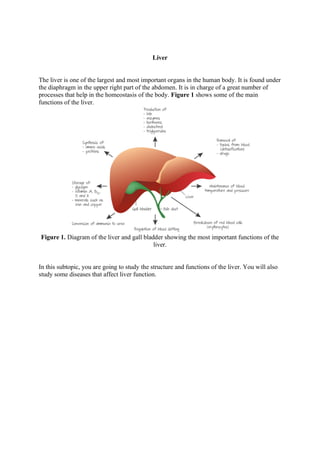
1. Liver
The liver is one of the largest and most important organs in the human body. It is found under
the diaphragm in the upper right part of the abdomen. It is in charge of a great number of
processes that help in the homeostasis of the body. Figure 1 shows some of the main
functions of the liver.
Figure 1. Diagram of the liver and gall bladder showing the most important functions of the
liver.
In this subtopic, you are going to study the structure and functions of the liver. You will also
study some diseases that affect liver function.
2. Blood Supply to The Liver
The liver is supplied with oxygenated blood from the heart through the hepatic artery. This
artery is a branch of the aorta. The blood then leaves the liver through the hepatic
vein, which carries deoxygenated blood. This vein joins the vena cava, which returns the
blood to the heart.
Figure 1. Blood supply to the liver.
The liver also receives deoxygenated blood coming from the spleen, stomach, pancreas, gall
bladder and intestines through the hepatic portal vein. This vein carries foods absorbed
mainly in the small intestine. It is rich in amino acids, glucose, vitamins, minerals and other
foods. The blood supplied by this blood vessel represents the majority of the blood received
by the liver (around 75% of the total blood supplied to the liver).
As the liver receives oxygenated blood from the hepatic artery and deoxygenated blood from
the hepatic portal vein, we say that the liver has a dual blood supply. Because the blood
from these two sources is mixed before entering the liver, its cells never receive fully
oxygenated blood.
In the liver, blood from the hepatic artery and the hepatic portal vein supply
the sinusoids that bathe the hepatocytes and Kupffer cells. As blood passes through the liver,
the hepatocytes monitor the contents of the blood and remove many toxic substances such as
alcohol and drugs before they can
reach the rest of the body. Enzymes metabolisem these toxins to render them harmless.
Many metabolic reactions take place in the liver. These reactions liberate heat, therefore
when blood passes through the liver it is warmed up. This helps to maintain the
body temperature in warm-blooded organisms.
3. Liver Structure
Liver lobules
The liver is a triangular shaped organ of approximately 1,500 to 2,000 g consisting of four
lobes. The internal structure of each lobe has around 100,000 lobules, each consisting of a
central venule coming from the hepatic vein surrounded by six venules coming from the
hepatic portal vein and six arterioles from the hepatic artery. These blood vessels are
connected by sinusoids (Figure 1).
Figure 1. Structure of a lobule in the liver.
Sinusoids
Sinusoids are tubes that resemble capillaries but have a discontinuous endothelium.
Figure 2. Capillaries and sinusoids.
4. Table 1. Differences between capillaries and sinusoids.
Hepatocytes and Kupffer cells
The lobules have mainly two types of cells, hepatocytes and Kupffer cells (as shown
in Figure 1). Hepatocytes perform most of the liver functions, especially storage and
metabolism. These cells are large (around 25 μm) and constitute around 80% of the total liver
cells. Their nucleus is round and found in the centre of the cell. These cells are capable of
regenerating when exposed to toxic substances.
Liver regeneration involves the replication of hepatocytes followed by the replication of other
liver cells. Once cell proliferation is completed, the newly divided cells undergo restructuring
and reformation of the extracellular matrix to complete the process. During regeneration,
liver function is only partially affected. Because human liver cells regenerate it has become
possible to use partial livers from living donors for transplantation, thereby increasing the
number of organs that are available for transplantation.
Plasma proteins are synthesised in hepatocytes mainly in the rough endoplasmic reticulum
(rER) and processed in Golgi complexes. As we have mentioned before, hepatocytes are also
involved in the degradation of toxins, such as detoxification of alcohol. Many of the
detoxification reactions occur in the smooth endoplasmic reticulum (sER). Hepatocytes also
have an exocrine function in the secretion of bile.
Kupffer cells are white blood cells (macrophages) that break down red blood cells. Therefore
they are involved in the recycling of erythrocytes, as you will see later in this subtopic. In the
lobules there are also canals (bile canaliculi) that carry bile to the bile duct that leads to the
gall bladder where bile is stored until it is used in the small intestine.
Temporary mounts of liver cells can be prepared from fresh liver tissue and observed under
light microscope. This can be done by mashing pieces of liver tissue in a mortar together
with 10 ml salt solution. Cells can be stained with a drop of methylene blue before placing
the coverslip on the slide. Part of the liver of a mouse under the light microscope is shown
in Figure 3(a) at a magnification of ×100 and Figure 3(b) at ×400.
5. Now that you know the structure of the liver, in the following sections we are going to study
some of its functions.
Figure 3b. Liver cells ×400
Figure 3a. Liver seen under light microscope ×100
6. Processing and Storage Nutrient
The liver is in charge of the processing and storage of many nutrients. As you have seen in
the previous section, blood enters the liver through the hepatic portal vein. This vein carries
most of the food digested and absorbed in the digestive tract.
Hepatocytes in the liver absorb most of the glucose and store it as glycogen. When the body
requires energy, this glycogen is broken down into glucose.
Fatty acids in the blood passing through the liver are absorbed by hepatocytes and
metabolised to produce energy in the form of ATP. Hepatocytes also synthesise lipids such as
triglycerides, cholesterol and phospholipids. These lipids can be bound to proteins forming
lipoproteins, which are now soluble in blood plasma and can therefore be transported in
blood to all the body. Much of the cholesterol produced by hepatocytes gets excreted from
the body as a component of bile.
Amino acids entering the liver are transformed into other amino acids or are used in the
synthesis of new proteins. Endoplasmic reticulum and Golgi apparatus in hepatocytes
produce plasma proteins. These plasma proteins include fibrinogen used in blood clotting,
and albumin that transports hormones and maintains the blood pH.
When amino acids are no longer necessary, hepatocytes remove the amine group from the
acid group (deamination). The acid group of the amino acid is used to produce energy or new
glucose molecules while the amine group is converted into ammonia. As ammonia is toxic, it
is transformed into urea, which is then eliminated by the kidneys in urine.
Figure 1 shows the processing of these molecules in the liver.
Figure 1. Metabolism of nutrients in the liver.
A very important function of the liver is detoxification. The liver gets rid of drugs, hormones
and other toxins. In some cases it breaks down the substances into harmless compounds. If it
cannot break them down, it attaches these substances to other organic groups (such a
glycine), which allows the kidneys to recognise them as unwanted waste material and are
therefore excreted.
7. The liver is in charge of metabolising alcohol. In Figure 2, it shows the chemical reactions
involved in this process. Ethanol is oxidised into acetaldehyde, a toxic substance, by the
hepatic enzyme alcohol dehydrogenase. Acetylaldehyde is converted into a less toxic
substance, acetate, by aldehyde dehydrogenase. Acetate is then broken down to acetyl-CoA
that can enter fatty acid metabolism or be used in the Krebs cycle. If acetaldehyde is not
broken down immediately, it can combine with proteins that induce liver injury. Excess of
alcohol can damage the liver, causing cirrhosis.
Figure 2. Alcohol detoxification.
The liver not only stores and processes nutrients, it has other important functions such as the
recycling of red blood cells and formation of bile, as you will see in the next sections.
Recyling Erythrocytes and Iron
Red blood cells or erythrocytes are cells modified to increase their capacity in the transport
of oxygen. In order to do this, they have a biconcave shape and have lost their nucleus and
organelles. These cells are rich in haemoglobin, a protein that binds oxygen (as HL students
will see in section haemoglobin and myoglobin). The biconcave shape increases their surface
area:volume ratio, thus increasing the absorption of oxygen. The lack of nucleus increases the
amount of hemoglobin in each cell. But at the same time this means that they cannot
reproduce, therefore they must be produced in the bone marrow from undifferentiated cells.
Erythrocytes are produced in the bone marrow and are liberated into the bloodstream. They
die after approximately 120 days circulating in blood. Dead erythrocytes are engulfed by
macrophages in the liver, spleen or bone marrow by phagocytosis. In the liver, these
macrophages are Kupffer cells.
In the Kupffer cells, the hemoglobin is split into globin chains and heme groups. Globin is
re-used in protein synthesis. The heme group is transformed into iron and bilirubin. Iron is
carried back to the bone marrow where it is used to produce new red blood cells. Bilirubin is
8. secreted into bile that will be used in the emulsification of fats. Figure 1 shows the function
of the liver in the recycling of erythrocytes.
Figure 1 . Recycling of erythrocytes and iron.
In the next section, you will study how the liver is involved in metabolic functions (formation
of cholesterol) and digestive functions (formation of bile salts).
Cholesterol and Bile Salt
Cholesterol
Cholesterol is one of the most well-known fats. You ingest cholesterol in your diet, but it is
not essential, as most cholesterol molecules are synthesised in the liver. As most animal cells
require cholesterol for membrane synthesis, a small portion is added to the membranes of
hepatocytes, and the rest is exported as lipoproteins or bile salts. Cholesterol is also a
precursor for other important molecules: the bile salts, steroid hormones (such as oestrogen
and progesterone), and vitamin D. Cholesterol synthesis is regulated according to its
concentration in cells. This depends on the amount ingested in diet, and the regulation is
performed by the hormones glucagon (inactivating its synthesis) and insulin (activating its
synthesis).
Figure 1 shows the structure of cholesterol.
Figure 1. Cholesterol molecule.
9. Cholesterol molecules, like triglycerides and phospholipids are insoluble in water. Therefore,
to be carried in blood to other tissues, they need to be carried as plasma lipoproteins.
Different combinations of lipids and proteins produce particles of different densities. High-
density lipoproteins (HDL) contain more protein, while low-density lipoproteins (LDL)
contain more lipids. LDLs and HDLs are both produced in the plasma; however a small
amount is synthesised in the liver. The function of LDLs is to transport cholesterol from the
liver to other organs. The function of HDLs is to transport cholesterol from tissues to the
liver.
As you have seen in the Atherosclerosis section, fats can deposit in arteries causing an
atheroma or plaque. This is mainly caused by white blood cells (foam cells) and LDL.
Molecules of LDL deposit in the blood vessels and can become oxidised. This will
cause atherosclerosis of the walls of the arteries (Figure 2).
Figure 2. Effect of LDL on blood vessels.
After some time this can cause cardiovascular disease and stroke. If the artery leading to the
heart (coronary artery) is clogged, the cells of the heart will not receive enough oxygen and
can therefore die. These cells are replaced by fibres causing coronary heart disease (CHD).
If the artery leading to the head (carotid artery) is affected, this can lead to a brain stroke.
Bile salts
10. Bile salts have a crucial role in digestion as they emulsify fats. This means they break fats
down into smaller droplets to increase their surface area. This allows enzymes (for example
pancreatic lipase) to work better.
Bile salts are synthesised by the liver from surplus cholesterol, and may be modified by
bacteria in the intestines. Bile salts are reabsorbed from the intestines into the liver, but lots
are lost in faeces. Approximately 600 mg of bile salts are synthesised daily to replace bile
acids lost in egestion. Bile salts aid in the digestion and absorption of dietary lipids and fat-
soluble vitamins.
The liver produces about one litre of bile per day. This fluid is carried by the bile canaliculi to
the bile duct, which carries it to the gall bladder to be stored. The composition of bile is
mainly water (97%), bile salts, cholesterol and fatty acids, bilirubin (from the breakdown of
erythrocytes) and inorganic salts.
In the next section, you will learn how liver damage affects the levels of bile and cholesterol.
Disesases Associated with Liver
Jaundice
Jaundice is a condition where the skin and white of the eyes turn yellow. It is caused by the
presence of bilirubin in extracellular fluid. As you have seen before, bilirubin is produced
from haemoglobin breakdown in erythrocyte recycling in the liver. The metabolism of
haemoglobin accounts for 65% to 80% of the total bilirubin production. Bilirubin in blood
binds reversibly to albumin (a plasma protein), forming conjugated bilirubin that travels to
the liver, which removes it from the plasma. When the liver is not able to remove the
bilirubin from blood, its level may rise (especially in the unconjugated form) and the skin and
eyes may begin to appear jaundiced.
Jaundice appears under several circumstances:
• Increased destruction of red blood cells.
• Immaturity in the conjugation of bilirubin (greater in premature babies).
• Genetic diseases (e.g. Gilbert syndrome).
• Defects in the secretion of conjugated bilirubin from hepatocytes (in liver damage).
• Defects in transit of bilirubin to intestines (e.g. with bile duct obstruction).
A high level of bilirubin in the blood is a sign of liver malfunction. Depending on the level of
exposure, the effects range from clinically unnoticeable to severe brain damage and even
death. Jaundice is usually a symptom of hepatitis or liver cancer. It can also be caused by the
use of drugs, genetic factors, malaria or anemia.
It is common for a baby's bilirubin level to be a bit high after birth as it might take some time
for the liver to function properly (Figure 1). Some of the causes are a mismatch between the
blood type of the mother and the child, lack of certain enzymes, or excess or abnormal blood
cells. Special blue lights are used on infants whose bilirubin levels are very high.
11. Figure 1. Premature baby being treated with ultraviolet light to cure jaundice.
Alcohol and cirrhosis
Cirrhosis is a disease where the damaged liver tissue is replaced by scar tissue, as shown
in Figure 2. Not only does this affect the functioning of liver cells, but also interferes with
the blood supply to these cells. The symptoms are weakness, fatigue, jaundice and bruising.
A liver biopsy will confirm the presence of scars. There is no cure for this disease. A liver
transplant can be the solution in extreme cases
Figure 2. Normal and cirrhotic liver.
Excessive alcohol or drug consumption may cause liver cirrhosis. Other causes of cirrhosis
include chronic viral hepatitis B or C, chronic bile duct obstruction, fatty liver disease, excess
of iron, cystic fibrosis and Wilson’s disease.
The consumption of alcohol has increased worldwide, as shown in Figure 3, therefore
increasing the amount of deaths due to liver cirrhosit.
12. Figure 3. Deaths due to liver cirrhosis per 100,000 people in 2010.
Source: Alcohol and Mortality: Global Alcohol-Attributable Deaths From Cancer, Liver
Cirrhosis, and Injury in 2010 .Jürgen Rehm and Kevin D. Shield. Alcohol Research: Current
Reviews, Volume 35, Issue Number 2.
The amount of alcohol consumed affects the possibility of liver cirrhosis in a direct
manner: the greater the consumption, the greater the chances of dying of liver cirrhosis. The
probability of dying of cirrhosis is greater in women than men at lower alcohol consumption,
but higher for men at greater alcohol consumption, as shown in Figure 4.
Figure 4. Increasing amounts of average daily alcohol consumption and relative risk of death
from cirrhosis.
Theory of Knowledge
Given the poor availability of organs for transplant, do you think an alcoholic should be
allowed a liver transplant?
There are many different opinions on to whether alcoholics should or shouldn't receive a
transplant. Here are only a few:
Why should alcoholics be entitled to receive a liver transplant?
• Everybody has the right to live.
13. • They can change their lifestyle and stop drinking.
• They might be the only support for a family.
• Many people care for them.
Why should they not receive a liver transplant?
• They caused the liver damage by their own choice of drinking.
• They must put up with the consequence of their own reckless attitude.
• Other people deserve the transplant more.
• They can relapse back into drinking and damage the new liver.