

Recommandé
Contenu connexe
Tendances
Tendances (20)
Similaire à Phylum sporozoa or acomplexa
Similaire à Phylum sporozoa or acomplexa (20)
Plus de Merlyn Denesia
Plus de Merlyn Denesia (20)
Dernier
Dernier (20)
Phylum sporozoa or acomplexa
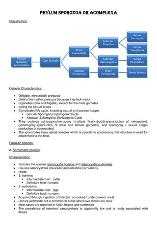
- 1. Phylum Sporozoa or Acomplexa Classification General Characteristics: Obligate, intracellular protozoa Distinct from other protozoa because they lack motor organelles (cilia and flagella), except for the male gametes during the sexual phase Complicated life cycle, including sexual and asexual stages Sexual- Sporogony/ Sporogonic Cycle Asexual- Schizogony/ Schizogonic Cycle They undergo schizogony/merogony (multiple fission/budding-production of merozoites), gametogony (production of male and female gametes), and sporogony ( sexual stage- production of sporozoites) The sporozoites have apical complex which is specific to sporozoans, this structure is used for attachment to the host. Parasitic Species: a. Sarcocystis species Characteristics: Includes the species Sarcocystis hominis and Sarcocystis suihominis Causes sarcocystosis (muscular and intestinal) in humans Hosts: S. hominis Intermediate host: cattle Definitive host: humans S. suihominis Intermediate host: pigs Definitive host: humans Acquired through ingestion of infected uncooked / undercooked meat Occurs worldwide but is common in areas where live stocks are raise Most cases are reported in Asian tropics and subtropics The prevalence of intestinal sarcocystosis is apparently low and is rarely associated with illness Phylum Sporozoa/ Apicomplexia Class Coccidia Order Eucoccidiida Suborder Eimeriina Genus Sarcocystis Genus Toxoplasma Suborder Haemosporina Genus Plasmodium Subclass Piroplasmea Order Piroplamida Genus Babesia
- 2. Life Cycle: The cysts with bradyzoites (infectice stage) ingested by humans will rupture in their intestines releasing the bradyzoites, these bradyzoites will differentiate into microgamete and macrogametes. The microgametes will fertilize the macrogametes resulting to oocysts. These oocysts will then be shed from the host in the feces. If these oocysts will be ingested by the intermediate hosts ( pigs and cattle) it will rupture in their blood vessels releasing sporozoites. These sporozoites will invade the muscle tissues of the intermediate hosts and develops into cysts containing bradyzoites which will then be passed into humans once ingested. Symptoms: 1. Muscular sarcocystosis Myositis Dyspnea Wheezing associated with eosinophilia 2. Intestinal sarcosystosis Nausea Stomach pain Diarrhea Diagnosis: Fecal flotation method IFA and ELISA Treatment: Albendazole Metronidazole Cotrimoxazole Corticosteroid b. Toxoplasma gondii Characteristics: Hosts: Definitive hosts: domestic cat and other felines Intermediate hosts: humans and mammals Causes toxoplasmosis Epidemiology: Transmission: food borne, zoonotic, transplacental, organ transplant 95% of the world has been infected with toxoplosama Infection is often highest in areas of the world that have hot, humid climates and lower altitudes.
- 3. Life Cycle: Sporocysts are shed from the feces of cats. These sporocsyts will undergo sporolation in the environment which will contaminate foods and water. If contaminated foods and/or water in ingested by livestock and humans it will undergo schizogony resulting into tachyzoites. Tachyzoites will invade the different organs of the body including the brain and will develop into bradyzoites (cysts). Tachyzoites can be transferred to another person through blood transfusion Tachyzoits can overcome the placental barrier in pregnant women and develop into bradyzoites (cysts) in the fetus’ brain. Symptoms: Immunocompetent – asymptomatic, (acute) cervical lymphadenopathy or flu-like illness Immunodeficeint- retinochoroiditis, pneumonitis, or other systemic disease. Patients with AIDS- intracerebral mass lessions Congenital infection can result in abortion, still birth, encephalitis, chorioretinitis, and hepatosplenomegaly Diagnosis: Observation of parasites in patient specimens, such as bronchoalveolar lavage material from immunocompromised patients, or lymph node biopsy. Isolation of parasites from blood or other body fluids, by intraperitoneal inoculation into mice or tissue culture. Detection of parasite genetic material by PCR, especially in detecting congenital infections in utero. Serologic testing is the routine method of diagnosis. c. Plasmodium species Characteristics: Malaria is the dominant protozoan disease From the Italian "mal' aria," meaning "bad air All are transmitted primarily by the female Anopheles mosquito; also shared needles, blood transfusions, and mother to child Hosts: Definitive host- Anopheles mosquito Intermediate host- Human 500 million new cases/year (most in Africa) Most frequent victims are children and young adults; ~ 2 million die/year Species involved:
- 4. P. falciparum P. Vivax P. malariae P. ovale Life Cycle: Exo-eryhtrocytic cycle (human)- when an infected anopheles mosquite bits a human, sporozoite (infective stage) is injected into the host. The sporozoite will then invade the host’s liver cells and undergo shizogony (multiple fission) which forms shizonts in the hepatic cells, once the schizonts rupture it will release merozoites. These merozoites invade the red blood cells. Others reinvade the liver cells resulting into another exo-erythrocytic cyle, some of them remain dormant in the liver cells (hypnozoites) which will become active later on. Erythrocytic cycle (human)- the merozoites that invade the RBC’s will form trophozoit/ring stage (feeding stage- they feed on the hemoglobin of the RBC’s) and will undergo another schizogony (multiple fission) which forms schizonts, if this schizonts will rupture (associated with paroxysm experienced by the patient) it will release merozoites which will reinvade the red blood cells initiating another erythrocytic cycle. Gametogony- some of the trophoziotes will form gametocysts which will differentiate into microgamete and macrogamete. These undergo no further development until taken by the mosquito. Sporogonic Cycle (mosquito)- If another female Anopheles mosquito bites an infected person it sucks blood containing the different stages of malarial parasites. All stages other than the gametes are digested in the stomach of the mosquito. Inside the mosquito the microgamete will fertilized the macrogamets resulting in the formation of ookinete. The ookinetes will transform into oocysts, the oocysts will then undergo meiosis. If these cysts will rupture it will release sporozites that migrates to the salivary gland of the mosquito. Epidemiology: Geographical distribution - malaria is present worldwide in tropical and subtropical areas. Relapse versus Recrudesence - P. vivax is, traditionally, the “relapsing” malaria. All others can recrudesce (“bloom”) due to incomplete therapy. Prevention - detect and treat infected individuals; mosquito control. Immunity - incomplete immunity follows infection. Some persons get reinfected over and over. o Sickle cell trait - the malaria parasite is not successful at utilizing the “S” haemoglobin. This trait does not confer immunity to infection, but does offer resistance to infection. o Duffy factor - represents the “portal of entry” antigen for P. vivax. Persons without the factor are immune to this species (but not the others). 1. Plasmodium falciparum Found worldwide in tropical and subtropical areas.
- 5. No selectivity in host erythrocytes (invades old and young RBC’s) The infected red blood cells do not enlarge and become distorted The trophozoite/ring is often seen in the host cells at the very edge or periphery of cell membrane Maurer’s dots are observed (reddish granules) Peripheral blood smears characteristically contain only young and forms and crescent shaped gametocytes Can cause severe malaria because it multiples rapidly in the blood, and can thus cause severe blood loss (anemia). The infected parasites can clog small blood vessels. When this occurs in the brain, cerebral malaria results, a complication that can be fatal. 2. Plasmodium vivax Found mostly in Asia, Latin America, and in some parts of Africa. Most prevalent human malaria parasite Selective (young immature erythrocytes) Infected RBC’s are usually enlarged and contains numerous schuffner’s dots (pinkish granules) The trophozoite is ring shaped but amoeboid in appearance The gametocytes are round Have dormant liver stages ("hypnozoites") that can activate and invade the blood ("relapse") several months or years after the infecting mosquito bite. 3. Plasmodium malariae Found worldwide, is the only human malaria parasite species that has a quartan cycle (three- day cycle). (The three other species have a tertian, two-day cycle.) Can infect only mature erythrocytes with relatively rigid cell membrane The parasite’s shape must conform to the size and shape of the cell, this requirement produces no RBC enlargement or distortion resulting in host cells’ “dark and band form” as well as a very compact dark staining forms In some chronically infected patients P. malariae can cause serious complications such as the nephrotic syndrome. 4. Plasmodium Ovale Found mostly in Africa (especially West Africa) and the islands of the western Pacific. Selectivity for young, pliable erythrocytes The host cell becomes enlarged and distorted, usually in an oval form The infected cell border is commonly fimbriated or ragged Shuffner’s dots appear as pale pinkish granules In comparison:
- 6. Uncomplicated Malaria (malaria attack lasts 6-10 hours) Stages: A cold stage (sensation of cold, shivering) A hot stage (fever, headaches, vomiting; seizures in young children) And finally a sweating stage (sweats, return to normal temperature, tiredness). Symptoms: Fever Chills Sweats Headaches Nausea and vomiting Body aches General malaise Mild jaundice Enlargement of the liver Increased respiratory rate Severe Malaria (occurs when infections are complicated by serious organ failures or abnormalities in the patient's blood or metabolism) Cerebral malaria, with abnormal behavior, impairment of consciousness, seizures, coma, or other neurologic abnormalities Severe anemia due to hemolysis (destruction of the red blood cells) Hemoglobinuria (hemoglobin in the urine) due to hemolysis Acute respiratory distress syndrome (ARDS), an inflammatory reaction in the lungs that inhibits oxygen exchange, which may occur even after the parasite counts have decreased in response to treatment Abnormalities in blood coagulation Low blood pressure caused by cardiovascular collapse Acute kidney failure Hyperparasitemia, where more than 5% of the red blood cells are infected by malaria parasites Metabolic acidosis (excessive acidity in the blood and tissue fluids), often in association with hypoglycemia Hypoglycemia (low blood glucose). Hypoglycemia may also occur in pregnant women with uncomplicated malaria, or after treatment with quinine. Other Manifestations: Neurologic defects may occasionally persist following cerebral malaria, especially in children. Such defects include trouble with movements (ataxia), palsies, speech difficulties, deafness, and blindness. Recurrent infections with P. falciparum may result in severe anemia. Malaria during pregnancy (especially P. falciparum) may cause severe disease in the mother, and may lead to premature delivery or delivery of a low-birth-weight baby. On rare occasions, P. vivax malaria can cause rupture of the spleen. Nephrotic syndrome (a chronic, severe kidney disease) can result from chronic or repeated infections with P. malariae. Hyperreactive malarial splenomegaly (also called "tropical splenomegaly syndrome") occurs infrequently and is attributed to an abnormal immune response to repeated malarial infections. The disease is marked by a very enlarged spleen and liver, abnormal immunologic findings, anemia, and a susceptibility to other infections (such as skin or respiratory infections). Diagnosis: Microscopy PCR IFA Serological Test RDT
- 7. Treatments: chloroquine atovaquone-proguanil artemether-lumefantrine mefloquine quinine quinidine doxycycline (used in combination with quinine) clindamycin (used in combination with quinine) primaquine is active against the dormant parasite liver forms (hypnozites) and prevents relapses d. Babesia sp. Babesia bigemina /Babesia microfti Causes babesiosis Occurs in the US Infects and destroys red blood cells Transmitted by ticks (under nymph stage) Transmission: bite of infected tick, blood transfusion, and congenital transmission Hosts: Definitive host –tick Intermediate host- humans Life Cycle: During a blood meal, a Babesia-infected tick introduces sporozoites into the mouse host. Sporozoites enter erythrocytes and undergo asexual reproduction (budding). In the blood, some parasites differentiate into male and female gametes. The definitive host is the tick. Once ingested by an appropriate tick, gametes unite and undergo a sporogonic cycle resulting in sporozoites. Humans enter the cycle when bitten by infected ticks. During a blood meal, a Babesia-infected tick introduces sporozoites into the human host. Sporozoites enter erythrocytes and undergo asexual replication (budding).Humans usually are dead-end hosts. However, human-to-human transmission is well recognized to occur via contaminated blood transfusions. Symptoms: presence of hemolytic anemia and nonspecific flu-like symptoms (e.g., fever, chills, body aches, weakness, fatigue). Splenomegaly Hepatomegaly Jaundice Severe cases Thrombocytopenia Myocardial infarction Renal failure Altered mental status Death.
- 8. Diagnosis: Microscopy Serological test Treatment: atovaquone PLUS azithromycin clindamycin PLUS quinine Phylum Ciliophora General Characteristics: Largest free living multi-cellular organism Have two kinds of nuclei, a large macronucleus and a smaller micronucleus Have cilia that are similar to but shorter than flagella The cilia are arranged in precise rows on the cell. They are moved in unison to propel the cell through its environment and to bring food particles to the mouth Reproduce asexually by mitosis and sexually by conjugation The only ciliate is a human parasite in Balantidium coli, The causative agent of a rare type of dysentery- balantidine dysentery (infection rate is 1%) a. Balantidium coli Found in the intestinal tract of arthropods and some vertebrates, including mammals Pathogen of humans, pigs, and monkeys Coarse cilia line the peristomal area Macronucleus is typically elongated and kidney shaped, while the micronucleus is spherical The trophozoite inhabits the cecum, and colon of humans and is the largest protozoan parasite to humans Infection occurs when contaminated water or food is ingested Excystation occurs in the small intestine and encystation occurs in the large intestine, it may also occur outside the host Life Cycle: Symptoms: Asymptomatic Persistent diarrhea Occasionally dysentery Abdominal pain Weight loss
- 9. Diagnosis: Stool Examination Endoscopy Treatment: Tetracycline Metronidazole Iodoquinol