ENCAPSULATING PERITONEAL SCLEROSIS (EPS)
•Télécharger en tant que PPT, PDF•
1 j'aime•1,807 vues

most common complication of peritoneal dialysis ENCAPSULATING PERITONEAL SCLEROSIS (EPS)

Recommandé
Recommandé
Contenu connexe
Tendances
Tendances (20)
Similaire à ENCAPSULATING PERITONEAL SCLEROSIS (EPS)
Similaire à ENCAPSULATING PERITONEAL SCLEROSIS (EPS) (20)
Plus de mohamed hassan abbass
Plus de mohamed hassan abbass (20)
Dernier
Dernier (20)
ENCAPSULATING PERITONEAL SCLEROSIS (EPS)
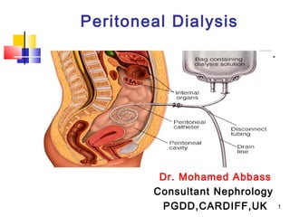
- 1. Peritoneal Dialysis Dr. Mohamed Abbass Consultant Nephrology PGDD,CARDIFF,UK 1
- 2. ENCAPSULATING PERITONEAL SCLEROSIS (EPS) EPS is an uncommon but serious complication of long term peritoneal dialysis partial or completes intestinal obstruction. 2
- 3. ENCAPSULATING PERITONEAL SCLEROSIS (EPS) The risk factor Duration of PD therapy, significant after 5 years and more after 10 years. Onset of PD at young age. No association of EPS with the type or number of episodes of PD peritonitis or with the type or strength of PD solutions 3
- 4. Pathophysiology In a healthy person, the peritoneum is very thin , so the intestinal loops move easily and food passes through In peritoneal dialysis patient, the membrane becomes thick and fibroses (sclerosed) The food is no longer passing through the blocked intestine which causes abdominal pain, nausea, vomiting and weight 4
- 5. Pathophysiology There are two phases: The early inflammatory phase Vague abdominal discomfort and Bloody effluent Rapid transport status Signs of inflammation (erythropoietin- resistant anemia and elevated CRP). Sclerosing phase: the membrane becomes thick and fibroses (sclerosed) over time till restrict the intestinal movement and become blocked 5
- 6. Clinical picture Most commonly symptoms occur after quitting PD. Symptoms of intestinal obstruction (abdominal pain, intermittent constipation, vomiting), weight loss, ascites (which can be haemorrhagic). An inflammatory process raising CRP. If patient still on PD; UF failure, Length of time on PD, Rapid transport status , Severe peritonitis. 6
- 7. Clinical course • Severe cases progress rapidly complete bowel obstruction severe malnutrition (on parenteral nutrition) death. • Moderate cases intermittent course with nutrition maintained small amounts frequently with oral nutritional supplements. • Mild cases improve all their symptoms over time and not require any dietary manipulations. 7
- 8. Diagnosis Should be early during inflammatory stage. Clinical features: HX of change to HD or transplanted after many years on PD Unexplained haemorrhagic ascites Unexplained high CRP with abdominal pain Unexplained weight loss. 8
- 9. Diagnosis Abdominal CT scan (with contrast) standard method Peritoneal thickening and calcification Thickened and Dilated bowel loops with bowel pulled into center of abdominal cavity (‘fist sign') MRI scan has been suggested as a useful tool. Abdominal X-ray is not helpful only may show 9
- 10. 10 Abdominal CT scan from a patient with EPS. Red arrows indicate thickened parietal peritoneum with calcification. Green arrows indicate thickened visceral peritoneum forming a cocoon containing loops of bowel.
- 11. Treatment Drug treatment: in inflammatory phase Corticosteroid : moderate dose, duration of treatment is unclear and should be titrated to the symptoms. Tamoxifen for its antifibrotic effects. Sirolimus :no evidence of any benefit. Surgery: in sclerosing phase