Membrane Structure Revision Questions
•Télécharger en tant que DOCX, PDF•
1 j'aime•433 vues

The document discusses cell membrane structure and models. It provides revision questions about phospholipid and cholesterol structure in membranes. Specifically, it asks students to: 1) Draw and label structures of phospholipids, membranes, and the plasma membrane. 2) Outline the Davson-Danielli and Singer-Nicholson models of membrane structure and the evidence used to support and refute each. 3) Explain how evidence from techniques like freeze-fracture and protein extraction refuted the Davson-Danielli model and supported the fluid mosaic model.

Recommandé
Contenu connexe
Tendances
Tendances (18)
En vedette
En vedette (13)
Similaire à Membrane Structure Revision Questions
Similaire à Membrane Structure Revision Questions (20)
Plus de Miltiadis Kitsos
Plus de Miltiadis Kitsos (15)
Dernier
Dernier (20)
Membrane Structure Revision Questions
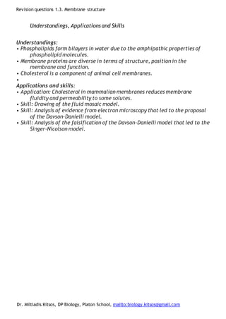
- 1. Revision questions 1.3. Membrane structure Dr. Miltiadis Kitsos, DP Biology, Platon School, mailto:biology.kitsos@gmail.com Understandings, Applications and Skills Understandings: • Phospholipids form bilayers in water due to the amphipathic properties of phospholipid molecules. • Membrane proteins are diverse in terms of structure, position in the membrane and function. • Cholesterol is a component of animal cell membranes. • Applications and skills: • Application: Cholesterol in mammalian membranes reduces membrane fluidity and permeability to some solutes. • Skill: Drawing of the fluid mosaic model. • Skill: Analysis of evidence from electron microscopy that led to the proposal of the Davson-Danielli model. • Skill: Analysis of the falsification of the Davson-Danielli model that led to the Singer-Nicolson model.
- 2. Revision questions 1.3. Membrane structure Dr. Miltiadis Kitsos, DP Biology, Platon School, mailto:biology.kitsos@gmail.com 1. Draw and label a diagram of a single phospholipid molecule. 2. Draw and label a phospholipid monolayer and a bilayer.
- 3. Revision questions 1.3. Membrane structure Dr. Miltiadis Kitsos, DP Biology, Platon School, mailto:biology.kitsos@gmail.com 3. Draw and label a diagram of the plasma membrane. 4. Outline the structure of the Davson-Danielli model of the cell membrane. Outline the evidence used to support it. …………………………………………………………………………………… …………………………………………………………………………………… …………………………………………………………………………………… …………………………………………………………………………………… …………………………………………………………………………………… …………………………………………………………………………………… …………………………………………………………………………………… …………………………………………………………………………………… …………………………………………………………………………………… …………………………………………………………………………………… …………………………………………………………………………………… …………………………………………………………………………………… …………………………………………………………………………………… …………………………………………………………………………………… 5. Outline the main points of the freeze-fracture technique and explain how evidence from this method, like this micrograph, were used to refute the Davson-Danielli model
- 4. Revision questions 1.3. Membrane structure Dr. Miltiadis Kitsos, DP Biology, Platon School, mailto:biology.kitsos@gmail.com …………………………………………………………………………………… …………………………………………………………………………………… …………………………………………………………………………………… …………………………………………………………………………………… …………………………………………………………………………………… …………………………………………………………………………………… …………………………………………………………………………………… …………………………………………………………………………………… …………………………………………………………………………………… …………………………………………………………………………………… 6. Improvements in biochemical techniques allowed proteins to be extracted from membranes. Outline how evidence from these methods were used to refute the Davson-Danielli model? …………………………………………………………………………………… …………………………………………………………………………………… …………………………………………………………………………………… …………………………………………………………………………………… …………………………………………………………………………………… …………………………………………………………………………………… …………………………………………………………………………………… …………………………………………………………………………………… …………………………………………………………………………………… ……………………………………………………………………………………
- 5. Revision questions 1.3. Membrane structure Dr. Miltiadis Kitsos, DP Biology, Platon School, mailto:biology.kitsos@gmail.com 7. In the following image you may see two integral proteins and some notes on their affinity with water. This evidence falsifies the Davson-Danielli model. Explain how Hydrophilic region Hydrophobic region
- 6. Revision questions 1.3. Membrane structure Dr. Miltiadis Kitsos, DP Biology, Platon School, mailto:biology.kitsos@gmail.com …………………………………………………………………………………… …………………………………………………………………………………… …………………………………………………………………………………… …………………………………………………………………………………… …………………………………………………………………………………… …………………………………………………………………………………… …………………………………………………………………………………… …………………………………………………………………………………… …………………………………………………………………………………… …………………………………………………………………………………… 8. Outline the Singer-Nicholson fluid mosaic model of the cell membrane. …………………………………………………………………………………… …………………………………………………………………………………… …………………………………………………………………………………… …………………………………………………………………………………… …………………………………………………………………………………… …………………………………………………………………………………… …………………………………………………………………………………… …………………………………………………………………………………… …………………………………………………………………………………… …………………………………………………………………………………… http://www.ib.bioninja.com.au/_Media/tracie_med.jpeg 9. List the six main functions of the membrane proteins. T :
- 7. Revision questions 1.3. Membrane structure Dr. Miltiadis Kitsos, DP Biology, Platon School, mailto:biology.kitsos@gmail.com R : A : C : I : E : 10. Outline the functions of glycoproteins found in the plasma membrane …………………………………………………………………………………… …………………………………………………………………………………… …………………………………………………………………………………… …………………………………………………………………………………… …………………………………………………………………………………… …………………………………………………………………………………… …………………………………………………………………………………… ……………………………………………………………………………………
- 8. Revision questions 1.3. Membrane structure Dr. Miltiadis Kitsos, DP Biology, Platon School, mailto:biology.kitsos@gmail.com 11. Cholesterol is a type of lipid, but it is not a fat or oil. What group does it belong to? …………………………………………………………………………………… 12. Outline the structure of cholesterol and its position in the membrane. You may sketch the relative position if you wish. …………………………………………………………………………………… …………………………………………………………………………………… …………………………………………………………………………………… …………………………………………………………………………………… …………………………………………………………………………………… …………………………………………………………………………………… …………………………………………………………………………………… …………………………………………………………………………………… 13. Explain how cholesterol affects the fluidity of the membrane. …………………………………………………………………………………… …………………………………………………………………………………… …………………………………………………………………………………… …………………………………………………………………………………… …………………………………………………………………………………… …………………………………………………………………………………… …………………………………………………………………………………… ……………………………………………………………………………………
- 9. Revision questions 1.3. Membrane structure Dr. Miltiadis Kitsos, DP Biology, Platon School, mailto:biology.kitsos@gmail.com References: Allott, Andrew. Biology: Course Companion. S.l.: Oxford UP, 2014. Print. Paine, Chris . “BioKnowledgy DP Notes 1.3 - Membrane structure” < http://www.slideshare.net/diverzippy/bioknowledgy-dp-notes-13-membrane- structure?ref=http://www.bioknowledgy.info/13-membrane-structure.html> Web. 25 Sep 2016.