SlideShare une entreprise Scribd logo
1  sur  38
Télécharger pour lire hors ligne
Recommended literature :
Berg-Tymoczky-Stryer: Biochemistry
Lehninger: Principles of Biochemistry
Biochemistry II
Proteins
Dr. Terez Barna
Function of proteins
• Catalysis – enzymes
• Structural support – collagen
• Transport – hemoglobin
• Trans-membrane transport – Na+/K+ ATPases
• Toxins – rattle snake venom, ricin
• Contractile function – actin, myosin
• Signaling molecules : hormones (insulin)
• Storage Proteins – Calreticulin and calnexin are
ubiquitous Ca2+ storage proteins
• Defensive proteins – antibodies
Central Dogma of Molecular Biology
DNA
mRNA
Protein
Transcription
Translation
Transcription
Translation
RNA Reading Direction corresponds to Protein Chemical
Directionality
NH2-terminus
COOH-terminus
5’ 3’
mRNA
1 2
3 4
5
Protein biosynthesis on ribosomes
CONDENSATION REACTION
FORMATION OF PEPTID BOND
6
N-terminális
C-terminális
POLYPEPTIDE CHAIN
Stereochemistry of
-amino acids
chiral center
Stereochemistry of
-amino acids
•Enantiomer
•Optically active
living organisms use L-amino acids
L-alanine D-alanine
5 6
7 8
Isoleucine –two chirality centers
*
*
Optically active substances rotate plane polarised light
Chirality explaines ligand binding specificity of proteins
Hydrophobic Amino Acids
9 10
11 12
Hydrophylic Amino Acids UV-VIS absorbance of aromatic side-chains of protein
Protonated/deprotonated forms of amino
acid
Formation of Disulfide-bridge (oxidation)
13 14
15 16
The functional insulin
contains three disulfide bridges
Post-translational modification of side
chains
O
NH
O
H
O
H
O
N
H2
O
O
H
O
O
-
O
-
P
Phosphoserine 4-Hydroxyproline
Folding into a unique conformation
Protein conformation
Variability of protein structure
17 18
19 20
Peptide bond is planar C N 1,27Å
C N 1,47Å
C N 1,27Å
C N 1,47Å
C-N 1,33Å
40% N−C' double bond character
Linus Pauling
C−C bond length in benzene is 1.39 A˚.
The Kekulé structure -
Bonds in bezene have partial double bond character
Single C-C bond length in
Hydrocarbons
Two configurations of the planar peptid bond are possible:
one in which C atoms are trans , the other in which they are cis
The trans form is intrinsically favored energetically – fewer repulsions between non-bonded atoms
Schematic diagram of an extended polypeptide chain
The Peptide Bond
Resonance of peptide bond
- Polarity, Dipole moment
- partial double-bond character
Trans-conformation of C
21 22
23 24
cis-trans isomers of proline
The cyclic side chains in proline diminishes the repulsions between atoms so the intrinsic stability
of cis isomer is comparable to that of the trans isomer
Linus Pauling: 1954 Noble prize
ªThe Structure of Proteins:
Two Hydrogen-Bonded Helical
Configurations of
the Polypeptide Chainº.
1951 Proceedings of the United States National Academy of Sciences
Linus Pauling predicted two regular
periodical structurea:
right handed -helix and
-pleeted sheet
Linus Pauling, has been awarded two undivided Nobel Prizes,
in 1954 in Chemistry and in 1962 in the Peace Prize.
Backbone Degrees of Freedom No rotation about the peptide bond.
 Limited range of conformations.
- Free rotation about the N(H)‒C bond:  (phi)
Free rotation about the C‒C(=O) bond:  (psi)
 and  = 180o when polypeptide is in its fully extended conformation and all peptide bonds
are in the same plane.
- +180o >  and  > ‒180o
25 26
27 28
Free rotation around C-atoms with  (fi) and 
(psi) angles
 (fi) and  (psi) angles are restricted to certain range -- Limited conformation
Some values are never observed because of the steric hindrance of the side chains
Torsion angle is positive if
the direction is clockwise (+)
Anticlockwise (-) N(H)‒C : bond rotation  (phi)
- C‒C(=O) bond rotation :  (psi)
- Due to the size and charge of the R groups,  and  are
rotationally hindered.
 Ramachandran Plot
( =  = 0o not allowed.)
 The conformational freedom and therefore
the torsion angles of a polypeptide backbone
are sterically constrained.
 Rotation around the C-N and C -C bonds
to form certain combinations of  and 
angles will cause the amide hydrogen, the
carbonyl oxygen, or the substituents of C  of
adjacent residues to collide.
Torsion Angles between Peptide Groups Describe Polypeptide Chain Conformations
The Ramachandran Diagram Indicates Allowed Conformations of Polypeptides
Ramachandran Plot: indicates allowed
conformations of polypeptides
No steric overlap


Not
allowed.
Allowed at the
extreme limits
• Sterically allowed values of  and .
• Sterically forbidden conformations,
in which the  and  values would bring
atoms closer than the corresponding
van der Waals distance (the distance of
closest contact between nonbonded atoms).
• Only three small regions of the
diagram are physically accessible to
most residues (allowed regions).
Left handed helix
29 30
31 32
 -Helix:
Periodicity = 3.6
Helical rise per amino acid
= 1.5 Å
Pitch of the helix = 5.4 Å
 -Sheet:
Periodicity = 2
Távolság = 5.4 Å
Two regular, periodic structures in the polypeptide chains
(Allowed regions in the Ramachadran plot)
Regular structures are composed of sequences of
residues with repeating of  and  values.
Secondary structure:
• Secondary structure refers to particularly stable arrangements
of amino acid residues giving rise to recurring structural patterns
defined by hydrogen bonds between backbone amide groups.
(side chain-main chain and side chain-side chain hydrogen bonds
are irrelevant).
• The most prominent are α-helices and β-sheets
Right-handed vs. Left handed α-helix
 α-helix can form in polypeptides
consisting of either L- or D-amino
acids. However, all residues must
be of one stereoisomeric series; a
D-amino acid will disrupt a regular
structure consisting of L-amino
acids, and vice versa.
 Naturally occurring L-amino acids
can form either right- or left-
handed helices, but extended left
handed helices have not been
observed in proteins.
N
N+4
 polypeptide backbone is wound around an imaginary axis with
R group protruding outwards.
• Stabilized by H-bonds between: C=O(n) and N-H(n+4).
atomic number / H-bridge:13
– Φ = –57O , Ψ = –47O
Properties of the Alpha Helix ( 3.613-helix)
strong hydrogen bonds with nearly optimum N..H…O distance of 2.8 Å.
1
2
3
13
33 34
35 36
Properties of the Alpha Helix ( 3.613-helix)
• 3.6 amino acid residues per helical turn
• p = hn = 5.4 Å (the distance, where the helix rises
along its axis per turn)
Properties of the Alpha Helix ( 3.613-helix)
average length: 12 amino acid (~three turns, ~18 Å
max. 80 Å
The rise along the axis after each
amino acid extention is 1.5 Å
alpha-helix has a dipole character
Amino terminus
Carboxyl terminus
Top view
R groups protrude outwards
-Helix
Amino acid side chains project outward and
downward from the helix ,thereby avoiding
steric interference with the polypeptide
backbone and with each other.
The core of the helix is tightly packed;
that is, its atoms are in van der Waals contact.
37 38
39 40
Among the theoretical helices 310, α and π helices are found in proteins
310 helix  vagy (4.416) helix
42
The globin structure containing eight alpha-helices
The globin fold
 sheet
• The backbone of the polypeptide chain is extended into a zigzag, and arranged side-by-side
to form a structure resembling a series of pleats.
• H-bonds are formed between adjacent segments. One peptide bonds make two H-bonds
with the neighboring polypeptide chains.
4.5 Å
C
N
C
7 Å
R1 R3
 Sheets in proteins contain 2 to as many
as 22 polypeptide strands, with an average
of 6 strands.
Each strand may contain up to 15 residues,
the average being 6 residues.
41 42
43 44
Parallel  sheet Antiparallel  sheet
Antiparallel  sheet: neighboring hydrogen-bonded
polypeptide chains run in opposite directions.
In  sheet the hydrogen-bonded chains extend in the
same direction.
Parallel  sheets are less stable than antiparallel  sheets,
because the hydrogen bonds of parallel sheets
are distorted compared to those of the antiparallel sheets.
In  sheet the peptide chains can be either parallel or antiparallel.
The two residues repeat distance is shorter for the parallel
conformation (6.5 Å, versus 7 Å for antiparallel).
6.5 Å
Beta barrel of retinol-binding protein
Properties of the Beta Sheet
 strands usually have a pronouced
right-handed twist, due to steric effects
arising from the L-amino acid configuration.
β-barrel strands topology in a β-barrel :
Connections between adjacent strands in 
sheets
• (a) Antiparallel strands may be connected by a small loop.
• (b) Parallel strands require a more extensive crossover connection
Turns and Loops
• Segments with regular secondary structure such as  helices or the strands of  sheets are
typically joined by stretches of polypeptide (turns and loops) that abruptly change direction.
• Such reverse turns or  turns( named because they often connect successive strands of
antiparallel  sheets) almost always occur at protein surfaces.
• Most reverse turns involve four
successive amino acid residues more or
less arranged in one of two ways, Type I
and Type II. Both turns are stabilized by a
hydrogen bond between backbone CO(i) and
the backbone NH (i+3).
• In Type II turns, the oxygen atom of residue
2 crowds the C atom of residue 3, which is
therefore usually Gly.
• Residue 2 of either type of turn is often Pro since it can assume the required
conformation.
45 46
47 48
Relative probabilities that a given amino acid will occur in the
secondary structure
Supersecondary structures or motif :
• The association of secondary structures in a particular geometrical arrangement (distinct,
stable folding pattern) with defined topology (connectivities) containing 10 to 40 residues in
length.
• Motifs are important in describing protein structure because they can be
repeated within many protein structures.
alpha-alpha corner
Short loop regions connecting
helices which are roughly
perpendicular to one another.
( a strictly definite alternation
of hydrophobic, hydrophilic and
glycine residues;)
Greek key motif:
antiparallel β sheet comprising
of four b strands with short and
longer loop connections
between some strands.
β-barrel structures contain
Greek key motifs named
after a Greek pottery.
An α helix connects
parallel β strands in β sheets.
Hydrophobic surfaces on the
helix and β sheet interact.
Residues in the first loop
(C-terminal end of the
β strand) often contribute to
the active site in enzymes.
Leucine zipper motif
Two identical subunits interact via
α helices, forming a short stretch
of coiled-coil.
The interaction is mediated by
hydrophobic interactions
between side-chains, notably
those of leucine residues.
Supersecondary structures:
helix-turn-helix
- two alpha helices connected by a short loop region
Recurring feature in proteins with DNA binding function
Recognition of the major groove in DNA
Helix-turn-helix motif binds to
Bent B-DNA
49 50
51 52
• A spatially separated, stable unit of tertiary structure, which is identified by some characteristic
patterns.
• Often linked by a flexible hinge region, these domains are compact and stable, with a hydrophobic core.
• Domains fold independently of the rest of the polypeptide, satisfying most of their residue–residue
contacts internally. Domain is a structural and functional unit of proteins.
• Proteins may have multiple domains and each domain has a distinct evolutionary origin and function
(DNA bind domain, coenzyme binding domain, Ca2+ binding domain).
See protein domain databases like Prosite, Conserved Domains Database (CDD), InterPro, Pfam.
Domains
Protein pyruvate kinase has
multidomain structure
Hemoglobin consists of
a single domain
Comparison of the structures of triosephosphate isomerase and dihydrofolate reductase
Tertiary Structure
The arrangement of multiple secondary structural elements leads to tertiary structure.
Protein tertiary structure is more reliably conserved than primary sequence.
•Two proteins with similar secondary structure elements but with different topology
(connectivities) leads to different tertiary structures – different fold.
Structural levels of proteins
3D structure of proteins
Tertiary
structure
Quaternary structure
53 54
55 56
Structural Classification of Proteins - SCOP database
aims at classifying proteins according to structural and evolutionary relationship.
Structural hierarchy: ClassFoldSuperfamilyFamily
Class: secondary structural elements
Fold: spatial arrangement of secondary structural elements
Superfamily: Made of several families, little sequence similarity, share motifs and functional
similarities  Probable evolutionary relationship
Family: Sequence similarity and/or similar structure/function  Strong evolutionary relationship
Classes:
1. All alpha proteins
2. All beta proteins
3. / Alpha and beta proteins
4.  and  relatively separated (a+b)
5. Multi-domain proteins (alpha and beta)
Folds consisting of two or more domains belonging to different classes
6. Membrane and cell surface proteins and peptides
7. Small proteins
Usually dominated by metal ligand, heme, and/or disulfide bridges
http://scop.mrc-lmb.cam.ac.uk/scop/
Structural Classification of proteins based on secondary structures
and their connectivities
1- all -helix
2- all  sheet
3- /
4-  and  relatively separated
5- multidomain protein
5
2,
3.
1
4.
4.
59
CATH: Protein Structure Classification database
http://cathwww.biochem.ucl.ac.uk/latest/index.html
60
C - Class - determined according to
secondary structure composition.
A - Architecture - describes the
overall shape of the domain structure.
T - Topology - major
structural similarities (similar connectivities
among the secondary elements).
H - Homology Superfamily – (Superfamily
in SCOP) Protein domains which share
a common ancestor.
CATH is a hierarchical classification of protein
domains, which clusters proteins at four major levels:
57 58
59 60
Classification of proteins at a higher hierarchy level:
Membrane protein
Globular protein
Fibrous protein
Unstructured protein
three-dimensional structure of part of the cytochrome bc1 complex
Membrane Protein Structure
Polar side chains
interacting with polar head
group of the lipids and
each other
Hydrophobic
side chain
Alfa- helices are the most common secondary structure elements in membrane proteins
Protein Data Bank (PDB: WWW.rcsb.org/pdb)
1. Crystallysing proteins
2. Using X-ray diffraction
3. Calculating electron density map
4. Model Building
Solving protein structure by X-ray diffraction
61 62
63 64
 3-D structure of a protein is determined by its amino acid sequence
(primary structure).
 The function of a protein depends on its structure.
 A protein exists multiple, thermodynamically stable conformation (lowest
G) native conformations
 A protein’s conformation is stabilized largely by weak interactions.
 Protein structures show common structural patterns (secondary structures,
supersecondary structures, domains.)
• Proteins undergo conformational change upon ligand binding
or during catalysis.
-
Structural levels of proteins
66
3-D structure of a protein is determined
by its amino acid sequence
Primary Structure: aminoacid sequence + the position of disulfide bonds
Protein Quaternary Structure
 More than one polypeptide chain associate together with a
specific geometry
 The spatial arrangement of the subunits is called the
quaternary structure
Hemoglobin,
22 tetramer
(dimer of 
protomers)
65 66
67 68
Quaternary Structure
Oligomers: composed of more than one polypeptide chain subunits
Protomer is the structural unit
of an oligomeric protein that are
symmetrically arranged.
Protein assemblies built on identical subunits are usually symmetric
The forces that drive folding (in native
structure)
Disulfide bridge
covalent: 70-100
kcal/mol)
Electrostatic interaction
(10 kcal/mol), affected
by pH
van der Waals force
(transient and weak:
0.1-0.2 kcal/mol)
Hydrophobic interaction
Hydrogen bond
2-5 kcal/mol
The forces that drive folding
69 70
71 72
van der Waals interaction
- caused by transient dipoles, the momentary random fluctuation
in the distribution of the electrons of any atoms
When any two atoms approach each other closely, they create a weak, nonspecific attractive force
(1 kJ/mol)
Típusai:
permanens dipólus
Permanens és indukált dipólus
.
mindkettő indukált dipólus, London féle diszperziós erők
van der Waals forces
van der Waals Erők
van der Waals radius
(nm):
H 0.1 nm
C 0.17 nm
N 0.15 nm
O 0.14 nm
P 0.19 nm
S 0.185 nm
Attraction
Repulsion
• van der Waals forces disappear with increasing distance (~1/ r6
)
• ~ 1 kJ/mol
• Repulsion stops two atoms approach themselves than their van
der Waals radius
energy
of
Interaction
Hydrophobic Effects
Water molecules tend to form
cages of relatively rigid
hydrogen bonded pentagons
and hexagons around
nonpolar molecules
Increasing entropy
73 74
75 76
Electrostatic forces (ionic interaction or salt bridge )
Coulomb law: q1 q2
r2 D
r : distance between A and B közötti távolság
q1 , q2 : charges
D : dielectric constant
F=
Strongest in vacuum
2. Hidrogen bridge
Bonds that Stabilize Folded Proteins
Folded proteins are stabilized mainly by weak noncovalent interactions
Cytocrome c
hyrophilic side chains Hydrophobic side chains
Protein conformations are stabilized by disulfide bonds (~210 kJ/mol),
weak (4~30 kJ/mol) noncovalent interactions (H-bonds, ionic and hydrophobic
interactions, van der Waals forces).
What drives the protein folding?
 Hydrophobic interactions: hydrophobic residues are largely buried in the
protein interior.
 H-bonds: the number of H-bonds within the protein is maximized.
 Ionic interactions and van der Waals forces also contribute.
77 78
79 80
PROTEIN FOLDING
• Is the one-dimensional sequence of a protein programmed to
achieve a definitive three-dimensional structure?
• How can we describe the process, which leads to the transition from
a random coil to a unique structure?
• Is this process spontaneous?
• Does protein folding occur only in the cell in vivo or in vitro, too?
RNAse : single chain of 124 residues
stabilized by 4 disulfide bonds,
catalysis the hydrolysis of RNA
The fully unfolded protein can
spontaneously refold back into
its native shape in vitro and regain
its catalytic function.
Disulfide bonds of RNAase reduced
with β-mercaptoethanol in 8 M urea.
All activity lost;
S-S bridges broken,
converted to –SH.
Christian Boehmer Anfinsen's Nobel-winning experiment (1962)
Denaturation of ribonuclease (RNAse)
Denaturation of RNAse
Spontaneous folding of denatured ribonuclease;
Ribonuclease will gain back its native structure and its enzyme activity;
Original, native conformation formed, with all correct S-S bonds
reestablished. Probability of correct S-S bonds by chance, 1%.
Timescale of the refolding ~ ½ h- 2h
Primary structure possesses sufficient information for proper folding.
The first proof for the “sequence determines structure”
Anfinsen was awarded Nobel prize in 1972.
Anfinsen's Nobel-winning experiment Protein Denaturation and Folding
• Denaturation: loss of 3-D structure initiates the loss
of biological function.
• Unfolding is a cooperative process: loss of structure
in one part of the protein destabilizes other parts.
• Heat: affects the weak interactions in a protein
(primarily H –bonds).
• Extremes of pH: disrupting net charge on the
protein; and H-bonds.
• Organic solvents (alcohol or acetone); urea, and
detergents: disrupting the hydrophobic interaction.
 Tm: melting point (50% folded and 50% denatured
polypeptide chain)
Denaturants: heat, pH, organic solvents, detergents
urea, guanidine chloride (GdnHCl)
81 82
83 84
Levintal’s Paradox (1968) – The protein folding problem
Theoretical experiments: We have a small protein (100 AA’s)
Each AA can assume try 3 conformations
Total possible conformations = 3100
= 5 x 1047
Examine each structure for 1 x 10-13
s (time scale of bond vibration)
Then total search requires 5 x 1034
s
This is 1.6 x 1027
years, a period longer than the age of the universe!
How long does a folding process of a linear polypeptide chain take in a random search
among all possible configurations to find the native conformation?
“How proteins fold to give such a unique structure” in which time scale, if the
folding is a random process?
In real time scale, in the cell, protein folding lasts msec, sec, !
Levinthal, 1969:
“We feel that protein folding is speeded and guided by the rapid formation of
local interactions, which then determine the further folding of the peptide.”
Folding is a spontaneous process
Energetics of folding
II. Law of Thermodynamics
determines the direction of
spontaneous processes:
ΔG 0
ΔG = ΔH – TΔS
ΔG = Gibbs free energy; ΔH = enthalpy;
T = temperature; ΔS = entropy
A folded protein consisting of 100 amino
acids is stabilized by G=GN-GD~-40kJ/mol
Folding is a spontaneous process.
Ordered arrays of water molecules surrounded the exposed
hydrophobic residues in bovine pancreatic ribonuclease A
Protein Stability : Weak Interactions and Flexibility
The folded protein is a thermodynamic compromise
•Stability is a net loss of free energy
(entropy + enthalpy)
Free energy difference between
the folded and unfolded states;
~21-42kJ/mole, marginally
stable.
•Folding decreases the confomrational
entropy of the proteins; but increase of
water entropy is much bigger
(hydrophobic effect).
Free-energy funnel
85 86
87 88
Thermodynamics of protein folding depicted
as a free-energy funnel
Thermodynamics of protein folding depicted
as a free-energy funnel.
• Hydrophobic effect is the primary driving force of
protein folding (the core of the folded protein is mostly
comprising of hydrophobic residues).
 Protein molecules have flexible regions with low
stability that bring about conformational change,
essential to function.
• Protein folding proceeds from a disordered state to
progressively more ordered conformations
corresponding to lower energy levels.
• Alternative conformations are represented by local
energy minima.
Folding traps
molten globule
Native state
Unfolded protein
Free-energy funnel
fast slow
D M N
Folding in two main steps:
D: denatured
M: molten globule
N: native
Denatured
Hydrophobe collapse
Native protein
Molten globule
Folding traps
• Folding is initiated by a spontaneous
collapse of the polypeptide into a
compact state, mediated by
hydrophobic interactions among
nonpolar residues---
• Molten globule: hydrophobic collapse:
may have secondary structure, but
many aa chain are not entirely fixed.
• May take a variety of routes to the same
end point
Vulnarability of the native structure
• A protein’s conformation can change in response to
the physical and chemical conditions.
• Changes in pH, salt concentration, temperature, or
other factors can unravel or denature a protein.
• These forces disrupt the hydrogen bonds, ionic
bonds, and disulfide bridges that maintain the
protein’s shape.
• Some proteins can return to their functional shape
after denaturation, but others cannot, especially in
the crowded environment of the cell.
• Usually denaturation is permanent
ΔG (folding) = -20 to -40 kJ/mole
for a 100 AA protein,
Stabilisation of the native state is not
larger than a couple of H-bonds
Significance:
evolution has favored flexibility
misfolding is a common occurence
89 90
91 92
Bonds that Stabilize Folded Proteins
Folded proteins are stabilized mainly by weak noncovalent interactions Protein folding is assisted by proteins in the cells
Protein disulfide isomerase
catalyze the formation of the proper disulfide bridges in the protein
Peptidil prolil isomerase
catalyze the cis-trans isomerisation of X-Pro
Molecular chaperons were first identified as "heat-shock proteins"
-interact with partially folded or improperly folded polypeptides and facilitate
achieving native structure (e.g.hsp60 and hsp70).
• The formation of correct disulfide pairings in nascent proteins is catalyzed by PDI.
• PDI preferentially binds with peptides that containing Cys residues. It has a broad substrate
specificity for the folding of diverse disulfide-containing proteins
• By shuffling disulfide bonds, PDI enables proteins to quickly find the thermodynamically
most stable pairing those that are accessible.
• PDI is especially important in accelerating disulfide inter-change in kinetically trapped
folding intermediate.
PDI
Protein Disulfide Isomerase (PDI) Peptidyl Prolyl Isomerase (PPI)
• Peptide bonds in proteins are nearly
always in the trans configuration, but
X-pro peptide bonds are 6% cis.
• Prolyl isomerization is the rate-
limiting in the folding of many
proteins in vitro.
• PPI accelerates cis-trans isomerization
more than 300 fold by twisting the
peptide bond so that the C,O, and N
atoms are no longer planar.
93 94
95 96
Free-energy folding landscape for chaperone-mediated protein folding The role of Molecular Chaperones
• Ensure correct folding, providing microenvironments in which folding can
occur by minimizing aggregation and other misfolding
• Minimize heat and stress damage to proteins (renaturation/degradation)
• Bind to nascent polypeptides to prevent premature folding and protect them
from the concentrated protein matrix in the cell
• Facilitate membrane translocation/import by preventing folding prior to
membrane translocation
• Facilitate assembly/disassembly of multiprotein complexes
• Unfold misfolded proteins before their degradation by the proteasome unit
Molecular chaperones
Hsp 70 system - prevents folding
of nascent chain
Chaperonins – reverse misfolded
structures
Molecular chaperones can be divided into three functional
subclasses based on their mechanism of action :
• ‘Folding’ chaperones (e.g., DnaK and GroEL) rely on ATP-
driven conformational changes to mediate the net
refolding/unfolding of their substrates.
• ‘Holding’ chaperones (e.g., IbpB) maintain partially
folded proteins on their surface to await availability of
folding chaperones upon stress response.
• ‘Disaggregating’ chaperone (e.g., ClpB) promotes the
solubilization of proteins that have become aggregated as
a result of stress.
97 98
99 100
Hsp100/ClpB
Classification of Chaperones (Heat shock proteins) according to
their molecular weight
Those proteins presented here are all ATP-dependent chaperones
Hsp60/GroEL
Hsp70/DnaK
Classification of chaperons according to their size
Only bacterial
ATP-dependent
Chaperones presented
here.
DOI 10.1007/s12192-015-0598-8
Small Hsps - Diverse "family" 10,000 - 30,000 MW preventing undesired protein–protein interactions and
assisting refolding of denatured proteins
HSP20 maintains denatured proteins in a folding-competent state and allows subsequent ATP-dependent
disaggregation through the HSP70/90 chaperone system.
alpha-crystallins belongs to small hsps family with a cellular function to bind to partially unfolded proteins
and maintain them in a refolding-competent state - holdase function (protect eye lens proteins from degradation)
Hsp40 (Hsp40/DnaJ), also termed J-domain-containing protein (J-protein) is a co-chaperone component
of the HSP70 system.
Hsp60 (e.g. GroEL in E. coli) ATPase, folding chaperone- ‚chaperonine’.
Hsp70 (e.g.DnaK in E. coli; BiP in ER in eukaryotes) ATPase; In animals and plants, Hsp70 functions as a
chaperone for newly synthesized proteins to prevent their accumulation as aggregates as well as to
ensure proper protein folding during their transfer to their final location.
Hsp90 is the most abundant in cytosolic heat shock protein family in both eukaryotic and prokaryotic
cells and is rapidly induced in response to various stress conditions.
Hsp100 (e.g. Clp) ATPase – ‚disaggregase’ under severe thermal stress, Hsp100 maintains the functional
integrity of certain key proteins by solubilizing the non-functional protein aggregates and enabling the
degradation of the irreversibly damaged proteins.
Lectin chaperone in the ER: Calnexin, calreticulin take part in the quality control of glycoproteins.
ERp57 a thiol-disulfide isomerase in the ER, exhibits molecular chaperone properties and associates with
calreticulin and calnexin.
Classification of heat shock proteins (Hsps)
Energetics of folding
II. Law of Thermodynamics
determines the direction of
spontaneous processes:
ΔG 0
ΔG = ΔH – TΔS
ΔG = Gibbs free energy; ΔH = enthalpy;
T = temperature; ΔS = entropy
A folded protein consisting of 100 amino
acids is stabilized by G=GN-GD~-40kJ/mol
Folding is a spontaneous process.
101 102
103 104
The protein quality control (PQC) system maintains protein homeostasis by
counteracting the accumulation of misfolded protein conformers.
Protein substrate degradation and refolding activities executed by ATP-
dependent proteases and chaperones constitute major strategies of the
proteostasis network.
Proteostasis
Hsp60 Chaperonin:
GroES
GroEL
Barrel structure consists of
-tetradecamer of 60 kDa chains ( GroEL subunit)
- heptamer of 10 kDa chains (GroES subunit that functions
as co-chaperonin partner)
Hsp60 Chaperonin : GroEL-GroES complex
bacterial folding chaperone
• An unfolded (U) or partially folded (I)
polypeptide binds to hydrophobic patches
on the apical ring of 7-subunits of GroEL.
• ATP binding to GroEL is followed by GroES
association and triggers conformational
change in the internal chamber.
ATP binding site
Domain motion upon ATP hydrolysis changes the nature of the
chamber surface (hydrophobichydrophilic)
105 106
107 108
GroEL-GroES: cage-like compartment for the folding of
single protein molecules
After ~15 seconds ATP hydrolysis
takes place, followed by binding of
ATP to the lower 7-subunit ring,
which causes release of the protein.
• ATP binding triggers a conformational
change that buries the 7-subunit
hydrophobic patches, releasing the
polypeptide into the central activity
(“Anfinsen cage”).
1). -DnaK, the major bacterial Hsp70
protein binds to regions of unfolded
protein that are rich in hydrophobic region,
preventing inappropriate aggregation.
-DnaK also stabilizes proteins for
subsequent folding by GroEL.
- DnaK cooperates with co-chaperone
DnaJ(a Hsp40 protein) and regulator GrpE
nucleotide-exchange factor protein.
2). also block the folding of certain
protein that must remain unfolded
until they have been translocated
across membranes.
The Hsp70 (DnaK) proteins bind to
and release polypeptides in a cycle
with Hsp40 (DnaJ) and ATP hydrolysis.
DnaJ functions in presenting unfolded proteins to DnaK and accelerate ATP hydrolysis
Bacterial cells containing inclusion bodies as visualized by electron microscopy.
The recombinant protein overproduced in bacterial cells cannot fold correctly
and forms large insoluble aggregates.
Inclusion body binding protein IbpB (belongs to sHSP) is able to bind non-
selectively aggregation-prone unfolded proteins – holding chaperone, does
not require ATP for its function
• IbpB (16-kDa) belongs to the
small heat shock (sHSP) protein family
the dodecameric structure
• It has an α-crystallin domain (white)
with the function of holding chaperone.
The amino-terminal 31 residues
in half of the 12 subunits (dodekameric
structure) of IbpB
are structurally disordered.
Structural disorderness is a prerequisite
for IbpB to bind its substrate proteins.
109 110
111 112
Biotechnological production of recombinant protein
without (A) and with (B) optimized amounts of
chaperones.
A) Without sufficient amounts of chaperones the
recombinant protein is highly prone to
aggregation and forms inclusion bodies.
B) The controlled co-overproduction of molecular
chaperones together with the target protein leads
to increased levels of the properly folded
recombinant protein.
Function of Small heat shock proteins
• Small heat shock proteins represent ATP-independent chaperones
that bind to misfolded proteins, preventing their uncontrolled
aggregation.
• Small heat shock proteins share the conserved α-crystallin domain
and largely disordered N- and C-terminal extensions. They form large
oligomers through multiple, weak interactions.
• They bind unfolded protein non-specifically and directed towards
refolding pathways by ATP-dependent Hsp70/Hsp100 chaperones or
sorted for degradation
Hsp100 is a family of disaggregating chaperones that can thread aggregated polypeptide chains through the central
pore of adenosine triphosphatase (ATPase) rings arranged in hexamer
Hsp70 recruits aggregated proteins and delivers them to Hsp100
Aggregated polypeptid chains
The molecular chaperone network in the cytoplasm of E. coli
DnaK-DnaJ
In bacteria, fungi, and plants,
disaggregation involves cooperation
between Hsp100 chaperones (ClpB in E. coli
and Hsp104 in S. cerevisiae) and the Hsp70
chaperone system (DnaK system in E. coli).
113 114
115 116
Quality control is based on glycosylation by the assistance of lectin chaperone Calnexin/calreticulin cycle and the quality control of glycoproteins in the ER
• Calnexin and calreticulin as ER chaperons are involved in the
folding and the quality control of newly synthesized
glycoproteins.
• These two chaperones recognize the monoglucosylated N-
linked glycans and unfolded protein regions and aid their
folding.
• ERp57, the protein disulphide isomerase that associates both
to calnexin and calreticulin mediates the formation of the
correct disulfide bridge pattern in the glycoproteins.
Steps 1-2. Glucosidase I and Glucosidase II cleave the terminal
glucosyl units of the N-glycosylated proteins.
Steps 3- 4. Glucosyl transferase (UGGT1)) reglucosylates only
those
glycoproteins that have exposed hydrophobic patches.
UGGT1 functions as folding sensor.
Steps 5. 6-protein in native state will leave ER
Steps 1-5: Some proteins require multiple rounds of association
with the chaperones, these proteins is reglucosylated by UGGT1.
The terminally misfolded proteins are targeted for ERAD (ER
associated degradation).
.
Diseases arise from protein misfolding
Diseases Protein involved
Alzheimer‘s disease Amyloid-β
Parkinson disease α-Synuclein
Diabetes type 2 Amylin
Amyotrophic lateral sclerosis Superoxide dismutase
Haemodialysis-related amyloidosis β2-microglobulin
Cystic fibrosis Cystic fibrosis transmembrane
regulator
Sickle cell anemia Hemoglobin
Hungtington disease Huntingtin
Creutzfeldt-Jakob disease Prion protein
Amyloidosis Ten other proteins
Cystic Fibrosis and α1-antitrypsin Deficiency (AAT) protein misfolding diseases
• ΔF508 CFTR and L342E α1-antitrypsin fail to fold correctly in ER and are
retained in ER.
.
117 118
119 120
Protein-misfolding diseases
α1-antitrypsin Deficiency (AAT):
• α1-antitrypsin is an acute-phase plasma protein produced by hepatocytes and functions as a protease
inhibitior. Mutant form of α1-antitrypsin (where Lys342 is replaced by Glu) fails to
complete proper folding and is retained in the ER of liver cells.
• It is associated with two major types of clinical disorders, chronic obstructive pulmonary
disease and hepatic cirrhosis. The lack of proteolytic inhibition in the lungs (mainly inhhibit
neutrophil elastase) , as a consequence of the reduced level of α1-antitrypsin, results in
proteolytic damage to the pulmonary connective tissue matrix.
Misfolded α1-antitrypsin is retained in the ER of hepatic cells
Loss of proteolytic inhibition
Cystic Fibrosis :
CFTR (cystic fibrosis transmembrane conductance regulator) is a chloride and bicarbonate ion channel that
regulates salt and fluid homeostasis and is localized located in the apical membranes of epithelial cells in multiple
exocrine organs.
The newly synthesized polypeptide chain of CFTR is glycosylated in ER and takes part in the ER quality control.
Phe508del CFTR mutant has misfolded conformation and partial biological activity. The ER chaperone system
recognizes the misfolded protein structure and the mutant CFTR is targeted for endoplasmic reticulum–associated
protein degradation (ERAD).
.
Protein-misfolding diseases
The process of CFTR maturation and
degradation requires association with
multiple chaperones and co-chaperones.
Disrupting the function of these
chaperone systems can allow mutant
CFTR to escape degradation.
Sickle-Cell Disease:
A chan Sickle-cell disease ge in
Primary Structure
• A slight change in primary structure can affect a
protein’s structure and ability to function
• Sickle-cell disease, an inherited blood disorder,
results from a single amino acid substitution in
the protein hemoglobin
123
Sickle cell anemia
Mutation in hemoglobin : Glutamate  Valine at 6th
position in  chain
glutamate (GAG codo)  valin (GTG kodon )
In deoxyhemoglobin (Deoxi S) Val6 is on the
surface  makes hydrophobic interaction with
Phe85 of the other hemoglobin β chain
Val6 mutation results in long fibril formation of deoxihemoglobin S molecules → this
aggreagation distorts the shape of the red blood cell → sickle blood cell can not move
through the capillary, the circulation of blood vessels is obstructed by sickled red blood cells
(Vaso-occlusion)
121 122
123 124
Sickle cell anemia pathophysiology is a consequence of this reduced
solubility, causing polymerization of hemoglobin S tetramers in red blood
cells upon partial deoxygenation and the impaired flow of these cells in
the microcirculation.
The demonstration of a molecular basis for a disease was a
significant turning point in medicine.
Sickle-Cell Disease:
A change in Primary Structure
126
Sickle cell protection from malaria in heterozygote individuals
•Malaria is a parasitic disease caused by Plasmodium falciparum
and transmitted by Anopheline mosquitoes and is highly widespread throughout
tropical and subtropical regions
The accumulation of the sickle cell gene in malarial regions of the
world became a convincing illustration of evolution by natural
selection.
Protein aggregates in neurodegenerative diseases
125 126
127 128
Amyloid fibril formation
Amyloid fiber
amyloid beta peptid - Alzheimer disease
alfa-synuclein -Parkinson disease
Neurodegenerative disorders (Alzheimer disease,
Parkinson disease) are characterized by the accumulation
of misfolded proteins (the formation of aggregates -
harmfull amyloid).
The misfolded protein is rich in -sheet conformation
and arrange into cross -sheet with oligomerisation
cross -sheet
Amyloid fibril formation
• In misfolded protein the production of β-sheets is usually
stabilized by protein oligomerization and eventially lead to
amiloid –like aggregation. It will induce tissue damage and
organ dysfunction.
• The amiloid fibril resists to proteolitic degradation.
chemical and pharmacological chaperones are under
invetigation as potential therapeutic agents.
131
• Mad cow disease (1996). Related diseases
include kuru and Creutzfeldt-Jakob disease in
humans and scrapie in sheep.
• Disease-causing agents appeared to lack nucleic
acids (proteinaceous infectious only -prion
protein: PrP)
• PrPsc interacts with cellular PrPc--- alter PrPc to
become PrPSc.
Prion diseases
Spongiform degeneration: cerebral section of a patient with
Creutzfeldt-Jakob disease (CJD)
N-terminal is an intrisically disordered
polypeptide region.
PrP exists in two conformations,
• PrPC - normal form ,42% -
helices, soluble, is a transmembrane
glycoprotein (neurons, lymphocytes);
its function is unknown; it binds Cu2+
,is monomeric and easily digested by
proteases.
PrPSc- has the same amino acid
sequence, altered conformation 43%
β-sheets, insoluble, forms
neurotoxic aggregates, resistant to
digestion by proteases. PrPSc
converts PrPC to PrPSc in chain reaction.
The prion diseases self propagating and transmissible
„ proteinaceous infectious particle”
Mad cow disease (BSE, bovine spongiform encephalopathy)
Kuru, Creutzfeldt-Jacob disease, scrapie
129 130
131 132
Classification of proteins according to their structures
Intrinsically disordered proteins
Globular proteins
Fibrous proteins
Structural function
Diverse function: transport, enzyme, regulatory
Protein-protein; protein-
DNA interacting protein
with regulatory function
Intrinsically disordered proteins
Tendency of amino acid to promote disorder
Tryptophan as the most order-promoting amino acid and Proline as the most disorder-promoting one.
They show low abundance of hydrophobic residues (Ile, Leu, Val, Trp, Tyr, Phe) compared to structured proteins.
Fibrous proteins
 Polypeptide chains are arranged in lone strands or sheet, highly elongated
molecules, form insoluble fibers
• Secondary structures:
A single type (helix or sheets)
Simple repetitive pattern
• Roles
Structural supports, protection
Located mainly in the extracellular matrix and are present in connective tissues
-keratin, collagen, silk fibroin have a protective, connective, or supportive role in
living organisms
The most abundant amino acids in fibrous protein
133 134
135 136
I-type of collagen C-terminal region
Every third residue is glycine Hyp: Hydroxyproline
Collagen represents up to 25 to 35 % of the total protein
content of the body.
Collagen helix
• The collagen polypeptide forms a left-
handed helical conformation with
three residues per turn.
• Three left handed collagen polypeptide
chains wind around each other with a
right-handed manner forming a typical
collagen triple helix
LH RH
Resistant to pressure and tension
Nagy mechanikai szilárdság
137 138
139 140
Collagen: the most abundant protein of mammals, main fibrous component of skin,
bone, tendon, cartilage, and teeth
Typical collagen polypeptide consists of monotonously
repeating triplets of sequence Gly-X-Y over a segment
of ~1000 residues, where X is often Pro and Y is often
Hyp. Hyl sometimes appears at the Y position.
The peptide groups are oriented such that the N..H of each
Gly makes a strong hydrogen bond with the carbonyl oxygen
of an X (Pro) residue on a neighboring chain
collagen triple helix
Three residues per turn
Collagen fibrils
periodic cross-striated collagen fibrils
Monomeric protocollagen chains trimerize,
the propeptides are cleaved off and
the collagen molecules self-assemble to
microfibrils and fibrils.
Oxidation of lysine and hydroxylysine
by lysyloxidase initiates the formation of
the various natural enzyme-derived crosslinks
Collagen
• every thrird residue Gly; proline content between 15 to 30%,
• contains two post-translationally modified residues:
Hyp (hydroxyproline)
Hyl: (hydroxylysine)
• prolyl hydroxylase catalysis the formation of Hyp, this enzyme requires ascorbic acid
(vitamin C) to maintain its activity - scurvy severe vitamin C deficiency
The synthesis of Hydroxi-Pro and hydroxi-Lys requires vitamin-C,
which is necessary for the functioning of prolyl hydroxylase
141 142
143 144
Vitamin C deprivation results in underhydoxylation of procollagen, which
accumulated and eventially degraded
Consequence of vitamin C deficiency : poor wound healing characteristic
of scurvy
Prolyl 4 hydroxylase; prolyl 3 hydroxylase, lysine hydrolxylase
Each enzyme has a binding site near iron centre, the role of ascorbate
to maintain iron in reduced state (Fe2+)
Hydroxylation of lysine, proline of procollagen are necessary for folding
into the triple helix (contribute to the stiffness of procollagen),
which can be secreted by fibreblast.
-Keratin and Hair
-keratin: left-handed superhelix of two right-handed  -helices.
from wool & hair, intermediate filaments in cytoskeleton, muscle protein (myosin & tropomyosin)
Mechanically durable and unreactive protein of vertebrates up to 85% of protein in horns, hair, nails & feathers
• -keratins occur in mammals;
• -keratins occur in birds and reptiles
• 30 keratin genes expressed in mammals
•  -keratins are classified as relatively acidic (Type I) or basic (Type II)
Keratins have complex quaternary structures
• keratins are dimers composed of a Type I and Type II subunit
• many dimers associate to form protofilaments
• protofilaments dimerize to form protofibrils
• protofibrils form tetramers called microfibrils
• microfibrils associate into macrofibrils
The central 310-residue segment of each keratin polypeptide chain has a 7-residue
pseudorepeat:
a-b-c-d-e-f-g, with nonpolar residues predominating at positions „a” and „d”.
Since an  -helix has 3.6 residues per turn, in keratin the hydrophobic „a” and „d” residues line
up along one side of each helix . The hydrophobic strip along one helix associates with the
hydrophobic region on another helix.
„a” and „d” amino acids most often: Phe, Ile, Val, Met, Ala
Keratin is a special example of -helix, it is
a coiled-coil structure
145 146
147 148
Permanent Waving
• Keratin is rich in Cys residues, which form disulfide bonds that crosslink adjacent
polypeptide chains.
• The keratins are classified as “hard” or “soft” according to whether they have a high
or low sulfur content (hard keratins: hair, horn, and nail).
• The disulfide bonds can be reductively cleaved by disulfide interchange with mercaptans .
Hair so treated can be curled and set in a “permanent wave” by applying an oxidizing agent
that reestablishes the disulfide bonds in the new “curled” conformation.
• Extended -conformation, forces involved:
H-bonds between different sheets, made
by: insects and spiders
• Silk does not stretch because it is already
highly extended
• Rich in Ala and Gly, allowing close packing
Ser Ala Ala
. /  /  /  /
. Gly Gly Gly
600-times repeat
•It forms antiparalel -sheet
•The insertion of Val and Tyr in the -sheet
causes flexibility of the structure
[ ]
Silk Fibroin
149 150

Contenu connexe

Similaire à Protein_structure_2022.pdf

Protein 3 dimensional structure and function
Protein 3 dimensional structure and functionProtein 3 dimensional structure and function
Protein 3 dimensional structure and functionDr. Armaan Singh
 
Principles of Protein Structure
Principles of Protein StructurePrinciples of Protein Structure
Principles of Protein StructureAsheesh Pandey
 
Chapter 4 (part 1) protein structure introduction- 2nd structure
Chapter 4 (part 1)   protein structure introduction- 2nd structureChapter 4 (part 1)   protein structure introduction- 2nd structure
Chapter 4 (part 1) protein structure introduction- 2nd structureAmmedicine Medicine
 
Structure of protein By KK Sahu Sir
Structure of protein By KK Sahu SirStructure of protein By KK Sahu Sir
Structure of protein By KK Sahu SirKAUSHAL SAHU
 
Proteins chp-4-bioc-361-version-oct-2012b
Proteins chp-4-bioc-361-version-oct-2012bProteins chp-4-bioc-361-version-oct-2012b
Proteins chp-4-bioc-361-version-oct-2012bJody Haddow
 
The Ramachandran plot
The Ramachandran plotThe Ramachandran plot
The Ramachandran plotK Tanay
 
Protein and protein Dystrophin
Protein and protein DystrophinProtein and protein Dystrophin
Protein and protein DystrophinHari Sharan Makaju
 
Structural organisation of protiens
Structural organisation of protiensStructural organisation of protiens
Structural organisation of protiensVikas CJ Vikki
 
Amino Acid and Proteins.pdf
Amino Acid and Proteins.pdfAmino Acid and Proteins.pdf
Amino Acid and Proteins.pdfRanjeettaram
 
Secondary structure of proteins.pptx
Secondary structure of proteins.pptxSecondary structure of proteins.pptx
Secondary structure of proteins.pptxNaveenKrishnaMK
 
Protein structure Lecture for M Sc biology students
Protein structure Lecture for M Sc biology students Protein structure Lecture for M Sc biology students
Protein structure Lecture for M Sc biology students Anuj Kumar
 
PROTEIN STRUCTURE PRESENTATION
PROTEIN STRUCTURE PRESENTATIONPROTEIN STRUCTURE PRESENTATION
PROTEIN STRUCTURE PRESENTATIONdevadevi666
 

Similaire à Protein_structure_2022.pdf (20)

Protein 3 dimensional structure and function
Protein 3 dimensional structure and functionProtein 3 dimensional structure and function
Protein 3 dimensional structure and function
 
219102 lecture 7
219102 lecture 7219102 lecture 7
219102 lecture 7
 
Principles of Protein Structure
Principles of Protein StructurePrinciples of Protein Structure
Principles of Protein Structure
 
Chapter 4 (part 1) protein structure introduction- 2nd structure
Chapter 4 (part 1)   protein structure introduction- 2nd structureChapter 4 (part 1)   protein structure introduction- 2nd structure
Chapter 4 (part 1) protein structure introduction- 2nd structure
 
Protein structure 3d
Protein structure 3dProtein structure 3d
Protein structure 3d
 
Protein structure
Protein structure Protein structure
Protein structure
 
Structure of protein By KK Sahu Sir
Structure of protein By KK Sahu SirStructure of protein By KK Sahu Sir
Structure of protein By KK Sahu Sir
 
Proteins chp-4-bioc-361-version-oct-2012b
Proteins chp-4-bioc-361-version-oct-2012bProteins chp-4-bioc-361-version-oct-2012b
Proteins chp-4-bioc-361-version-oct-2012b
 
protein
protein protein
protein
 
The Ramachandran plot
The Ramachandran plotThe Ramachandran plot
The Ramachandran plot
 
Protein
ProteinProtein
Protein
 
Protein and protein Dystrophin
Protein and protein DystrophinProtein and protein Dystrophin
Protein and protein Dystrophin
 
Structural organisation of protiens
Structural organisation of protiensStructural organisation of protiens
Structural organisation of protiens
 
Atindra-protein.pptx
Atindra-protein.pptxAtindra-protein.pptx
Atindra-protein.pptx
 
Amino Acid and Proteins.pdf
Amino Acid and Proteins.pdfAmino Acid and Proteins.pdf
Amino Acid and Proteins.pdf
 
proteins.pdf
proteins.pdfproteins.pdf
proteins.pdf
 
Secondary structure of proteins.pptx
Secondary structure of proteins.pptxSecondary structure of proteins.pptx
Secondary structure of proteins.pptx
 
Protein structure Lecture for M Sc biology students
Protein structure Lecture for M Sc biology students Protein structure Lecture for M Sc biology students
Protein structure Lecture for M Sc biology students
 
Protein
ProteinProtein
Protein
 
PROTEIN STRUCTURE PRESENTATION
PROTEIN STRUCTURE PRESENTATIONPROTEIN STRUCTURE PRESENTATION
PROTEIN STRUCTURE PRESENTATION
 

Dernier

VIP Call Girl Sector 25 Gurgaon Just Call Me 9899900591
VIP Call Girl Sector 25 Gurgaon Just Call Me 9899900591VIP Call Girl Sector 25 Gurgaon Just Call Me 9899900591
VIP Call Girl Sector 25 Gurgaon Just Call Me 9899900591adityaroy0215
 
Call Girl Raipur 📲 9999965857 ヅ10k NiGhT Call Girls In Raipur
Call Girl Raipur 📲 9999965857 ヅ10k NiGhT Call Girls In RaipurCall Girl Raipur 📲 9999965857 ヅ10k NiGhT Call Girls In Raipur
Call Girl Raipur 📲 9999965857 ヅ10k NiGhT Call Girls In Raipurgragmanisha42
 
💚😋Kolkata Escort Service Call Girls, ₹5000 To 25K With AC💚😋
💚😋Kolkata Escort Service Call Girls, ₹5000 To 25K With AC💚😋💚😋Kolkata Escort Service Call Girls, ₹5000 To 25K With AC💚😋
💚😋Kolkata Escort Service Call Girls, ₹5000 To 25K With AC💚😋Sheetaleventcompany
 
ooty Call Girls 👙 6297143586 👙 Genuine WhatsApp Number for Real Meet
ooty Call Girls 👙 6297143586 👙 Genuine WhatsApp Number for Real Meetooty Call Girls 👙 6297143586 👙 Genuine WhatsApp Number for Real Meet
ooty Call Girls 👙 6297143586 👙 Genuine WhatsApp Number for Real MeetCall Girls Service
 
Mangalore Call Girls 👙 6297143586 👙 Genuine WhatsApp Number for Real Meet
Mangalore Call Girls 👙 6297143586 👙 Genuine WhatsApp Number for Real MeetMangalore Call Girls 👙 6297143586 👙 Genuine WhatsApp Number for Real Meet
Mangalore Call Girls 👙 6297143586 👙 Genuine WhatsApp Number for Real MeetCall Girls Service
 
VIP Call Girl Sector 88 Gurgaon Delhi Just Call Me 9899900591
VIP Call Girl Sector 88 Gurgaon Delhi Just Call Me 9899900591VIP Call Girl Sector 88 Gurgaon Delhi Just Call Me 9899900591
VIP Call Girl Sector 88 Gurgaon Delhi Just Call Me 9899900591adityaroy0215
 
VIP Call Girls Noida Sia 9711199171 High Class Call Girl Near Me
VIP Call Girls Noida Sia 9711199171 High Class Call Girl Near MeVIP Call Girls Noida Sia 9711199171 High Class Call Girl Near Me
VIP Call Girls Noida Sia 9711199171 High Class Call Girl Near Memriyagarg453
 
Hubli Call Girls 👙 6297143586 👙 Genuine WhatsApp Number for Real Meet
Hubli Call Girls 👙 6297143586 👙 Genuine WhatsApp Number for Real MeetHubli Call Girls 👙 6297143586 👙 Genuine WhatsApp Number for Real Meet
Hubli Call Girls 👙 6297143586 👙 Genuine WhatsApp Number for Real MeetCall Girls Service
 
VIP Call Girl Sector 32 Noida Just Book Me 9711199171
VIP Call Girl Sector 32 Noida Just Book Me 9711199171VIP Call Girl Sector 32 Noida Just Book Me 9711199171
VIP Call Girl Sector 32 Noida Just Book Me 9711199171Call Girls Service Gurgaon
 
Call Girls Service Faridabad 📲 9999965857 ヅ10k NiGhT Call Girls In Faridabad
Call Girls Service Faridabad 📲 9999965857 ヅ10k NiGhT Call Girls In FaridabadCall Girls Service Faridabad 📲 9999965857 ヅ10k NiGhT Call Girls In Faridabad
Call Girls Service Faridabad 📲 9999965857 ヅ10k NiGhT Call Girls In Faridabadgragmanisha42
 
Call Girls Hyderabad Just Call 9907093804 Top Class Call Girl Service Available
Call Girls Hyderabad Just Call 9907093804 Top Class Call Girl Service AvailableCall Girls Hyderabad Just Call 9907093804 Top Class Call Girl Service Available
Call Girls Hyderabad Just Call 9907093804 Top Class Call Girl Service AvailableDipal Arora
 
VIP Call Girls Noida Jhanvi 9711199171 Best VIP Call Girls Near Me
VIP Call Girls Noida Jhanvi 9711199171 Best VIP Call Girls Near MeVIP Call Girls Noida Jhanvi 9711199171 Best VIP Call Girls Near Me
VIP Call Girls Noida Jhanvi 9711199171 Best VIP Call Girls Near Memriyagarg453
 
👯‍♀️@ Bangalore call girl 👯‍♀️@ Jaspreet Russian Call Girls Service in Bangal...
👯‍♀️@ Bangalore call girl 👯‍♀️@ Jaspreet Russian Call Girls Service in Bangal...👯‍♀️@ Bangalore call girl 👯‍♀️@ Jaspreet Russian Call Girls Service in Bangal...
👯‍♀️@ Bangalore call girl 👯‍♀️@ Jaspreet Russian Call Girls Service in Bangal...Gfnyt
 
Vip sexy Call Girls Service In Sector 137,9999965857 Young Female Escorts Ser...
Vip sexy Call Girls Service In Sector 137,9999965857 Young Female Escorts Ser...Vip sexy Call Girls Service In Sector 137,9999965857 Young Female Escorts Ser...
Vip sexy Call Girls Service In Sector 137,9999965857 Young Female Escorts Ser...Call Girls Noida
 
raisen Call Girls 👙 6297143586 👙 Genuine WhatsApp Number for Real Meet
raisen Call Girls 👙 6297143586 👙 Genuine WhatsApp Number for Real Meetraisen Call Girls 👙 6297143586 👙 Genuine WhatsApp Number for Real Meet
raisen Call Girls 👙 6297143586 👙 Genuine WhatsApp Number for Real MeetCall Girls Service
 
Enjoyment ★ 8854095900 Indian Call Girls In Dehradun 🍆🍌 By Dehradun Call Girl ★
Enjoyment ★ 8854095900 Indian Call Girls In Dehradun 🍆🍌 By Dehradun Call Girl ★Enjoyment ★ 8854095900 Indian Call Girls In Dehradun 🍆🍌 By Dehradun Call Girl ★
Enjoyment ★ 8854095900 Indian Call Girls In Dehradun 🍆🍌 By Dehradun Call Girl ★indiancallgirl4rent
 
Jaipur Call Girls 9257276172 Call Girl in Jaipur Rajasthan
Jaipur Call Girls 9257276172 Call Girl in Jaipur RajasthanJaipur Call Girls 9257276172 Call Girl in Jaipur Rajasthan
Jaipur Call Girls 9257276172 Call Girl in Jaipur Rajasthanindiancallgirl4rent
 
Ozhukarai Call Girls 👙 6297143586 👙 Genuine WhatsApp Number for Real Meet
Ozhukarai Call Girls 👙 6297143586 👙 Genuine WhatsApp Number for Real MeetOzhukarai Call Girls 👙 6297143586 👙 Genuine WhatsApp Number for Real Meet
Ozhukarai Call Girls 👙 6297143586 👙 Genuine WhatsApp Number for Real MeetCall Girls Service
 
Dehradun Call Girls Service 08854095900 Real Russian Girls Looking Models
Dehradun Call Girls Service 08854095900 Real Russian Girls Looking ModelsDehradun Call Girls Service 08854095900 Real Russian Girls Looking Models
Dehradun Call Girls Service 08854095900 Real Russian Girls Looking Modelsindiancallgirl4rent
 
Call Girls Service In Goa 💋 9316020077💋 Goa Call Girls By Russian Call Girl...
Call Girls Service In Goa  💋 9316020077💋 Goa Call Girls  By Russian Call Girl...Call Girls Service In Goa  💋 9316020077💋 Goa Call Girls  By Russian Call Girl...
Call Girls Service In Goa 💋 9316020077💋 Goa Call Girls By Russian Call Girl...russian goa call girl and escorts service
 

Dernier (20)

VIP Call Girl Sector 25 Gurgaon Just Call Me 9899900591
VIP Call Girl Sector 25 Gurgaon Just Call Me 9899900591VIP Call Girl Sector 25 Gurgaon Just Call Me 9899900591
VIP Call Girl Sector 25 Gurgaon Just Call Me 9899900591
 
Call Girl Raipur 📲 9999965857 ヅ10k NiGhT Call Girls In Raipur
Call Girl Raipur 📲 9999965857 ヅ10k NiGhT Call Girls In RaipurCall Girl Raipur 📲 9999965857 ヅ10k NiGhT Call Girls In Raipur
Call Girl Raipur 📲 9999965857 ヅ10k NiGhT Call Girls In Raipur
 
💚😋Kolkata Escort Service Call Girls, ₹5000 To 25K With AC💚😋
💚😋Kolkata Escort Service Call Girls, ₹5000 To 25K With AC💚😋💚😋Kolkata Escort Service Call Girls, ₹5000 To 25K With AC💚😋
💚😋Kolkata Escort Service Call Girls, ₹5000 To 25K With AC💚😋
 
ooty Call Girls 👙 6297143586 👙 Genuine WhatsApp Number for Real Meet
ooty Call Girls 👙 6297143586 👙 Genuine WhatsApp Number for Real Meetooty Call Girls 👙 6297143586 👙 Genuine WhatsApp Number for Real Meet
ooty Call Girls 👙 6297143586 👙 Genuine WhatsApp Number for Real Meet
 
Mangalore Call Girls 👙 6297143586 👙 Genuine WhatsApp Number for Real Meet
Mangalore Call Girls 👙 6297143586 👙 Genuine WhatsApp Number for Real MeetMangalore Call Girls 👙 6297143586 👙 Genuine WhatsApp Number for Real Meet
Mangalore Call Girls 👙 6297143586 👙 Genuine WhatsApp Number for Real Meet
 
VIP Call Girl Sector 88 Gurgaon Delhi Just Call Me 9899900591
VIP Call Girl Sector 88 Gurgaon Delhi Just Call Me 9899900591VIP Call Girl Sector 88 Gurgaon Delhi Just Call Me 9899900591
VIP Call Girl Sector 88 Gurgaon Delhi Just Call Me 9899900591
 
VIP Call Girls Noida Sia 9711199171 High Class Call Girl Near Me
VIP Call Girls Noida Sia 9711199171 High Class Call Girl Near MeVIP Call Girls Noida Sia 9711199171 High Class Call Girl Near Me
VIP Call Girls Noida Sia 9711199171 High Class Call Girl Near Me
 
Hubli Call Girls 👙 6297143586 👙 Genuine WhatsApp Number for Real Meet
Hubli Call Girls 👙 6297143586 👙 Genuine WhatsApp Number for Real MeetHubli Call Girls 👙 6297143586 👙 Genuine WhatsApp Number for Real Meet
Hubli Call Girls 👙 6297143586 👙 Genuine WhatsApp Number for Real Meet
 
VIP Call Girl Sector 32 Noida Just Book Me 9711199171
VIP Call Girl Sector 32 Noida Just Book Me 9711199171VIP Call Girl Sector 32 Noida Just Book Me 9711199171
VIP Call Girl Sector 32 Noida Just Book Me 9711199171
 
Call Girls Service Faridabad 📲 9999965857 ヅ10k NiGhT Call Girls In Faridabad
Call Girls Service Faridabad 📲 9999965857 ヅ10k NiGhT Call Girls In FaridabadCall Girls Service Faridabad 📲 9999965857 ヅ10k NiGhT Call Girls In Faridabad
Call Girls Service Faridabad 📲 9999965857 ヅ10k NiGhT Call Girls In Faridabad
 
Call Girls Hyderabad Just Call 9907093804 Top Class Call Girl Service Available
Call Girls Hyderabad Just Call 9907093804 Top Class Call Girl Service AvailableCall Girls Hyderabad Just Call 9907093804 Top Class Call Girl Service Available
Call Girls Hyderabad Just Call 9907093804 Top Class Call Girl Service Available
 
VIP Call Girls Noida Jhanvi 9711199171 Best VIP Call Girls Near Me
VIP Call Girls Noida Jhanvi 9711199171 Best VIP Call Girls Near MeVIP Call Girls Noida Jhanvi 9711199171 Best VIP Call Girls Near Me
VIP Call Girls Noida Jhanvi 9711199171 Best VIP Call Girls Near Me
 
👯‍♀️@ Bangalore call girl 👯‍♀️@ Jaspreet Russian Call Girls Service in Bangal...
👯‍♀️@ Bangalore call girl 👯‍♀️@ Jaspreet Russian Call Girls Service in Bangal...👯‍♀️@ Bangalore call girl 👯‍♀️@ Jaspreet Russian Call Girls Service in Bangal...
👯‍♀️@ Bangalore call girl 👯‍♀️@ Jaspreet Russian Call Girls Service in Bangal...
 
Vip sexy Call Girls Service In Sector 137,9999965857 Young Female Escorts Ser...
Vip sexy Call Girls Service In Sector 137,9999965857 Young Female Escorts Ser...Vip sexy Call Girls Service In Sector 137,9999965857 Young Female Escorts Ser...
Vip sexy Call Girls Service In Sector 137,9999965857 Young Female Escorts Ser...
 
raisen Call Girls 👙 6297143586 👙 Genuine WhatsApp Number for Real Meet
raisen Call Girls 👙 6297143586 👙 Genuine WhatsApp Number for Real Meetraisen Call Girls 👙 6297143586 👙 Genuine WhatsApp Number for Real Meet
raisen Call Girls 👙 6297143586 👙 Genuine WhatsApp Number for Real Meet
 
Enjoyment ★ 8854095900 Indian Call Girls In Dehradun 🍆🍌 By Dehradun Call Girl ★
Enjoyment ★ 8854095900 Indian Call Girls In Dehradun 🍆🍌 By Dehradun Call Girl ★Enjoyment ★ 8854095900 Indian Call Girls In Dehradun 🍆🍌 By Dehradun Call Girl ★
Enjoyment ★ 8854095900 Indian Call Girls In Dehradun 🍆🍌 By Dehradun Call Girl ★
 
Jaipur Call Girls 9257276172 Call Girl in Jaipur Rajasthan
Jaipur Call Girls 9257276172 Call Girl in Jaipur RajasthanJaipur Call Girls 9257276172 Call Girl in Jaipur Rajasthan
Jaipur Call Girls 9257276172 Call Girl in Jaipur Rajasthan
 
Ozhukarai Call Girls 👙 6297143586 👙 Genuine WhatsApp Number for Real Meet
Ozhukarai Call Girls 👙 6297143586 👙 Genuine WhatsApp Number for Real MeetOzhukarai Call Girls 👙 6297143586 👙 Genuine WhatsApp Number for Real Meet
Ozhukarai Call Girls 👙 6297143586 👙 Genuine WhatsApp Number for Real Meet
 
Dehradun Call Girls Service 08854095900 Real Russian Girls Looking Models
Dehradun Call Girls Service 08854095900 Real Russian Girls Looking ModelsDehradun Call Girls Service 08854095900 Real Russian Girls Looking Models
Dehradun Call Girls Service 08854095900 Real Russian Girls Looking Models
 
Call Girls Service In Goa 💋 9316020077💋 Goa Call Girls By Russian Call Girl...
Call Girls Service In Goa  💋 9316020077💋 Goa Call Girls  By Russian Call Girl...Call Girls Service In Goa  💋 9316020077💋 Goa Call Girls  By Russian Call Girl...
Call Girls Service In Goa 💋 9316020077💋 Goa Call Girls By Russian Call Girl...
 

Protein_structure_2022.pdf

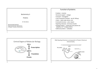
  • 1. Recommended literature : Berg-Tymoczky-Stryer: Biochemistry Lehninger: Principles of Biochemistry Biochemistry II Proteins Dr. Terez Barna Function of proteins • Catalysis – enzymes • Structural support – collagen • Transport – hemoglobin • Trans-membrane transport – Na+/K+ ATPases • Toxins – rattle snake venom, ricin • Contractile function – actin, myosin • Signaling molecules : hormones (insulin) • Storage Proteins – Calreticulin and calnexin are ubiquitous Ca2+ storage proteins • Defensive proteins – antibodies Central Dogma of Molecular Biology DNA mRNA Protein Transcription Translation Transcription Translation RNA Reading Direction corresponds to Protein Chemical Directionality NH2-terminus COOH-terminus 5’ 3’ mRNA 1 2 3 4
  • 2. 5 Protein biosynthesis on ribosomes CONDENSATION REACTION FORMATION OF PEPTID BOND 6 N-terminális C-terminális POLYPEPTIDE CHAIN Stereochemistry of -amino acids chiral center Stereochemistry of -amino acids •Enantiomer •Optically active living organisms use L-amino acids L-alanine D-alanine 5 6 7 8
  • 3. Isoleucine –two chirality centers * * Optically active substances rotate plane polarised light Chirality explaines ligand binding specificity of proteins Hydrophobic Amino Acids 9 10 11 12
  • 4. Hydrophylic Amino Acids UV-VIS absorbance of aromatic side-chains of protein Protonated/deprotonated forms of amino acid Formation of Disulfide-bridge (oxidation) 13 14 15 16
  • 5. The functional insulin contains three disulfide bridges Post-translational modification of side chains O NH O H O H O N H2 O O H O O - O - P Phosphoserine 4-Hydroxyproline Folding into a unique conformation Protein conformation Variability of protein structure 17 18 19 20
  • 6. Peptide bond is planar C N 1,27Å C N 1,47Å C N 1,27Å C N 1,47Å C-N 1,33Å 40% N−C' double bond character Linus Pauling C−C bond length in benzene is 1.39 A˚. The Kekulé structure - Bonds in bezene have partial double bond character Single C-C bond length in Hydrocarbons Two configurations of the planar peptid bond are possible: one in which C atoms are trans , the other in which they are cis The trans form is intrinsically favored energetically – fewer repulsions between non-bonded atoms Schematic diagram of an extended polypeptide chain The Peptide Bond Resonance of peptide bond - Polarity, Dipole moment - partial double-bond character Trans-conformation of C 21 22 23 24
  • 7. cis-trans isomers of proline The cyclic side chains in proline diminishes the repulsions between atoms so the intrinsic stability of cis isomer is comparable to that of the trans isomer Linus Pauling: 1954 Noble prize ªThe Structure of Proteins: Two Hydrogen-Bonded Helical Configurations of the Polypeptide Chainº. 1951 Proceedings of the United States National Academy of Sciences Linus Pauling predicted two regular periodical structurea: right handed -helix and -pleeted sheet Linus Pauling, has been awarded two undivided Nobel Prizes, in 1954 in Chemistry and in 1962 in the Peace Prize. Backbone Degrees of Freedom No rotation about the peptide bond.  Limited range of conformations. - Free rotation about the N(H)‒C bond:  (phi) Free rotation about the C‒C(=O) bond:  (psi)  and  = 180o when polypeptide is in its fully extended conformation and all peptide bonds are in the same plane. - +180o >  and  > ‒180o 25 26 27 28
  • 8. Free rotation around C-atoms with  (fi) and  (psi) angles  (fi) and  (psi) angles are restricted to certain range -- Limited conformation Some values are never observed because of the steric hindrance of the side chains Torsion angle is positive if the direction is clockwise (+) Anticlockwise (-) N(H)‒C : bond rotation  (phi) - C‒C(=O) bond rotation :  (psi) - Due to the size and charge of the R groups,  and  are rotationally hindered.  Ramachandran Plot ( =  = 0o not allowed.)  The conformational freedom and therefore the torsion angles of a polypeptide backbone are sterically constrained.  Rotation around the C-N and C -C bonds to form certain combinations of  and  angles will cause the amide hydrogen, the carbonyl oxygen, or the substituents of C  of adjacent residues to collide. Torsion Angles between Peptide Groups Describe Polypeptide Chain Conformations The Ramachandran Diagram Indicates Allowed Conformations of Polypeptides Ramachandran Plot: indicates allowed conformations of polypeptides No steric overlap   Not allowed. Allowed at the extreme limits • Sterically allowed values of  and . • Sterically forbidden conformations, in which the  and  values would bring atoms closer than the corresponding van der Waals distance (the distance of closest contact between nonbonded atoms). • Only three small regions of the diagram are physically accessible to most residues (allowed regions). Left handed helix 29 30 31 32
  • 9.  -Helix: Periodicity = 3.6 Helical rise per amino acid = 1.5 Å Pitch of the helix = 5.4 Å  -Sheet: Periodicity = 2 Távolság = 5.4 Å Two regular, periodic structures in the polypeptide chains (Allowed regions in the Ramachadran plot) Regular structures are composed of sequences of residues with repeating of  and  values. Secondary structure: • Secondary structure refers to particularly stable arrangements of amino acid residues giving rise to recurring structural patterns defined by hydrogen bonds between backbone amide groups. (side chain-main chain and side chain-side chain hydrogen bonds are irrelevant). • The most prominent are α-helices and β-sheets Right-handed vs. Left handed α-helix  α-helix can form in polypeptides consisting of either L- or D-amino acids. However, all residues must be of one stereoisomeric series; a D-amino acid will disrupt a regular structure consisting of L-amino acids, and vice versa.  Naturally occurring L-amino acids can form either right- or left- handed helices, but extended left handed helices have not been observed in proteins. N N+4  polypeptide backbone is wound around an imaginary axis with R group protruding outwards. • Stabilized by H-bonds between: C=O(n) and N-H(n+4). atomic number / H-bridge:13 – Φ = –57O , Ψ = –47O Properties of the Alpha Helix ( 3.613-helix) strong hydrogen bonds with nearly optimum N..H…O distance of 2.8 Å. 1 2 3 13 33 34 35 36
  • 10. Properties of the Alpha Helix ( 3.613-helix) • 3.6 amino acid residues per helical turn • p = hn = 5.4 Å (the distance, where the helix rises along its axis per turn) Properties of the Alpha Helix ( 3.613-helix) average length: 12 amino acid (~three turns, ~18 Å max. 80 Å The rise along the axis after each amino acid extention is 1.5 Å alpha-helix has a dipole character Amino terminus Carboxyl terminus Top view R groups protrude outwards -Helix Amino acid side chains project outward and downward from the helix ,thereby avoiding steric interference with the polypeptide backbone and with each other. The core of the helix is tightly packed; that is, its atoms are in van der Waals contact. 37 38 39 40
  • 11. Among the theoretical helices 310, α and π helices are found in proteins 310 helix  vagy (4.416) helix 42 The globin structure containing eight alpha-helices The globin fold  sheet • The backbone of the polypeptide chain is extended into a zigzag, and arranged side-by-side to form a structure resembling a series of pleats. • H-bonds are formed between adjacent segments. One peptide bonds make two H-bonds with the neighboring polypeptide chains. 4.5 Å C N C 7 Å R1 R3  Sheets in proteins contain 2 to as many as 22 polypeptide strands, with an average of 6 strands. Each strand may contain up to 15 residues, the average being 6 residues. 41 42 43 44
  • 12. Parallel  sheet Antiparallel  sheet Antiparallel  sheet: neighboring hydrogen-bonded polypeptide chains run in opposite directions. In  sheet the hydrogen-bonded chains extend in the same direction. Parallel  sheets are less stable than antiparallel  sheets, because the hydrogen bonds of parallel sheets are distorted compared to those of the antiparallel sheets. In  sheet the peptide chains can be either parallel or antiparallel. The two residues repeat distance is shorter for the parallel conformation (6.5 Å, versus 7 Å for antiparallel). 6.5 Å Beta barrel of retinol-binding protein Properties of the Beta Sheet  strands usually have a pronouced right-handed twist, due to steric effects arising from the L-amino acid configuration. β-barrel strands topology in a β-barrel : Connections between adjacent strands in  sheets • (a) Antiparallel strands may be connected by a small loop. • (b) Parallel strands require a more extensive crossover connection Turns and Loops • Segments with regular secondary structure such as  helices or the strands of  sheets are typically joined by stretches of polypeptide (turns and loops) that abruptly change direction. • Such reverse turns or  turns( named because they often connect successive strands of antiparallel  sheets) almost always occur at protein surfaces. • Most reverse turns involve four successive amino acid residues more or less arranged in one of two ways, Type I and Type II. Both turns are stabilized by a hydrogen bond between backbone CO(i) and the backbone NH (i+3). • In Type II turns, the oxygen atom of residue 2 crowds the C atom of residue 3, which is therefore usually Gly. • Residue 2 of either type of turn is often Pro since it can assume the required conformation. 45 46 47 48
  • 13. Relative probabilities that a given amino acid will occur in the secondary structure Supersecondary structures or motif : • The association of secondary structures in a particular geometrical arrangement (distinct, stable folding pattern) with defined topology (connectivities) containing 10 to 40 residues in length. • Motifs are important in describing protein structure because they can be repeated within many protein structures. alpha-alpha corner Short loop regions connecting helices which are roughly perpendicular to one another. ( a strictly definite alternation of hydrophobic, hydrophilic and glycine residues;) Greek key motif: antiparallel β sheet comprising of four b strands with short and longer loop connections between some strands. β-barrel structures contain Greek key motifs named after a Greek pottery. An α helix connects parallel β strands in β sheets. Hydrophobic surfaces on the helix and β sheet interact. Residues in the first loop (C-terminal end of the β strand) often contribute to the active site in enzymes. Leucine zipper motif Two identical subunits interact via α helices, forming a short stretch of coiled-coil. The interaction is mediated by hydrophobic interactions between side-chains, notably those of leucine residues. Supersecondary structures: helix-turn-helix - two alpha helices connected by a short loop region Recurring feature in proteins with DNA binding function Recognition of the major groove in DNA Helix-turn-helix motif binds to Bent B-DNA 49 50 51 52
  • 14. • A spatially separated, stable unit of tertiary structure, which is identified by some characteristic patterns. • Often linked by a flexible hinge region, these domains are compact and stable, with a hydrophobic core. • Domains fold independently of the rest of the polypeptide, satisfying most of their residue–residue contacts internally. Domain is a structural and functional unit of proteins. • Proteins may have multiple domains and each domain has a distinct evolutionary origin and function (DNA bind domain, coenzyme binding domain, Ca2+ binding domain). See protein domain databases like Prosite, Conserved Domains Database (CDD), InterPro, Pfam. Domains Protein pyruvate kinase has multidomain structure Hemoglobin consists of a single domain Comparison of the structures of triosephosphate isomerase and dihydrofolate reductase Tertiary Structure The arrangement of multiple secondary structural elements leads to tertiary structure. Protein tertiary structure is more reliably conserved than primary sequence. •Two proteins with similar secondary structure elements but with different topology (connectivities) leads to different tertiary structures – different fold. Structural levels of proteins 3D structure of proteins Tertiary structure Quaternary structure 53 54 55 56
  • 15. Structural Classification of Proteins - SCOP database aims at classifying proteins according to structural and evolutionary relationship. Structural hierarchy: ClassFoldSuperfamilyFamily Class: secondary structural elements Fold: spatial arrangement of secondary structural elements Superfamily: Made of several families, little sequence similarity, share motifs and functional similarities  Probable evolutionary relationship Family: Sequence similarity and/or similar structure/function  Strong evolutionary relationship Classes: 1. All alpha proteins 2. All beta proteins 3. / Alpha and beta proteins 4.  and  relatively separated (a+b) 5. Multi-domain proteins (alpha and beta) Folds consisting of two or more domains belonging to different classes 6. Membrane and cell surface proteins and peptides 7. Small proteins Usually dominated by metal ligand, heme, and/or disulfide bridges http://scop.mrc-lmb.cam.ac.uk/scop/ Structural Classification of proteins based on secondary structures and their connectivities 1- all -helix 2- all  sheet 3- / 4-  and  relatively separated 5- multidomain protein 5 2, 3. 1 4. 4. 59 CATH: Protein Structure Classification database http://cathwww.biochem.ucl.ac.uk/latest/index.html 60 C - Class - determined according to secondary structure composition. A - Architecture - describes the overall shape of the domain structure. T - Topology - major structural similarities (similar connectivities among the secondary elements). H - Homology Superfamily – (Superfamily in SCOP) Protein domains which share a common ancestor. CATH is a hierarchical classification of protein domains, which clusters proteins at four major levels: 57 58 59 60
  • 16. Classification of proteins at a higher hierarchy level: Membrane protein Globular protein Fibrous protein Unstructured protein three-dimensional structure of part of the cytochrome bc1 complex Membrane Protein Structure Polar side chains interacting with polar head group of the lipids and each other Hydrophobic side chain Alfa- helices are the most common secondary structure elements in membrane proteins Protein Data Bank (PDB: WWW.rcsb.org/pdb) 1. Crystallysing proteins 2. Using X-ray diffraction 3. Calculating electron density map 4. Model Building Solving protein structure by X-ray diffraction 61 62 63 64
  • 17.  3-D structure of a protein is determined by its amino acid sequence (primary structure).  The function of a protein depends on its structure.  A protein exists multiple, thermodynamically stable conformation (lowest G) native conformations  A protein’s conformation is stabilized largely by weak interactions.  Protein structures show common structural patterns (secondary structures, supersecondary structures, domains.) • Proteins undergo conformational change upon ligand binding or during catalysis. - Structural levels of proteins 66 3-D structure of a protein is determined by its amino acid sequence Primary Structure: aminoacid sequence + the position of disulfide bonds Protein Quaternary Structure  More than one polypeptide chain associate together with a specific geometry  The spatial arrangement of the subunits is called the quaternary structure Hemoglobin, 22 tetramer (dimer of  protomers) 65 66 67 68
  • 18. Quaternary Structure Oligomers: composed of more than one polypeptide chain subunits Protomer is the structural unit of an oligomeric protein that are symmetrically arranged. Protein assemblies built on identical subunits are usually symmetric The forces that drive folding (in native structure) Disulfide bridge covalent: 70-100 kcal/mol) Electrostatic interaction (10 kcal/mol), affected by pH van der Waals force (transient and weak: 0.1-0.2 kcal/mol) Hydrophobic interaction Hydrogen bond 2-5 kcal/mol The forces that drive folding 69 70 71 72
  • 19. van der Waals interaction - caused by transient dipoles, the momentary random fluctuation in the distribution of the electrons of any atoms When any two atoms approach each other closely, they create a weak, nonspecific attractive force (1 kJ/mol) Típusai: permanens dipólus Permanens és indukált dipólus . mindkettő indukált dipólus, London féle diszperziós erők van der Waals forces van der Waals Erők van der Waals radius (nm): H 0.1 nm C 0.17 nm N 0.15 nm O 0.14 nm P 0.19 nm S 0.185 nm Attraction Repulsion • van der Waals forces disappear with increasing distance (~1/ r6 ) • ~ 1 kJ/mol • Repulsion stops two atoms approach themselves than their van der Waals radius energy of Interaction Hydrophobic Effects Water molecules tend to form cages of relatively rigid hydrogen bonded pentagons and hexagons around nonpolar molecules Increasing entropy 73 74 75 76
  • 20. Electrostatic forces (ionic interaction or salt bridge ) Coulomb law: q1 q2 r2 D r : distance between A and B közötti távolság q1 , q2 : charges D : dielectric constant F= Strongest in vacuum 2. Hidrogen bridge Bonds that Stabilize Folded Proteins Folded proteins are stabilized mainly by weak noncovalent interactions Cytocrome c hyrophilic side chains Hydrophobic side chains Protein conformations are stabilized by disulfide bonds (~210 kJ/mol), weak (4~30 kJ/mol) noncovalent interactions (H-bonds, ionic and hydrophobic interactions, van der Waals forces). What drives the protein folding?  Hydrophobic interactions: hydrophobic residues are largely buried in the protein interior.  H-bonds: the number of H-bonds within the protein is maximized.  Ionic interactions and van der Waals forces also contribute. 77 78 79 80
  • 21. PROTEIN FOLDING • Is the one-dimensional sequence of a protein programmed to achieve a definitive three-dimensional structure? • How can we describe the process, which leads to the transition from a random coil to a unique structure? • Is this process spontaneous? • Does protein folding occur only in the cell in vivo or in vitro, too? RNAse : single chain of 124 residues stabilized by 4 disulfide bonds, catalysis the hydrolysis of RNA The fully unfolded protein can spontaneously refold back into its native shape in vitro and regain its catalytic function. Disulfide bonds of RNAase reduced with β-mercaptoethanol in 8 M urea. All activity lost; S-S bridges broken, converted to –SH. Christian Boehmer Anfinsen's Nobel-winning experiment (1962) Denaturation of ribonuclease (RNAse) Denaturation of RNAse Spontaneous folding of denatured ribonuclease; Ribonuclease will gain back its native structure and its enzyme activity; Original, native conformation formed, with all correct S-S bonds reestablished. Probability of correct S-S bonds by chance, 1%. Timescale of the refolding ~ ½ h- 2h Primary structure possesses sufficient information for proper folding. The first proof for the “sequence determines structure” Anfinsen was awarded Nobel prize in 1972. Anfinsen's Nobel-winning experiment Protein Denaturation and Folding • Denaturation: loss of 3-D structure initiates the loss of biological function. • Unfolding is a cooperative process: loss of structure in one part of the protein destabilizes other parts. • Heat: affects the weak interactions in a protein (primarily H –bonds). • Extremes of pH: disrupting net charge on the protein; and H-bonds. • Organic solvents (alcohol or acetone); urea, and detergents: disrupting the hydrophobic interaction.  Tm: melting point (50% folded and 50% denatured polypeptide chain) Denaturants: heat, pH, organic solvents, detergents urea, guanidine chloride (GdnHCl) 81 82 83 84
  • 22. Levintal’s Paradox (1968) – The protein folding problem Theoretical experiments: We have a small protein (100 AA’s) Each AA can assume try 3 conformations Total possible conformations = 3100 = 5 x 1047 Examine each structure for 1 x 10-13 s (time scale of bond vibration) Then total search requires 5 x 1034 s This is 1.6 x 1027 years, a period longer than the age of the universe! How long does a folding process of a linear polypeptide chain take in a random search among all possible configurations to find the native conformation? “How proteins fold to give such a unique structure” in which time scale, if the folding is a random process? In real time scale, in the cell, protein folding lasts msec, sec, ! Levinthal, 1969: “We feel that protein folding is speeded and guided by the rapid formation of local interactions, which then determine the further folding of the peptide.” Folding is a spontaneous process Energetics of folding II. Law of Thermodynamics determines the direction of spontaneous processes: ΔG 0 ΔG = ΔH – TΔS ΔG = Gibbs free energy; ΔH = enthalpy; T = temperature; ΔS = entropy A folded protein consisting of 100 amino acids is stabilized by G=GN-GD~-40kJ/mol Folding is a spontaneous process. Ordered arrays of water molecules surrounded the exposed hydrophobic residues in bovine pancreatic ribonuclease A Protein Stability : Weak Interactions and Flexibility The folded protein is a thermodynamic compromise •Stability is a net loss of free energy (entropy + enthalpy) Free energy difference between the folded and unfolded states; ~21-42kJ/mole, marginally stable. •Folding decreases the confomrational entropy of the proteins; but increase of water entropy is much bigger (hydrophobic effect). Free-energy funnel 85 86 87 88
  • 23. Thermodynamics of protein folding depicted as a free-energy funnel Thermodynamics of protein folding depicted as a free-energy funnel. • Hydrophobic effect is the primary driving force of protein folding (the core of the folded protein is mostly comprising of hydrophobic residues).  Protein molecules have flexible regions with low stability that bring about conformational change, essential to function. • Protein folding proceeds from a disordered state to progressively more ordered conformations corresponding to lower energy levels. • Alternative conformations are represented by local energy minima. Folding traps molten globule Native state Unfolded protein Free-energy funnel fast slow D M N Folding in two main steps: D: denatured M: molten globule N: native Denatured Hydrophobe collapse Native protein Molten globule Folding traps • Folding is initiated by a spontaneous collapse of the polypeptide into a compact state, mediated by hydrophobic interactions among nonpolar residues--- • Molten globule: hydrophobic collapse: may have secondary structure, but many aa chain are not entirely fixed. • May take a variety of routes to the same end point Vulnarability of the native structure • A protein’s conformation can change in response to the physical and chemical conditions. • Changes in pH, salt concentration, temperature, or other factors can unravel or denature a protein. • These forces disrupt the hydrogen bonds, ionic bonds, and disulfide bridges that maintain the protein’s shape. • Some proteins can return to their functional shape after denaturation, but others cannot, especially in the crowded environment of the cell. • Usually denaturation is permanent ΔG (folding) = -20 to -40 kJ/mole for a 100 AA protein, Stabilisation of the native state is not larger than a couple of H-bonds Significance: evolution has favored flexibility misfolding is a common occurence 89 90 91 92
  • 24. Bonds that Stabilize Folded Proteins Folded proteins are stabilized mainly by weak noncovalent interactions Protein folding is assisted by proteins in the cells Protein disulfide isomerase catalyze the formation of the proper disulfide bridges in the protein Peptidil prolil isomerase catalyze the cis-trans isomerisation of X-Pro Molecular chaperons were first identified as "heat-shock proteins" -interact with partially folded or improperly folded polypeptides and facilitate achieving native structure (e.g.hsp60 and hsp70). • The formation of correct disulfide pairings in nascent proteins is catalyzed by PDI. • PDI preferentially binds with peptides that containing Cys residues. It has a broad substrate specificity for the folding of diverse disulfide-containing proteins • By shuffling disulfide bonds, PDI enables proteins to quickly find the thermodynamically most stable pairing those that are accessible. • PDI is especially important in accelerating disulfide inter-change in kinetically trapped folding intermediate. PDI Protein Disulfide Isomerase (PDI) Peptidyl Prolyl Isomerase (PPI) • Peptide bonds in proteins are nearly always in the trans configuration, but X-pro peptide bonds are 6% cis. • Prolyl isomerization is the rate- limiting in the folding of many proteins in vitro. • PPI accelerates cis-trans isomerization more than 300 fold by twisting the peptide bond so that the C,O, and N atoms are no longer planar. 93 94 95 96
  • 25. Free-energy folding landscape for chaperone-mediated protein folding The role of Molecular Chaperones • Ensure correct folding, providing microenvironments in which folding can occur by minimizing aggregation and other misfolding • Minimize heat and stress damage to proteins (renaturation/degradation) • Bind to nascent polypeptides to prevent premature folding and protect them from the concentrated protein matrix in the cell • Facilitate membrane translocation/import by preventing folding prior to membrane translocation • Facilitate assembly/disassembly of multiprotein complexes • Unfold misfolded proteins before their degradation by the proteasome unit Molecular chaperones Hsp 70 system - prevents folding of nascent chain Chaperonins – reverse misfolded structures Molecular chaperones can be divided into three functional subclasses based on their mechanism of action : • ‘Folding’ chaperones (e.g., DnaK and GroEL) rely on ATP- driven conformational changes to mediate the net refolding/unfolding of their substrates. • ‘Holding’ chaperones (e.g., IbpB) maintain partially folded proteins on their surface to await availability of folding chaperones upon stress response. • ‘Disaggregating’ chaperone (e.g., ClpB) promotes the solubilization of proteins that have become aggregated as a result of stress. 97 98 99 100
  • 26. Hsp100/ClpB Classification of Chaperones (Heat shock proteins) according to their molecular weight Those proteins presented here are all ATP-dependent chaperones Hsp60/GroEL Hsp70/DnaK Classification of chaperons according to their size Only bacterial ATP-dependent Chaperones presented here. DOI 10.1007/s12192-015-0598-8 Small Hsps - Diverse "family" 10,000 - 30,000 MW preventing undesired protein–protein interactions and assisting refolding of denatured proteins HSP20 maintains denatured proteins in a folding-competent state and allows subsequent ATP-dependent disaggregation through the HSP70/90 chaperone system. alpha-crystallins belongs to small hsps family with a cellular function to bind to partially unfolded proteins and maintain them in a refolding-competent state - holdase function (protect eye lens proteins from degradation) Hsp40 (Hsp40/DnaJ), also termed J-domain-containing protein (J-protein) is a co-chaperone component of the HSP70 system. Hsp60 (e.g. GroEL in E. coli) ATPase, folding chaperone- ‚chaperonine’. Hsp70 (e.g.DnaK in E. coli; BiP in ER in eukaryotes) ATPase; In animals and plants, Hsp70 functions as a chaperone for newly synthesized proteins to prevent their accumulation as aggregates as well as to ensure proper protein folding during their transfer to their final location. Hsp90 is the most abundant in cytosolic heat shock protein family in both eukaryotic and prokaryotic cells and is rapidly induced in response to various stress conditions. Hsp100 (e.g. Clp) ATPase – ‚disaggregase’ under severe thermal stress, Hsp100 maintains the functional integrity of certain key proteins by solubilizing the non-functional protein aggregates and enabling the degradation of the irreversibly damaged proteins. Lectin chaperone in the ER: Calnexin, calreticulin take part in the quality control of glycoproteins. ERp57 a thiol-disulfide isomerase in the ER, exhibits molecular chaperone properties and associates with calreticulin and calnexin. Classification of heat shock proteins (Hsps) Energetics of folding II. Law of Thermodynamics determines the direction of spontaneous processes: ΔG 0 ΔG = ΔH – TΔS ΔG = Gibbs free energy; ΔH = enthalpy; T = temperature; ΔS = entropy A folded protein consisting of 100 amino acids is stabilized by G=GN-GD~-40kJ/mol Folding is a spontaneous process. 101 102 103 104
  • 27. The protein quality control (PQC) system maintains protein homeostasis by counteracting the accumulation of misfolded protein conformers. Protein substrate degradation and refolding activities executed by ATP- dependent proteases and chaperones constitute major strategies of the proteostasis network. Proteostasis Hsp60 Chaperonin: GroES GroEL Barrel structure consists of -tetradecamer of 60 kDa chains ( GroEL subunit) - heptamer of 10 kDa chains (GroES subunit that functions as co-chaperonin partner) Hsp60 Chaperonin : GroEL-GroES complex bacterial folding chaperone • An unfolded (U) or partially folded (I) polypeptide binds to hydrophobic patches on the apical ring of 7-subunits of GroEL. • ATP binding to GroEL is followed by GroES association and triggers conformational change in the internal chamber. ATP binding site Domain motion upon ATP hydrolysis changes the nature of the chamber surface (hydrophobichydrophilic) 105 106 107 108
  • 28. GroEL-GroES: cage-like compartment for the folding of single protein molecules After ~15 seconds ATP hydrolysis takes place, followed by binding of ATP to the lower 7-subunit ring, which causes release of the protein. • ATP binding triggers a conformational change that buries the 7-subunit hydrophobic patches, releasing the polypeptide into the central activity (“Anfinsen cage”). 1). -DnaK, the major bacterial Hsp70 protein binds to regions of unfolded protein that are rich in hydrophobic region, preventing inappropriate aggregation. -DnaK also stabilizes proteins for subsequent folding by GroEL. - DnaK cooperates with co-chaperone DnaJ(a Hsp40 protein) and regulator GrpE nucleotide-exchange factor protein. 2). also block the folding of certain protein that must remain unfolded until they have been translocated across membranes. The Hsp70 (DnaK) proteins bind to and release polypeptides in a cycle with Hsp40 (DnaJ) and ATP hydrolysis. DnaJ functions in presenting unfolded proteins to DnaK and accelerate ATP hydrolysis Bacterial cells containing inclusion bodies as visualized by electron microscopy. The recombinant protein overproduced in bacterial cells cannot fold correctly and forms large insoluble aggregates. Inclusion body binding protein IbpB (belongs to sHSP) is able to bind non- selectively aggregation-prone unfolded proteins – holding chaperone, does not require ATP for its function • IbpB (16-kDa) belongs to the small heat shock (sHSP) protein family the dodecameric structure • It has an α-crystallin domain (white) with the function of holding chaperone. The amino-terminal 31 residues in half of the 12 subunits (dodekameric structure) of IbpB are structurally disordered. Structural disorderness is a prerequisite for IbpB to bind its substrate proteins. 109 110 111 112
  • 29. Biotechnological production of recombinant protein without (A) and with (B) optimized amounts of chaperones. A) Without sufficient amounts of chaperones the recombinant protein is highly prone to aggregation and forms inclusion bodies. B) The controlled co-overproduction of molecular chaperones together with the target protein leads to increased levels of the properly folded recombinant protein. Function of Small heat shock proteins • Small heat shock proteins represent ATP-independent chaperones that bind to misfolded proteins, preventing their uncontrolled aggregation. • Small heat shock proteins share the conserved α-crystallin domain and largely disordered N- and C-terminal extensions. They form large oligomers through multiple, weak interactions. • They bind unfolded protein non-specifically and directed towards refolding pathways by ATP-dependent Hsp70/Hsp100 chaperones or sorted for degradation Hsp100 is a family of disaggregating chaperones that can thread aggregated polypeptide chains through the central pore of adenosine triphosphatase (ATPase) rings arranged in hexamer Hsp70 recruits aggregated proteins and delivers them to Hsp100 Aggregated polypeptid chains The molecular chaperone network in the cytoplasm of E. coli DnaK-DnaJ In bacteria, fungi, and plants, disaggregation involves cooperation between Hsp100 chaperones (ClpB in E. coli and Hsp104 in S. cerevisiae) and the Hsp70 chaperone system (DnaK system in E. coli). 113 114 115 116
  • 30. Quality control is based on glycosylation by the assistance of lectin chaperone Calnexin/calreticulin cycle and the quality control of glycoproteins in the ER • Calnexin and calreticulin as ER chaperons are involved in the folding and the quality control of newly synthesized glycoproteins. • These two chaperones recognize the monoglucosylated N- linked glycans and unfolded protein regions and aid their folding. • ERp57, the protein disulphide isomerase that associates both to calnexin and calreticulin mediates the formation of the correct disulfide bridge pattern in the glycoproteins. Steps 1-2. Glucosidase I and Glucosidase II cleave the terminal glucosyl units of the N-glycosylated proteins. Steps 3- 4. Glucosyl transferase (UGGT1)) reglucosylates only those glycoproteins that have exposed hydrophobic patches. UGGT1 functions as folding sensor. Steps 5. 6-protein in native state will leave ER Steps 1-5: Some proteins require multiple rounds of association with the chaperones, these proteins is reglucosylated by UGGT1. The terminally misfolded proteins are targeted for ERAD (ER associated degradation). . Diseases arise from protein misfolding Diseases Protein involved Alzheimer‘s disease Amyloid-β Parkinson disease α-Synuclein Diabetes type 2 Amylin Amyotrophic lateral sclerosis Superoxide dismutase Haemodialysis-related amyloidosis β2-microglobulin Cystic fibrosis Cystic fibrosis transmembrane regulator Sickle cell anemia Hemoglobin Hungtington disease Huntingtin Creutzfeldt-Jakob disease Prion protein Amyloidosis Ten other proteins Cystic Fibrosis and α1-antitrypsin Deficiency (AAT) protein misfolding diseases • ΔF508 CFTR and L342E α1-antitrypsin fail to fold correctly in ER and are retained in ER. . 117 118 119 120
  • 31. Protein-misfolding diseases α1-antitrypsin Deficiency (AAT): • α1-antitrypsin is an acute-phase plasma protein produced by hepatocytes and functions as a protease inhibitior. Mutant form of α1-antitrypsin (where Lys342 is replaced by Glu) fails to complete proper folding and is retained in the ER of liver cells. • It is associated with two major types of clinical disorders, chronic obstructive pulmonary disease and hepatic cirrhosis. The lack of proteolytic inhibition in the lungs (mainly inhhibit neutrophil elastase) , as a consequence of the reduced level of α1-antitrypsin, results in proteolytic damage to the pulmonary connective tissue matrix. Misfolded α1-antitrypsin is retained in the ER of hepatic cells Loss of proteolytic inhibition Cystic Fibrosis : CFTR (cystic fibrosis transmembrane conductance regulator) is a chloride and bicarbonate ion channel that regulates salt and fluid homeostasis and is localized located in the apical membranes of epithelial cells in multiple exocrine organs. The newly synthesized polypeptide chain of CFTR is glycosylated in ER and takes part in the ER quality control. Phe508del CFTR mutant has misfolded conformation and partial biological activity. The ER chaperone system recognizes the misfolded protein structure and the mutant CFTR is targeted for endoplasmic reticulum–associated protein degradation (ERAD). . Protein-misfolding diseases The process of CFTR maturation and degradation requires association with multiple chaperones and co-chaperones. Disrupting the function of these chaperone systems can allow mutant CFTR to escape degradation. Sickle-Cell Disease: A chan Sickle-cell disease ge in Primary Structure • A slight change in primary structure can affect a protein’s structure and ability to function • Sickle-cell disease, an inherited blood disorder, results from a single amino acid substitution in the protein hemoglobin 123 Sickle cell anemia Mutation in hemoglobin : Glutamate  Valine at 6th position in  chain glutamate (GAG codo)  valin (GTG kodon ) In deoxyhemoglobin (Deoxi S) Val6 is on the surface  makes hydrophobic interaction with Phe85 of the other hemoglobin β chain Val6 mutation results in long fibril formation of deoxihemoglobin S molecules → this aggreagation distorts the shape of the red blood cell → sickle blood cell can not move through the capillary, the circulation of blood vessels is obstructed by sickled red blood cells (Vaso-occlusion) 121 122 123 124
  • 32. Sickle cell anemia pathophysiology is a consequence of this reduced solubility, causing polymerization of hemoglobin S tetramers in red blood cells upon partial deoxygenation and the impaired flow of these cells in the microcirculation. The demonstration of a molecular basis for a disease was a significant turning point in medicine. Sickle-Cell Disease: A change in Primary Structure 126 Sickle cell protection from malaria in heterozygote individuals •Malaria is a parasitic disease caused by Plasmodium falciparum and transmitted by Anopheline mosquitoes and is highly widespread throughout tropical and subtropical regions The accumulation of the sickle cell gene in malarial regions of the world became a convincing illustration of evolution by natural selection. Protein aggregates in neurodegenerative diseases 125 126 127 128
  • 33. Amyloid fibril formation Amyloid fiber amyloid beta peptid - Alzheimer disease alfa-synuclein -Parkinson disease Neurodegenerative disorders (Alzheimer disease, Parkinson disease) are characterized by the accumulation of misfolded proteins (the formation of aggregates - harmfull amyloid). The misfolded protein is rich in -sheet conformation and arrange into cross -sheet with oligomerisation cross -sheet Amyloid fibril formation • In misfolded protein the production of β-sheets is usually stabilized by protein oligomerization and eventially lead to amiloid –like aggregation. It will induce tissue damage and organ dysfunction. • The amiloid fibril resists to proteolitic degradation. chemical and pharmacological chaperones are under invetigation as potential therapeutic agents. 131 • Mad cow disease (1996). Related diseases include kuru and Creutzfeldt-Jakob disease in humans and scrapie in sheep. • Disease-causing agents appeared to lack nucleic acids (proteinaceous infectious only -prion protein: PrP) • PrPsc interacts with cellular PrPc--- alter PrPc to become PrPSc. Prion diseases Spongiform degeneration: cerebral section of a patient with Creutzfeldt-Jakob disease (CJD) N-terminal is an intrisically disordered polypeptide region. PrP exists in two conformations, • PrPC - normal form ,42% - helices, soluble, is a transmembrane glycoprotein (neurons, lymphocytes); its function is unknown; it binds Cu2+ ,is monomeric and easily digested by proteases. PrPSc- has the same amino acid sequence, altered conformation 43% β-sheets, insoluble, forms neurotoxic aggregates, resistant to digestion by proteases. PrPSc converts PrPC to PrPSc in chain reaction. The prion diseases self propagating and transmissible „ proteinaceous infectious particle” Mad cow disease (BSE, bovine spongiform encephalopathy) Kuru, Creutzfeldt-Jacob disease, scrapie 129 130 131 132
  • 34. Classification of proteins according to their structures Intrinsically disordered proteins Globular proteins Fibrous proteins Structural function Diverse function: transport, enzyme, regulatory Protein-protein; protein- DNA interacting protein with regulatory function Intrinsically disordered proteins Tendency of amino acid to promote disorder Tryptophan as the most order-promoting amino acid and Proline as the most disorder-promoting one. They show low abundance of hydrophobic residues (Ile, Leu, Val, Trp, Tyr, Phe) compared to structured proteins. Fibrous proteins  Polypeptide chains are arranged in lone strands or sheet, highly elongated molecules, form insoluble fibers • Secondary structures: A single type (helix or sheets) Simple repetitive pattern • Roles Structural supports, protection Located mainly in the extracellular matrix and are present in connective tissues -keratin, collagen, silk fibroin have a protective, connective, or supportive role in living organisms The most abundant amino acids in fibrous protein 133 134 135 136
  • 35. I-type of collagen C-terminal region Every third residue is glycine Hyp: Hydroxyproline Collagen represents up to 25 to 35 % of the total protein content of the body. Collagen helix • The collagen polypeptide forms a left- handed helical conformation with three residues per turn. • Three left handed collagen polypeptide chains wind around each other with a right-handed manner forming a typical collagen triple helix LH RH Resistant to pressure and tension Nagy mechanikai szilárdság 137 138 139 140
  • 36. Collagen: the most abundant protein of mammals, main fibrous component of skin, bone, tendon, cartilage, and teeth Typical collagen polypeptide consists of monotonously repeating triplets of sequence Gly-X-Y over a segment of ~1000 residues, where X is often Pro and Y is often Hyp. Hyl sometimes appears at the Y position. The peptide groups are oriented such that the N..H of each Gly makes a strong hydrogen bond with the carbonyl oxygen of an X (Pro) residue on a neighboring chain collagen triple helix Three residues per turn Collagen fibrils periodic cross-striated collagen fibrils Monomeric protocollagen chains trimerize, the propeptides are cleaved off and the collagen molecules self-assemble to microfibrils and fibrils. Oxidation of lysine and hydroxylysine by lysyloxidase initiates the formation of the various natural enzyme-derived crosslinks Collagen • every thrird residue Gly; proline content between 15 to 30%, • contains two post-translationally modified residues: Hyp (hydroxyproline) Hyl: (hydroxylysine) • prolyl hydroxylase catalysis the formation of Hyp, this enzyme requires ascorbic acid (vitamin C) to maintain its activity - scurvy severe vitamin C deficiency The synthesis of Hydroxi-Pro and hydroxi-Lys requires vitamin-C, which is necessary for the functioning of prolyl hydroxylase 141 142 143 144
  • 37. Vitamin C deprivation results in underhydoxylation of procollagen, which accumulated and eventially degraded Consequence of vitamin C deficiency : poor wound healing characteristic of scurvy Prolyl 4 hydroxylase; prolyl 3 hydroxylase, lysine hydrolxylase Each enzyme has a binding site near iron centre, the role of ascorbate to maintain iron in reduced state (Fe2+) Hydroxylation of lysine, proline of procollagen are necessary for folding into the triple helix (contribute to the stiffness of procollagen), which can be secreted by fibreblast. -Keratin and Hair -keratin: left-handed superhelix of two right-handed  -helices. from wool & hair, intermediate filaments in cytoskeleton, muscle protein (myosin & tropomyosin) Mechanically durable and unreactive protein of vertebrates up to 85% of protein in horns, hair, nails & feathers • -keratins occur in mammals; • -keratins occur in birds and reptiles • 30 keratin genes expressed in mammals •  -keratins are classified as relatively acidic (Type I) or basic (Type II) Keratins have complex quaternary structures • keratins are dimers composed of a Type I and Type II subunit • many dimers associate to form protofilaments • protofilaments dimerize to form protofibrils • protofibrils form tetramers called microfibrils • microfibrils associate into macrofibrils The central 310-residue segment of each keratin polypeptide chain has a 7-residue pseudorepeat: a-b-c-d-e-f-g, with nonpolar residues predominating at positions „a” and „d”. Since an  -helix has 3.6 residues per turn, in keratin the hydrophobic „a” and „d” residues line up along one side of each helix . The hydrophobic strip along one helix associates with the hydrophobic region on another helix. „a” and „d” amino acids most often: Phe, Ile, Val, Met, Ala Keratin is a special example of -helix, it is a coiled-coil structure 145 146 147 148
  • 38. Permanent Waving • Keratin is rich in Cys residues, which form disulfide bonds that crosslink adjacent polypeptide chains. • The keratins are classified as “hard” or “soft” according to whether they have a high or low sulfur content (hard keratins: hair, horn, and nail). • The disulfide bonds can be reductively cleaved by disulfide interchange with mercaptans . Hair so treated can be curled and set in a “permanent wave” by applying an oxidizing agent that reestablishes the disulfide bonds in the new “curled” conformation. • Extended -conformation, forces involved: H-bonds between different sheets, made by: insects and spiders • Silk does not stretch because it is already highly extended • Rich in Ala and Gly, allowing close packing Ser Ala Ala . / / / / . Gly Gly Gly 600-times repeat •It forms antiparalel -sheet •The insertion of Val and Tyr in the -sheet causes flexibility of the structure [ ] Silk Fibroin 149 150