2. which would trigger cell death; instead, it was sequestered
subcellularly.
SecretedzinccanenterintothepostsynapticneuronsviaCa-A/K,
voltage-sensitive calcium channels (VSCC) (Siddiq and Tsirka,
2004), or be imported via zinc transporter proteins, such as the ZnT
and the Zrt, Irt-like Protein (ZIP) family of transporters (Liuzzi and
Cousins,2004).ZIPfamilymemberswerecharacterizedinyeastand
rodent tissues, but not in the mammalian CNS.
We address here how tPA mediates controlled zinc import
and where zinc localizes in neurons during tPA-mediated seques-
tration. tPA interacts with the zinc transporter ZIP4, triggering
intravesicular zinc accumulation that results in neuroprotection.
Materials and Methods
Animals. Adult C57BL6 (wild-type, wt) and tPA-deficient (tPAϪ/Ϫ
)
mice were used according to protocols approved by the Stony Brook
University IACUC and the Division of Laboratory Animal Resources
(DLAR). Kainate was injected into the hippocampus at the following
coordinates from bregma: anteroposterior Ϫ2.5 mm, mediolateral 1.7
mm, and dorsoventral Ϫ1.6 mm (Tsirka et al., 1995). The animals were
deeply anesthetized and killed by transcardial perfusion with PBS or 4%
paraformaldehyde in PBS for samples analyzed by RT-PCR or immunohis-
tochemistry, respectively. The brain tissue was removed and sectioned in 20
m sections on a Leica cryostat when used in immunohistochemistry.
Reverse transcriptase PCR. Trizol (Invitrogen) was used to extract total
RNA from mouse brain tissues and reverse transcriptase PCRs (RT-
PCRs) were performed as previously described (Takimoto et al., 1999).
Oligo dTTP was incubated with 1 g of total RNA at 65°C for 5 min. cDNAs
were then synthesized, as described in the Superscript TM II Transcriptase
first strand synthesis protocol from Invitrogen. Two microliters of this RT
reaction was used as a template in each 50 l total volume of PCR with
forwardandreverseprimers(seebelow).PCreactionswereperformedusing
Sigma Taq polymerase. The following primer sets were used for PCR ampli-
fication: mZIP1 forward, 5Ј-cacagccaccatggggccct-3Ј; mZIP1 reverse, 5Ј-ct-
tagatttggacaaagaga-3Ј; mZIP2 forward, 5Ј-cttcttgggagcagggttgatgc-3Ј;
mZIP2 reverse, 5Ј-cgccactgtggcctgtagtcc-3Ј; mZIP3 forward, 5Ј-cgcaccgctc-
caagaaggtcctgtccctctgc-3Ј;mZIP3reverse,5Ј-gctcacggtcacagccaacttggccg-3Ј;
mZIP4 forward, 5Ј-agaagtcagcacctctacaaggaacgc-3Ј; mZIP4 reverse,
5Ј-gctggctcagacccagggtc-3Ј.
Primary neuronal cultures. Hippocampal neuronal cultures were pre-
pared from embryonic day 17–19 (E17–19) mice as previously described
(Rogove and Tsirka, 1998; Siao and Tsirka, 2002). These are pure hip-
pocampal neuronal cultures where the glial contamination is ϳ2–3%.
Organotypic hippocampal slice cultures. Hippocampi were isolated
from postnatal day 9 wt and tPAϪ/Ϫ
mice and sectioned into 400 m
slices. Slices were placed on a membrane insert (Millipore, Inc.) in media
containing 50% MEM, 25% HBSS, 25% normal horse serum and placed
in an incubator with a 5% CO2 humidified atmosphere at 34°C. After 7 d
in culture, slices were treated for 2 h with 50 M zinc in HBSS in which
Fluozin-3 was added 30 min before washing the slices and fixing in 4%
PFA in PBS for imaging.
mZIP4 stable cell lines. The mZIP4 cDNA was cloned into pCDNA3.1
tagged with HA (the mZIP plasmids were a generous gift from Drs G. An-
drews and J. Dufner-Beattie, University of Kansas Medical Center). To es-
tablish mZIP4 stable cell lines, HA-tagged mZIP4 was transfected into
HEK293 cells using Lipofectamine from Invitrogen followed by selection
with 10 g/ml puromycin. Isolated stable cells were then grown in DMEM
with10%FBS.Asanegativecontrolforsubsequentexperiments,HA-tagged
angiotensin-receptor 1 was similarly transfected in HEK293 cells.
tPA cloning and expression. The full-length wild-type mouse tPA cDNA
was cloned in pMT/Bip/V5-His vector using AvaI and Pme I (gift from
Dr. Dan Lawrence, University of Michigan, Ann Arbor, MI). tPA mu-
tants were generated using the Quick Change Site-Directed Mutagenesis
kit from Stratagene based on the wild-type mouse tPA sequence. The
mutants were: Ser481 to Ala (generating catalytically inactive tPA), finger
domain deletion (deleting the sequence between amino acids Arg10 and
Cys46), growth factor domain deletion (deleting the sequence between
amino acids Phe60 and Cys87), and kringle 2 domain deletion (deleting
the sequence between amino acids Cys183 and Cys265). The tPA con-
structs were transfected into Drosophila S2 cells and stable clones were
obtained following selection with 25 g/ml Blasticidin. To induce tPA
overexpression, copper sulfate was added to the S2 medium (Schneider’s
Drosophila Medium containing 10% heat-inactivated FBS) to a final con-
centration of 500 M. Three days after induction, the supernatants were
collected and analyzed by Western blot (immunoblot) and zymography
for the presence of the induced recombinant proteins.
Immunoblotting. Immunoblots were performed on total cell lysates of
the mZIP4 HEK cells. Equivalent concentrations of total protein were
resolved by SDS-PAGE. For quantitative analysis, membranes were pre-
incubated with Odyssey blocking buffer (LI-COR Biosciences) in PBS
(1:1), and probed with primary antibodies in Odyssey blocking buffer in
PBS ϩ 0.2% Tween 20. Primary antibodies included anti-HA (Sigma),
anti-ZIP4 (Alpha Diagnostic International), anti-phospho p42/p44 (p-
ERK1/2), anti-p42/p44 (total ERK1/2), anti-phospho p38 (p-p38), anti-
total p38, anti-phospho SAPK/JNK (p-JNK), anti-SAPK/JNK (total
JNK), anti-phospho AKT (p-AKT) and anti-total AKT (Cell Signaling
Technology). Membranes were probed with anti-rabbit IR dye 800
(Rockland) and anti-mouse Alexa Fluor 680 (Invitrogen) diluted in
PBS-T. The Odyssey Infrared Imaging System (LI-COR Biosciences) was
used to detect the proteins of interest for quantification. Quantification
was performed with the Odyssey application Software 2.1.
Immunoprecipitation. mZIP4 transfected HEK cells were preincubated
with 10 g/ml tPA (Activase) for different time periods up to 30 min at
37°C followed by ice-cold PBS rinse, and lysed at 4°C in RIPA buffer that
contained phosphatase inhibitors (20 mM Tris-HCl pH 7.5, 100 mM
NaCl, 10 mM EDTA, 10% glycerol, 1% Triton X-100). Protein concen-
trations were measured using Lowry assay reagents (Bio-Rad Laborato-
ries). Immunoprecipitation were performed using 0.1–1 mg of cellular
proteins. Lysates were precleared using 20 l of protein A/G beads for 2 h
at 4°C. The supernatant was incubated with anti-tPA, anti-HA, or anti-
ZIP4 antibodies and protein A/G beads overnight at 4°C. Beads were
pelleted at 1000 ϫ g for 1 min, washed in PBS, and resuspended in 1ϫ
SDS with or without -mercaptoethanol and boiled to further detect the
presence of the HA tag, or were analyzed without boiling for tPA activity
by zymography, respectively. Antibodies used for immunoprecipitation
analysis included anti-HA (Sigma H9658) and anti-tPA (Santa Cruz Bio-
technology SC-5241, or American Diagnostica Inc.).
Detection of tPA activity by gel zymography. Ten percent polyacrylamide-
SDS gels were copolymerized with 3 g/ml casein and 50 g/ml plasmin-
ogen. After electrophoresis, the SDS was removed by incubating the gels
with 2.5% Triton X-100 for 30 min. The gels were washed with H2O
before incubating in 0.1 M Tris pH 8.1 overnight at room temperature,
staining with Coomassie brilliant blue, and destaining until clear zones of
lysis became visible (Siconolfi and Seeds, 2001). The gels were dried and
scanned and the images processed using ImageJ.
Amidolytic assay. tPA activity was assayed as described previously
(Andrade-Gordon and Strickland, 1986).
65
ZnCl2 overlay. 65
ZnCl2 overlay was performed as described previ-
ously (Serrano et al., 1988) with modifications. Briefly, tPA and various
tPA domain mutants (⌬growth factor and ⌬finger) were analyzed via
SDS-Page and transferred on a PVDF membrane. The PVDF membrane
was soaked in 0.05% Tween 20 in PBS for 3 h at RT, followed by 2 h
incubation in 10 mM Na PIPES, pH 6.9, 50 mM NaCl, and 0.5 mM MnCl2
and 5 mM dithiothreitol. The membrane was then incubated overnight
following the addition of 5 M
65
ZnCl2 (1 Ci/ml), washed with the
above buffer minus zinc for 1 min, then two 30 s washes with distilled
water, and finally air dried on filter paper and exposed for 2–4 h on
Kodak XAR-5 film.
Immunocytochemistry/immunohistochemistry. Parental and mZIP4-
transfected HEK cells were plated on coverslips coated with 0.2% gelatin.
Once the desired cell density was obtained, the cells were fixed on the
coverslips with 4% paraformaldehyde for 10 min at RT and permeabil-
ized with 0.1% Triton-100 in PBS for 10 min at RT. After blocking with
goat serum, the cells were incubated overnight with anti-HA antibody
(1:1000, Sigma) to detect ZIP4. For the experiments on ZIP4 localization
following tPA treatment, the mZIP4-transfected HEK cells were pre-
treated with 10 g/ml tPA and with increasing concentrations of zinc (10
Emmetsberger et al. • tPA-Mediated Intracellular Zinc Sequestration via ZIP4 J. Neurosci., May 12, 2010 • 30(19):6538–6547 • 6539
3. M, 50 M, and 100 M) for 2 h. Cells were
permeabilized and blocked, and actin and ZIP4
expression was detected with Alexa Fluor 488
phalloidin (1:1000, Invitrogen) and anti-HA
antibody (1:1000, Sigma), respectively. Anti-
LAMP-1 (1:300, Cell Signaling Technology)
was used as a lysosomal marker.
For immunohistochemistry 20 m sec-
tions of brain tissue through the hippocam-
pus were used to visualize the expression of
ZIP4 using an anti-ZIP4 antibody (1:100, Al-
pha Diagnostic International). Neurons
were detected using anti-NeuN (1:500, Mil-
lipore Bioscience Research Reagents). A bio-
tinylated anti-rabbit secondary antibody was
used followed by anti-mouse Alexa Fluor 488
andStreptavidin-conjugated555. Sections were
mounted with Fluoromount-G plus DAPI
and analyzed via fluorescent microscopy.
Zinc import assay. Cells were plated onto 96-
well plates the day before the assay. The cells
were washed three times with prewarmed
Locke’s buffer (154 mM NaCl, 5.6 mM KCl, 3.6
mM NaHCO3, 1.2 mM MgSO4, 5.6 mM glucose,
2.5 mM CaCl2, 10 mM HEPES, pH 7.5). A 5 M
final concentration of FluoZin-3 cell permeant
AM ester (Invitrogen) in Locke’s buffer was
added for 30 min to load the cells with
FluoZin-3. This was followed by the addition
of tPA to the cells. After a 15 min incubation
period with tPA, zinc was added to the cells and
incubated for 2 h. The zinc import assay was
terminated by washing the cells with Locke’s
buffer. To examine the intracellular zinc lev-
els, cellular fluorescence was read at excita-
tion 480 nm and emission 510 nm on a
fluorescent plate reader. All results presented
were performed at least in triplicate in 3–4 sep-
arate experiments. The data were analyzed by
one-way ANOVA with the Bonferroni’s multi-
ple comparison test.
Two-photon microscopy. Primary neurons
grown on coverslips in 6-well tissue culture
plates. Zinc import assay was performed as
above and the cells were fixed in 4% parafor-
maldehyde, 4% sucrose in PBS for 15 min at
37°C. Cells were permeabilized in 0.25% Tri-
ton X-100, blocked with 10% normal goat
serum-PBS at 37°C for 1 h, incubated with pri-
mary antibody for cytochrome C (PharMin-
gen, BD Biosciences) in 3% goat serum-PBS at
4°C for 24–48 h, incubated with fluorescent
secondary antibody goat anti-mouse Cy3 (In-
vitrogen) in PBS for 20 min, and mounted with
Vectashield containing 4Ј,6-diamidino-2-
phenylindole (DAPI) (Vector Laboratories).
At least 4 cells in each group (n ϭ 3) were im-
aged using a Zeiss 510 two-photon microscope,
and representative cells from two separate ex-
periments are shown.
X-ray fluorescence microscopy. Primary neurons were plated on 200
mesh formvar-coated London Finer gold grids (Electron Microscopy
Sciences), which were further coated in PDL (100 g/ml) and laminin
(50 g). Following the zinc import assay, the cells were washed with PBS
and fixed in 4% paraformaldehyde in PBS for 10 min. Residual PBS was
removed by several washes in 20 mM PIPES, pH 7.2/200 mM sucrose, and
the cells air dried. This method of cell fixation and preparation for x-ray
fluorescence minimized disruption of metal ion topology (Glesne et al.,
2006; Finney et al., 2007) and did not significantly alter typical cellular
zinc content relative to fixation by plunge freezing in liquid nitrogen.
Phase contrast optical images were obtained on a DMXRE microscope
(Leica) using Nomarski differential interface contrast microscopy and
10ϫ and 40ϫ objectives.
Samples were imaged with the scanning x-ray microprobe at beamline
2-ID-E at the Advanced Photon Source (Argonne, IL). X-rays of 10-keV
incident energy were focused using Fresnel zone plate optics (Xradia) to
a spot size of 0.3 ϫ 0.5 M. Cells were scanned in steps of 0.4 M, and
fluorescence spectra were collected in 1 s dwell times using a three-
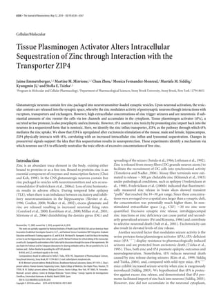
Figure1. ZIP4increasesinexpressioninthehippocampusduringexcitotoxicevents.A,Representativeimageoftheexpression
of ZIP isoforms from wt and tPAϪ/Ϫ
cortices (Cx) and hippocampi (Hc) was analyzed by RT-PCR. B, Quantification of ZIP isoform
expression by densitometry using ImageJ software and comparison using one-way ANOVA. Error bars represent SEM (n ϭ 3
independentexperiments).C,WtandtPAϪ/Ϫ
animalswereinjectedunilaterallywithkainate(0.75nmol)orPBSandthemRNA
levels of the ZIP isoforms in the cortex and hippocampus (ipsilateral and contralateral to the injection site) were examined.
D, Quantification of ZIP isoform expression after kainate injection by densitometry using ImageJ software and comparison using
one-wayANOVAfollowedbyBonferroni’sposthoctest.ErrorbarsrepresentSEMwhere*pϽ0.05,**pϽ0.01(nϭ3indepen-
dentexperiments).E,WtandtPAϪ/Ϫ
animalswereunilaterallyinfusedfor24hwithkainate(KA;0.75nmol)intothehippocam-
pus,followingwhichthebrainswerefixed,sectioned,immunostainedusinganti-ZIP4(redfluorescence)andanti-NeuN(green,a
neuronal marker), and imaged by confocal microscopy. DAPI (blue) denotes nuclei. The CA3 region is marked. Boxed regions are
shown at higher magnification in the panels in the inset. DG, Dentate gyrus. F, Comparison of fluorescent intensity of ZIP/NeuN
positive cells in the CA3 regions of the ipsilateral and contralateral sides. Quantification was performed by analyzing four 40ϫ
imagesoftheCA3regionandmeasuringthefluorescentintensityusingAxioVision4.6imagingsoftware.ErrorbarsrepresentSEM,
where**pϽ0.0098byone-tailedStudent’sttest(nϭ3independentexperiments).Scalebar,20m.G,ExpressionofZIP1and
ZIP4inculturedprimaryhippocampalneuronsusingRT-PCR.-Actinwasusedascontrol.Whitearrowheadindicatescellsonthe
ipsilateral side that are positive for nuclear DAPI and NeuN staining and cytoplasmic ZIP4 staining, which presents as yellow or
white nuclei surrounded by a red halo.
6540 • J. Neurosci., May 12, 2010 • 30(19):6538–6547 Emmetsberger et al. • tPA-Mediated Intracellular Zinc Sequestration via ZIP4
4. element UltraLE Ge-detector (Canberra). Image processing and deter-
mination of metal content within individual cells (g/cm2
) was
performed with MAPS software (Vogt et al., 2003).
Results
Neuronal activity modulates mZIP4 expression
ZIP family members have been reported to be expressed in many
tissues (Vogt et al., 2003). Using RT-PCR, we confirmed that
mouse (m)ZIP1, 2, 3, and 4 are all expressed in the mouse brain
(Fig. 1A,C). mZIP 2 and 3 exhibited similar levels of expression
in the cortex and in the hippocampus, whereas mZIP4, and to a
lesser extent, mZIP1, were expressed at higher levels in the hip-
pocampus, which exhibits neuronal excitability during TLE. No
significant differences in levels of expression of the mZIPs were
observed for wild-type (wt) versus tPAϪ/Ϫ
mice (Fig. 1B).
Since the physiological requirement for zinc transport could
vary dramatically according to localized neuronal activity, and
since relatively little is known about how the ZIPs are regulated,
we hypothesized that cells might control their capacity for zinc
import by using transcriptional mechanisms. We thus examined
whether there were ZIP family members that changed their level
of expression as a consequence of CNS stimulation. Wt (C57BL6)
mice were injected unilaterally in the hippocampus with kainate,
a glutamate analog that induces excitation and seizures, and the
brains harvested for RT-PCR analysis 1 d later. No significant
changes in expression level were observed for mZIP2, or 3 (Fig.
1C); however, the levels of mZIP1 and 4 increased significantly in
hippocampus on the side of excitotoxin injection (ipsilateral
side), but not on the contralateral side, nor on the ipsilateral side
after injection of PBS (Fig. 1D). Interestingly the change in ZIP4
expression was the most significant. ZIP1 expression has previ-
ously been associated with neurons (Belloni-Olivi et al., 2009).
This finding was confirmed, as we used RT-PCR on isolated pri-
mary hippocampal neurons to evaluate the mZIP1 expression as
well that of mZIP4 (Fig. 1G).
The change in expression of ZIP4 after stimulation was con-
firmed using immunohistochemistry with an anti-ZIP4 antibody
(Fig. 1E; supplemental Fig. 1, available at www.jneurosci.org as
supplemental material). Upregulation of ZIP4 expression was
evident on the injected side of the hippocampus in the KA-
injected wt and tPAϪ/Ϫ
mice, especially in
the CA3 subregion (quantification in Fig.
1F), and its expression colocalized with
the neuronal marker NeuN (Fig. 1E) and
the microglial marker 5D4 (data not
shown). Thus, neuronal expression of
ZIP4 increases with neuronal activity,
identifying ZIP4 as a potential candidate
in the response to high level release of zinc
during excitotoxicity, an event that also
upregulates tPA (Qian et al., 1993).
mZIP4 and tPA form a physical
complex on the cell surface
Since ZIP4 was the zinc transporter
whose expression was affected to the
greatest extent by neuronal injury, we
focused subsequent investigation on
this protein. We had reported previ-
ously that tPA increases import of zinc;
this finding therefore suggested that tPA
should mediate a direct or indirect effect
on ZIP4 function. tPA is known to exert
part of its actions through noncatalytic,
protein–protein interactions involving cell surface binding part-
ners such as annexin II (Siao and Tsirka, 2002). We thus began by
exploring the possibility that tPA might physically interact with
ZIP4.
To assess the interaction between ZIP4 and tPA, exogenous
tPA was added to the cultured cells. We used 10 g/ml tPA, a
concentration which has previously been used and shown to
abolish zinc-induced cell death (Kim et al., 1999). Control
HEK293 cells and HEK293 cells stably expressing HA-tagged
mZIP4 (supplemental Fig. 1, available at www.jneurosci.org as
supplemental material) were cooled on ice and exposed to hu-
man tPA in culture medium for 5 min. The cells were washed,
lysed in RIPA buffer to preserve interacting protein complexes,
immunoprecipitated using anti-tPA antibody, and immunoblot-
ted using anti-HA antibody to detect coprecipitated HA-tagged
ZIP4. In the absence of exogenously added tPA, very little ZIP4
was detected; in the presence of tPA, ZIP4 was robustly pulled-
down (Fig. 2A). In the reverse experiment, the control and HA-
ZIP4-expressing HEK293 cells were incubated with a range of
concentrations of mouse tPA and immunoprecipitated with
anti-HA antibody, following which tPA activity was measured
using zymography (Fig. 2B; zymography was required because
the anti-human tPA antibody does not recognize mouse tPA
well). Larger amounts of tPA were pulled down from the lysates
of ZIP4-expressing cells than from the control HEK 293 cells at
every concentration of tPA used. Intriguingly, more tPA activity
was present even in the sample in which no exogenous tPA had
been added, indicating that endogenous tPA was being pulled
down by the ZIP4.
To ascertain that the ZIP4/tPA interaction is physiologically
relevant in vivo, wt mice were intrahippocampally injected with
PBS or kainate and after 24 h their brains were harvested and
lysed. ZIP4 was immunoprecipitated and the associated tPA ac-
tivity was assessed via zymography. Higher levels of tPA activity
associated with the immunoprecipitated ZIP4 were detected in
the injected side (ipsi) after the kainate injection (Fig. 2C). These
data indicate that tPA and ZIP4 form a physical complex on the
cell surface, providing a possible mechanism through which tPA
might facilitate ZIP4-mediated zinc import.
Figure 2. tPA forms a complex with ZIP4 on the cell surface. A, Lysates from HA-mZIP4-expressing HEK293 cells preincubated
with or without 10 g/ml tPA on ice were immunoprecipitated using anti-tPA antibody and immunoblotted using anti-HA
antibodytodetectHA-taggedZIP4.B,LysatesofcontrolHEK293cellsandHA-mZIP4-expressingHEK293cellswerepreincubated
withvariedconcentrationsoftPA,immunoprecipitatedwithanti-HA,andassayedfortPAactivityusingzymography.Ϫdenotes
noadditionoftPA.C,Control(injectedwithPBS)andkainate-injectedbrainswereseparatedintheinjected(ipsi)anduninjected
(contra) sides and lysed. The lysates were immunoprecipitated with anti-mZIP4, and assayed for tPA activity using zymography.
Emmetsberger et al. • tPA-Mediated Intracellular Zinc Sequestration via ZIP4 J. Neurosci., May 12, 2010 • 30(19):6538–6547 • 6541
5. Domain analysis of the physical and functional interaction of
tPA with ZIP4 and zinc
Given the physical association between mZIP4 and tPA, we next
examined whether the interaction was mediated by a specific
region of the tPA protein. tPA consists of five domains: a
fibronectin-3 finger domain (F) at the N terminus, followed by an
epidermal growth factor (EGF)-like domain (GF), two kringle
domains (K1 and K2) and a carboxy-terminal light (L) catalytic
domain (van Zonneveld et al., 1986). We previously reported that
the catalytic activity of tPA was dispensable for this pathway,
since a catalytically inactive tPA mutant (S481A tPA) promoted
zinc intracellular import just as well as the wild-type, catalytically
active protein (Siddiq, 2003; Siddiq and Tsirka, 2004).
Recombinant tPA proteins lacking individual or multiple do-
mains, all of which were catalytically active, were expressed and
purified and assayed for the ability to be pulled-down in associ-
ation with ZIP4. All of the mutant proteins demonstrated re-
duced binding, with deletion of the second kringle domain (⌬K2)
having the greatest impact (Fig. 3A,B). As a negative control in
this set of experiments HA-tagged Angiotensin Receptor 1
(AT1R) was used (transfected into HEK293 cells similarly to
ZIP4). This protein was not found to interact with tPA nor to
enhance zinc import (data not shown). The ability of the wild-
type and mutant proteins to stimulate zinc import was then as-
sessed. Wild-type and catalytically inactive S481A tPA facilitated
zinc import with equal efficiency (Fig. 3C), in agreement with our
previous results (Siddiq and Tsirka, 2004), and the mutant pro-
teins were less effective. Surprisingly though, the mutant protein
lacking the growth factor domain (⌬GF) was less potent than
⌬K2. The correlation derived between zinc import and ZIP4
binding by the different tPA recombinant proteins has a coeffi-
cient of 0.8504 (Fig. 3D).
An additional factor to consider, however, is that tPA not only
binds ZIP4, but also binds zinc; thus the observed facilitation of
zinc import could result either or both from increased zinc bound
to the transporter, or to internalization of zinc bound to tPA. To
address this issue, we next examined the ability of the mutant tPA
proteins to bind zinc using a published 65
ZnCl2
Ϫ
overlay assay.
Relatively normal binding was observed for the ⌬F, whereas all
binding was lost for the ⌬GF mutant (supplemental Fig. 2, avail-
able at www.jneurosci.org as supplemental material).
Together, these findings indicate that the ability of tPA to
promote zinc import via interaction with ZIP4 depends both on
the ability to bind to ZIP4 and independently on the ability to
bind zinc.
Increased import of zinc in the presence of tPA is associated
with changes in zinc and ZIP4 subcellular localization
We previously demonstrated that the levels of intracellular zinc
increase in neuronal cells in the presence of tPA (Siddiq, 2003;
Siddiq and Tsirka, 2004). Examining this relationship using an-
otherapproach,x-rayspectroscopyrevealedthattheadditionoftPA
Figure 3. tPA protein domains important for ZIP4 physical and functional interaction.
A, HA-mZIP4-expressing HEK293 cells were incubated on ice with 10 g/ml wt tPA or varied
tPA domain mutants, washed, lysed, immunoprecipitated with anti-HA, and assessed for tPA
activity via zymography. LK2, Mutant encoding only light catalytic chain and second kringle
domain;⌬F,mutantlackingfingerdomain;⌬GF,mutantlackinggrowthfactordomain;⌬K2,
mutant lacking second kringle domain. B, tPA zymographic activity was quantified by densi-
tometryusingImageJsoftware.TheinactivemutantS481AtPAwasusedasanegativecontrol
(data not shown) and its densitometric value (since it is catalytically inactive) was subtracted
from the values of all the other recombinant proteins, following which the binding for each
mutant was normalized to that observed for the wild-type protein. Error bars represent SEM
where *p Ͻ 0.05, ***p Ͻ 0.001by ANOVA followed by Bonferroni’s post hoc test (n ϭ 3
independent experiments). C, HEK293 cells expressing mZIP4 were preloaded with FluoZin-3
and incubated with wt tPA, tPA domain mutants, or catalytically inactive (S481A). Ctr, Control
mZIP4-HEK293 cells incubated with media that did not contain recombinant tPA. Zinc import
wasquantifiedbyfluorometricanalysis(ratioA480/A510).D,Correlationbetweenzincimport
and ZIP4 binding efficiency by different tPA recombinant proteins.
Figure4. ZincuptakeisaugmentedinthepresenceoftPA.HEKcellsandmZIP4-expressing
HEK cells preloaded with FluoZin-3 were incubated with zinc in the presence or absence of 10
g/ml tPA. The amount of zinc uptake was quantified by fluorometry (ratio A480/A510).
6542 • J. Neurosci., May 12, 2010 • 30(19):6538–6547 Emmetsberger et al. • tPA-Mediated Intracellular Zinc Sequestration via ZIP4
6. to the culture medium for 30 min increased zinc uptake by 25%
(from 4 to 5 g/cm2
) in the cytoplasm of wild-type hippocampal
neurons, and by 250% (from 1 to 3.5 g/cm2
) in neurites, which
have many presynaptic vesicles that can store zinc (supplemen-
tal Fig. 3, available at www.jneurosci.org as supplemental mate-
rial). Additional measurements for other elements, such as
phosphorus, sodium, calcium and potassium, were also obtained
to confirm basic cellular viability and functions, and were found
not to change (data not shown). This experiment confirmed our
previous observations that tPA facilitates the import of zinc into
neurons, and adds the new observation that uptake is promoted
primarily into neurites.
To extend this finding, HEK293 cells and ZIP4-expressing
HEK293 cells were loaded with the fluorescent zinc reporter,
FluoZin-3, and exposed to 50 M zinc in
the presence or absence of tPA. As shown
in Figure 4, tPA increased zinc uptake in
the mZIP4-expressing cells. Similar re-
sults were obtained with primary hip-
pocampal neurons, which endogenously
express mZIP4 (Fig. 5). The neurons were
loadedwithFluoZin-3andincubatedwithin-
creasingconcentrationsofzincinthepresence
or absence of 10 g/ml tPA. The levels of in-
tracellular zinc (evident as change in the
amount of fluorescence within the cells) in-
creasedwhenthecellswereincubatedwithin-
creasing concentrations of zinc, and a further
increase was seen when tPA was also
present (Fig. 5A). The increase in fluores-
cence was specific, as TPEN [tetrakis(2-
pyridylmethyl)ethylenediamine], a chelator
of free zinc, eliminated the uptake-
dependent fluorescence.
To further explore the localization of
intracellularly imported zinc, we used
FluoZin-3 loading and two-photon mi-
croscopy. The FluoZin-3-loaded hip-
pocampal neurons were immunostained
for cytochrome C (cyt C, as a marker of
mitochondria, a site of potential zinc ac-
cumulation) and counterstained with
DAPI (to visualize nuclei). Nontreated
control cells displayed baseline levels of
cytoplasmic zinc present in the soma,
axon, and dendrites. tPA treatment by it-
self did not affect the basal levels of zinc
import (although there was consistently a
minor decrease in FluoZin-3 fluorescence
detected with tPA-alone controls). Cyt C
staining (Fig. 5B, visualized as red) ap-
peared normal in these cells, as mitochon-
dria were dispersed throughout the soma.
Treatment with zinc increased the
amount of FluoZin-3 fluorescence in the
cells, in agreement with our previous data
(supplemental Fig. 3, available at www.
jneurosci.org as supplemental material;
Fig. 4; Siddiq and Tsirka, 2004), and in-
duced colocalization with cyt C, visualized
as yellow (arrows). This colocalization
presented in many cells within the soma.
When cells were incubated with zinc and
tPA, the staining was more punctate (arrows), spread throughout
the cell and included areas of the axons and dendrites, possibly
indicative of vesicular distribution of zinc.
To determine the physiological significance of tPA on zinc im-
port wt and tPAϪ/Ϫ
hippocampal slice cultures were prepared and
treated with 50 M zinc. Zinc uptake was analyzed using the marker
FluoZin-3(Fig.5C)andthefluorescenceuptakewasquantified.The
CA3 region in particular took up the most zinc. Higher zinc uptake
was measured in the wt slice compared with the tPAϪ/Ϫ
slice (the
tPAϪ/Ϫ
slices had ϳ60% of the fluorescence of the wt slices) sup-
porting the evidence that tPA facilitates zinc import.
ZIP4 functions as a carrier protein to import zinc by binding it
on the cell surface and internalizing it using endocytosis. Zinc-
bound ZIP4 traffics to recycling endosomes, releases the zinc, and
Figure5. tPAenhanceszincuptakeinhippocampalneurons.A,HippocampalneuronsloadedwithFluoZin-3wereincubated0,
10, 35, and 50 M zinc and/or tPA (10 g/ml). Quantification of intracellular zinc was performed by fluorometric analysis
(A480/A510)at2h,andthevaluesareexpressedasarbitraryunits.Themembrane-permeablezincchelatorTPENwasthenadded
and the cells reimaged. The import of zinc is presented in arbitrary units. B, Two-photon imaging of wt hippocampal neurons
(naive,treatedwithtPA,with50M zinc,orwithbothzincandtPA)loadedwithFluoZin-3(green)andcounterstainedagainstcyt
C(red).ArrowsindicatecoincidentfluorescenceofFluoZin3andcytC.ErrorbarsrepresentSEM,where**pϽ0.01,***pϽ0.001
bytwo-wayANOVAfollowedbyBonferroni’sposthoctest(4neuronsofeachexperimentalconditionwerequantifiedfromnϭ3
independent experiments). C, Organotypic hippocampal slices from wild-type and tPAϪ/Ϫ
mice treated with 50 M zinc and
loadedwithFluozin-3(green)toassesszincuptake.Scalebar,20m(nϭ3independentexperiments,3slices/experiment).The
Fluozin-3 fluorescence intensity of the whole slice was quantified, divided by the slice surface area and was analyzed using
AxioVision 4.6 image software. Wt slices exhibited 0.58 arbitrary units of fluorescence/surface area compared with 0.35 units for
the tPAϪ/Ϫ
slices ( p ϭ 0.2232 by one-tailed Student’s t test).
Emmetsberger et al. • tPA-Mediated Intracellular Zinc Sequestration via ZIP4 J. Neurosci., May 12, 2010 • 30(19):6538–6547 • 6543
7. then returns to the plasma membrane. ZIP4 trafficking is regu-
lated by unknown mechanisms that sense zinc levels; ZIP4 cy-
cling to the plasma membrane is enhanced in zinc deficiency
(Kim et al., 2004). We next examined whether tPA affects zinc-
loaded ZIP4 trafficking, using the HA-ZIP4-expressing HEK293
cells. Under control conditions, ZIP4 was observed at the plasma
membrane (red, Fig. 6A) at or near cortical actin (green), as
shown in representative images (quantification is described in
Fig. 6B). A similar localization was observed when either tPA or a
low concentration (10 M) of zinc was added. However, addition
of both tPA and 10 M zinc caused a tenfold increase in endocy-
tosis, resulting in internalization of a significant fraction of the
peripheral ZIP4 into cytoplasmic vesicles, presumably early en-
dosomes, while the remaining ZIP4 plasma membrane staining
became punctate. Small amounts of endocytosis were observed
when 50 or 100 M zinc was added to the cultures; in contrast,
addition of tPA with 50 M zinc resulted in complete endocytosis of
ZIP4 into vesicles, and addition of tPA with 100 M zinc resulted in
accumulation of the endocytosed ZIP4 into perinuclear recycling
endosomes.Thus,tPAincreaseszinc-loadedZIP4endocytosis,facil-
itatingzincentry,anddirectingtheenteringzincintovesicles,rather
than into the cytoplasm as occurs when it enters through Ca-A/K,
voltage-sensitive calcium channels (VSCC).
It has been suggested that during seizures, the high levels of
zinc released are taken back up and sequestered in lysosomal
vesicles (Hwang et al., 2008), as a byproduct of ZIP4 degradation
by lysosomal and proteosomal pathways (Mao et al., 2007). This
led us to evaluate whether tPA enhanced the sequestration of zinc
and mZIP4 to lysosomes as a method of protection against zinc
toxicity.ImmunostainingagainstmZIP4revealedminimallocaliza-
tion with the lysosomal marker LAMP-1 in control, untreated cells
and in cells treated with tPA alone (Fig. 6C). When the cells were
treated with 50 M zinc, there was an increase in mZIP4 and
LAMP-1 colocalization; significant colocalization between mZIP4
and LAMP-1 was also evident in cells treated with zinc and tPA (Fig.
6C). These data suggest that in addition to the physical interaction
between the zinc transporter (ZIP4) and tPA (Figs. 2, 3), the pres-
enceoftPAbothenhancedtheimportofzincintothecells(Figs.4,5)
and affected the localization of the mZIP4 transporter, which traf-
ficked from the cell surface to lysosomal compartments.
tPA facilitation of zinc import by ZIP4 shifts intracellular
signaling networks toward prosurvival pathways, providing
protection against zinc toxicity
Very little is known about the signaling pathways that may be in-
volved or affected by ZIP4/zinc trafficking. However, ZIP family
Figure6. tPAincreasesvesicularuptakeofZIP4intolysosomesinZIP4-expressingHEK293cells.ConfocalimagesofmZIP4-expressingHEK293cellsincubatedwithincreasingconcentrationsof
zincinthepresenceorabsenceoftPA.A,ThepresenceofmZIP4wasvisualizedwithananti-HAantibody(red);actinwasvisualizedusingphalloidin(green),andnucleiweremarkedwithDAPI(blue).
B,FluorescentintensityofintracellularZIP4wasquantifiedusingtheLSMimagingsoftware.Theexperimentswererepeatedatleastfivetimesand30cellsquantifiedforeachcondition.Errorbars
represent SEM, where *p Ͻ 0.05, ***p Ͻ 0.001 by ANOVA followed by Bonferroni’s post hoc test. Scale bar, 20 M. C, mZIP4 cells were incubated with 50 M zinc or zinc and tPA. The cells were
stained for ZIP4 (red) using the anti-HA antibody, and the lysosomal marker LAMP-1 (green). n ϭ 3 individual experiments.
6544 • J. Neurosci., May 12, 2010 • 30(19):6538–6547 Emmetsberger et al. • tPA-Mediated Intracellular Zinc Sequestration via ZIP4
8. members have been associated with intracellular signaling changes:
ZIP1internalizationhasbeenlinkedtochangesintheactincytoskel-
eton (Khadeer et al., 2005), ZIP7 to tyrosine kinase activation in
cancer cells (Hogstarnd et al., 2009), and ZIP13 to growth factor/
BMP signaling (Fukada et al., 2008); these reports suggest that ZIP4
could mediate global changes in cellular responses. To explore
this possibility in the context of zinc toxicity, we probed several sig-
naling pathways known to be involved in pro- and antiapoptotic
outcomes for neurons. The mitogen-activated protein kinase
(MAPK) pathway (ERK1/2, p38 kinase and c-Jun N terminal kinase
(JNK)) has been shown to contribute to neuronal cell death in a
variety of in vitro and in vivo neurotoxicity models (Murray et al.,
1998; Jeon et al., 2000; Eom et al., 2001; Koh, 2001; Seo et al., 2001;
Jiang et al., 2005; Namiki et al., 2007). Under resting conditions,
phospho-p38 (p-p38) and p-ERK were
barely detectable in mZIP4-expressing
HEK293 cells, and modest amounts of
p-JNK were observed (Fig. 7A). Upon zinc
addition, p-ERK, p-JNK, and p-p38 in-
creased substantially, while p-AKT stayed
the same. However, when tPA was added
along with zinc, most of the p-ERK, p-JNK,
and p-p38 zinc-induced increases were
bluntedoreliminated,andp-AKTmodestly
increased (Fig. 7B). Together, zinc exposure
seemed to trigger proapoptotic pathways
that were ameliorated by the presence of
tPA.ThesedatasuggestedthattPAmaypro-
tect against zinc toxicity through regulated
importofzincthatleadstothesequestration
into vesicles and modulation of the activa-
tion of MAPKs involved with the apoptotic
process associated with zinc toxicity.
Discussion
We report here that excitotoxic stimula-
tion of neurons upregulates the expres-
sion of the zinc transporter ZIP4 in the
mouse hippocampus, and that ZIP4 phys-
ically interacts with tPA, enhances the im-
port of zinc into cells, and promotes a
prosurvival state. In our previous work,
we had inhibited general endocytosis as
well as the low-density lipoprotein
receptor-related protein (LRP, which acts
as a scavenger receptor for tPA), and de-
termined that these mechanisms were not
responsible for zinc import in the pres-
ence of tPA.
During the course of this study we ob-
served that the expression of the ZIP4
gene in the hippocampus was increased
after kainate injection. This is an interest-
ing finding since the ZIP4 protein is rec-
ognized to be an important transporter
for dietary zinc absorption in the intestine
(Eide, 2006); Dufner-Beattie et al., 2003).
In the intestines and liver ZIP4 mRNA is
upregulated in conditions of zinc defi-
ciency and the ZIP4 protein is transported
to the plasma membrane. When zinc is
present, ZIP4 is regulated in various ways:
at the mRNA level, at the plasma mem-
brane by endocytosis though recycling en-
dosomes, and by ubiquitination and subsequent degradation
(Kim et al., 2004; Mao et al., 2007). In another study using whole
braintissue,littletonoZIP4expression(Dufner-Beattieetal.,2003)
was observed, however different subregions of the brain were not
examined. As hippocampal neurons are postmitotic, it is feasible
that following seizures, the levels of zinc are regulated differently
than in replicating cells. Another possibility is that since tPA
partly acts as a zinc chelator, its presence may mimic zinc defi-
ciency, which would then trigger an upregulation of ZIP4 on the
surface of cells.
One potential caveat of our experiments is the fact that the
concentration of tPA used was quite high (10 g/ml). The ratio-
nale behind using such a concentration is that it was previously
Figure 7. Proapoptotic signals are altered in the presence of tPA. A, Quantitative immunoblot analyses of proteins involved in
celldeathduetozinctoxicitywereperformedonHEKcelllysates.mZIP4HEKcellsweretreatedwithvariousconcentrationsofzinc
withorwithouttPAandtheexpressionofERK,p-ERK,JNK,p-JNK,p38,p-p38,AKT,andp-AKTassessed.B,Quantitativemeasure-
mentsofkinasephosphorylationwereperformedusingtheOdyssey2.1softwareandthedatawereplottedasaratioofthepixel
volume of the phosphorylated kinase over the pixel volume of total kinase. For statistical analysis, zinc treatment was compared
with zinc plus tPA treatment. Error bars represent SEM, where *p Ͻ 0.05, **p Ͻ 0.01, ***p Ͻ 0.001 by ANOVA followed by
Bonferroni’s post hoc test (n ϭ 3 independent experiments).
Emmetsberger et al. • tPA-Mediated Intracellular Zinc Sequestration via ZIP4 J. Neurosci., May 12, 2010 • 30(19):6538–6547 • 6545
9. used to show that zinc toxicity is eliminated by the presence of
tPA (Kim et al., 1999; Siddiq and Tsirka, 2004). Moreover, as tPA
is thought to be secreted from the presynaptic neurons into the
synaptic cleft, it is possible that the local concentration of tPA in
the synaptic cleft is much higher than the concentration used in
our work. In experiments modeling secretion of tPA into the
extracellular/synaptic space other investigators have used con-
centrations up to 60 g/ml (Nicole et al., 2001; Pang et al., 2004).
Utilization of tPA domain deletion mutants indicated that
zinc binds to tPA through the growth factor domain. Not all
import was abolished, however, due to other routes of zinc entry
into the cells; in neurons such routes of entry include the Ca2ϩ
-
permeable AMPA/KA channels, Zn exchangers, and NMDA re-
ceptors (Weiss et al., 1993, 2000; Siddiq and Tsirka, 2004). It is
possible that zinc binds to the growth factor domain of tPA and
that ZIP4 interacts with the kringle domain of tPA through its
histidine-rich cluster in the cytoplasmic region (Mao et al., 2007),
and that through these interactions tPA facilitates zinc import
into cells. Our data would also suggest that the catalytic domain/
activity for tPA is not critical for ZIP binding, as is also the case for
zinc import. Following zinc release from presynaptic boutons,
zinc may initially bind to and is partially chelated by the growth
factor domain of tPA, this consequently induces an increase of
ZIP4 at the plasma membrane. Subsequently, zinc-bound-tPA
interacts with ZIP4 (bound or not bound to zinc) through the
kringle domain of tPA with the extracellular region of the trans-
porter facilitating zinc and ZIP4 vesicular internalization. Al-
though it is not necessary for zinc to be imported into neurons for
ZIP4 to be protective in vivo, since the increase in ZIP4 expression
appears to be primarily neuronal, it is likely that this zinc import
renders ZIP4 neuroprotective.
Under conditions of increased cytosolic levels of zinc, mito-
chondria have been demonstrated to function as possible zinc-
accumulating organelles (Sensi et al., 2003). Other studies have
linked zinc-storage to late endosomes/lysosomes in neuron syn-
aptic vesicles (Danscher and Stoltenberg, 2005; Smith and Lee,
2007; Smith et al., 2007). Hwang et al. (2008) demonstrated that
in the presence of kainate and high concentrations of zinc, the
majority of zinc-containing vesicles in hippocampal neurons are
indeed lysosomes. This led us to consider whether the interaction
between tPA, zinc and ZIP4 mitigates toxicity by altering zinc
homeostasis, including the sequestration of zinc to lysosomal
vesicles subsequent to seizures. Consistent with the data from
Hwang et al., ZIP4 containing vesicles colocalized with the lyso-
somal marker LAMP-1 demonstrating that ZIP4, and presum-
ably zinc, is sequestered to lysosomes. Zinc toxicity is associated
with high intracellular concentrations of unbound chelatable
zinc. It is conceivable that lysosomes act to harbor zinc and that
the intracellular release of zinc from lysosomes is regulated by the
binding of zinc to tPA. In addition, tPA can chelate zinc (Siddiq
and Tsirka, 2004), although the concentration of tPA required to
chelate all secreted zinc would be prohibitively high.
Interestingly, perturbation of the actin cytoskeleton was ob-
served upon tPA treatment, which points toward an active re-
sponse of the cells to the high zinc challenge. The protein kinases
that were analyzed, ERK, JNK and p38, become phosphorylated
and activated under excitotoxic conditions. Similar findings were
observed when mZIP4-transfected HEK cells were incubated
with increasing concentrations of zinc, although when tPA was
present, there was a significant reduction of the phosphorylated
forms of the kinases.
Mice deficient in the ZnT3 transporter lack vesicular zinc, a
finding that would infer that neurons would not be able to secrete
zinc once stimulated. However, zinc accumulates in ZnT3-null
mice after injection with kainate either as a result of zinc released
from internal stores, or because neurons do take up zinc (Lee et
al., 2000). ZIP4 may function as an additional transporter in-
volved in neuronal uptake of zinc, active even when vesicular zinc
is absent. It is possible that under these conditions zinc is being
released by surrounding cells and that it is taken up via various
routes of entry, such as ZIP4, Ca2ϩ
-permeable AMPA/KA chan-
nels, Zn exchangers, and NMDA receptors. Alternatively the neu-
rons may possess an internal zinc release mechanism from
intracellular compartments (Lee et al., 2000). It is feasible that
under pathological conditions, neuronal stress could result in the
zinc release observed in the ZnT3-null mice. Our data revealed
that ZIP4 was expressed at low basal levels under normal condi-
tions, but when an insult occurred, ZIP4 expression increased in
the hippocampus. It is possible that in the absence of vesicular
zinc, ZIP4 is upregulated at the cell surface to compensate for the
lack of synaptic zinc. Zinc taken up by neurons via ZIP4 may not
be entering synaptic vesicles but instead sequestered to zinc-
storage compartments.
In conclusion, our results have shown that tPA facilitates the
partial sequestration of zinc into lysosomal compartments via
ZIP4, thereby regulating the intracellular concentrations of zinc
and acting in a neuroprotective manner by modulating intracel-
lular signaling events that could lead to cell death.
References
Andrade-Gordon P, Strickland S (1986) Interaction of heparin with plas-
minogen activators and plasminogen: effects on the activation of plasmin-
ogen. Biochemistry 25:4033–4040.
Belloni-Olivi L, Marshall C, Laal B, Andrews GK, Bressler J (2009) Localiza-
tion of zip1 and zip4 mRNA in the adult rat brain. J Neurosci Res
87:3221–3230.
Choi DW, Koh JY (1998) Zinc and brain injury. Annu Rev Neurosci
21:347–375.
Cornford EM, Nguyen EV, Landaw EM (2000) Acute upregulation of
blood-brain barrier glucose transporter activity in seizures. Am J Physiol
Heart Circ Physiol 279:H1346–H1354.
Coulter D (2000) Mossy fiber zinc and temporal lobe epilepsy: pathological
association with altered “epileptic” gamma-aminobutyric acid A recep-
tors in dentate granule cells. Epilepsia 41 [Suppl 6]:S96–S99.
Danscher G, Stoltenberg M (2005) Zinc-specific autometallographic in vivo
selenium methods: tracing of zinc-enriched (ZEN) terminals, ZEN path-
ways, and pools of zinc ions in a multitude of other ZEN cells. J Histochem
Cytochem 53:141–153.
Dufner-Beattie J, Wang F, Kuo YM, Gitschier J, Eide D, Andrews GK (2003)
The acrodermatitis enteropathica gene ZIP4 encodes a tissue-specific,
zinc-regulated zinc transporter in mice. J Biol Chem 278:33474–33481.
Eide DJ (2006) Zinc transporters and the cellular trafficking of zinc. Bio-
chim Biophys Acta 1763:711–722.
Eom SJ, Kim EY, Lee JE, Kang HJ, Shim J, Kim SU, Gwag BJ, Choi EJ (2001)
Zn(2ϩ) induces stimulation of the c-Jun N-terminal kinase signaling path-
way through phosphoinositide 3-kinase. Mol Pharmacol 59:981–986.
Finney L, Mandava S, Ursos L, Zhang W, Rodi D, Vogt S, Legnini D, Maser J,
Ikpatt F, Olopade OI, Glesne D (2007) X-ray fluorescence microscopy
reveals large-scale relocalization and extracellular translocation of cellular
copper during angiogenesis. Proc Natl Acad Sci U S A 104:2247–2252.
Frederickson CJ, Hernandez MD, Goik SA, Morton JD, McGinty JF (1988)
Loss of zinc staining from hippocampal mossy fibers during kainic acid
induced seizures: a histofluorescence study. Brain Res 446:383–386.
Frederickson CJ, Giblin LJ, Krezel A, McAdoo DJ, Mueller RN, Zeng Y, Balaji
RV, Masalha R, Thompson RB, Fierke CA, Sarvey JM, de Valdenebro M,
Prough DS, Zornow MH (2006a) Concentrations of extracellular free
zinc (pZn)e in the central nervous system during simple anesthetization,
ischemia and reperfusion. Exp Neurol 198:285–293.
Frederickson CJ, Giblin LJ 3rd, Balaji RV, Rengarajan B, Masalha R,
Frederickson CJ, Zeng Y, Lopez EV, Koh JY, Chorin U, Besser L, Hershfinkel
M, Li Y, Thompson RB, Krezel A (2006b) Synaptic release of zinc from
6546 • J. Neurosci., May 12, 2010 • 30(19):6538–6547 Emmetsberger et al. • tPA-Mediated Intracellular Zinc Sequestration via ZIP4
10. brain slices: factors governing release, imaging, and accurate calculation of
concentration. J Neurosci Methods 154:19–29.
Fukada T, Civic N, Furuichi T, Shimoda S, Mishima K, Higashiyama H, Idaira
Y, Asada Y, Kitamura H, Yamasaki S, Hojyo S, Nakayama M, Ohara O,
Koseki H, Dos Santos HG, Bonafe L, Ha-Vinh R, Zanki A, Unger S,
Kraenzlin ME, et al. (2008) The zinc transporter SLC39A13/ZIP13 is re-
quired for connective tissue development; its involvement in BMP/TGF-
beta signaling pathways. PLoS One 3:e3642.
Glesne D, Vogt S, Maser J, Legnini D, Huberman E (2006) Regulatory prop-
erties and cellular redistribution of zinc during macrophage differentia-
tion of human leukemia cells. J Struct Biol 155:2–11.
Hogstrand C, Kille P, Nicholson RI, Taylor KM (2009) Zinc transporters and
cancer: a potential role for ZIP7 as a hub for tyrosine kinase activation.
Trends Mol Med 15:101–111.
Hwang JJ, Lee SJ, Kim TY, Cho JH, Koh JY (2008) Zinc and 4-hydroxy-2-
nonenal mediate lysosomal membrane permeabilization induced by
H2O2 in cultured hippocampal neurons. J Neurosci 28:3114–3122.
Jeon SH, Kim YS, Bae CD, Park JB (2000) Activation of JNK and p38 in rat
hippocampus after kainic acid induced seizure. Exp Mol Med 32:227–230.
Jiang W, Van Cleemput J, Sheerin AH, Ji SP, Zhang Y, Saucier DM, Corcoran
ME, Zhang X (2005) Involvement of extracellular regulated kinase and
p38 kinase in hippocampal seizure tolerance. J Neurosci Res 81:581–588.
Khadeer MA, Sahu SN, Bai G, Abdulla S, Gupta A (2005) Expression of the
zinc transporter ZIP1 in osteoclasts. Bone 37:296–304.
Kim BE, Wang F, Dufner-Beattie J, Andrews GK, Eide DJ, Petris MJ (2004)
Zn2ϩ
-stimulated endocytosis of the mZIP4 zinc transporter regulates its
location at the plasma membrane. J Biol Chem 279:4523–4530.
Kim YH, Park JH, Hong SH, Koh JY (1999) Nonproteolytic neuroprotec-
tion by human recombinant tissue plasminogen activator. Science 284:
647–650.
Klitenick MA, Frederickson CJ, Manton WI (1983) Acid-vapor decomposi-
tion for determination of zinc in brain tissue by isotope dilution mass
spectrometry. Anal Chem 55:921–923.
Koh JY (2001) Zinc and disease of the brain. Mol Neurobiol 24:99–106.
Kornblum HI, Araujo DM, Annala AJ, Tatsukawa KJ, Phelps ME, Cherry SR
(2000) In vivo imaging of neuronal activation and plasticity in the rat
brain by high resolution positron emission tomography (microPET). Nat
Biotechnol 18:655–660.
Lee JY, Cole TB, Palmiter RD, Koh JY (2000) Accumulation of zinc in de-
generating hippocampal neurons of ZnT3-null mice after seizures: evi-
dence against synaptic vesicle origin. J Neurosci 20:RC79.
Liuzzi JP, Cousins RJ (2004) Mammalian zinc transporters. Annu Rev Nutr
24:151–172.
Lothman EW, Stringer JL, Bertram EH (1992) The dentate gyrus as a con-
trol point for seizures in the hippocampus and beyond. Epilepsy Res
[Suppl] 7:301–313.
Mao X, Kim BE, Wang F, Eide DJ, Petris MJ (2007) A histidine-rich cluster
mediates the ubiquitination and degradation of the human zinc trans-
porter, hZIP4, and protects against zinc cytotoxicity. J Biol Chem 282:
6992–7000.
Millan E, Abou-Khalil B, Delbeke D, Konrad P (2001) Frontal localization
of absence seizures demonstrated by ictal positron emission tomography.
Epilepsy Behav 2:54–60.
Mirrione MM, Schiffer WK, Siddiq M, Dewey SL, Tsirka SE (2006) PET
imaging of glucose metabolism in a mouse model of temporal lobe epi-
lepsy. Synapse 59:119–121.
Murray B, Alessandrini A, Cole AJ, Yee AG, Furshpan EJ (1998) Inhibition
of the p44/42 MAP kinase pathway protects hippocampal neurons in a
cell-culture model of seizure activity. Proc Natl Acad Sci U S A 95:
11975–11980.
Namiki K, Nakamura A, Furuya M, Mizuhashi S, Matsuo Y, Tokuhara N,
Sudo T, Hama H, Kuwaki T, Yano S, Kimura S, Kasuya Y (2007) In-
volvement of p38alpha in kainate-induced seizure and neuronal cell dam-
age. J Recept Signal Transduct Res 27:99–111.
Nicole O, Docagne F, Ali C, Margaill I, Carmeliet P, MacKenzie ET, Vivien D,
Buisson A (2001) The proteolytic activity of tissue-plasminogen activa-
tor enhances NMDA receptor-mediated signaling. Nat Med 7:59–64.
Pang PT, Teng HK, Zaitsev E, Woo NT, Sakata K, Zhen S, Teng KK, Yung
WH, Hempstead BL, Lu B (2004) Cleavage of proBDNF by tPA/plasmin
is essential for long-term hippocampal plasticity. Science 306:487–491.
Pei YQ, Koyama I (1986) Features of seizures and behavioral changes in-
duced by intrahippocampal injection of zinc sulfate in the rabbit: a new
experimental model of epilepsy. Epilepsia 27:183–188.
Qian Z, Gilbert ME, Colicos MA, Kandel ER, Kuhl D (1993) Tissue-
plasminogen activator is induced as an immediate-early gene during sei-
zure, kindling and long-term potentiation. Nature 361:453–457.
Rogove AD, Tsirka SE (1998) Neurotoxic responses by microglia elicited by
excitotoxic injury in the mouse hippocampus. Curr Biol 8:19–25.
Sensi SL, Ton-That D, Sullivan PG, Jonas EA, Gee KR, Kaczmarek LK, Weiss
JH (2003) Modulation of mitochondrial function by endogenous Zn2ϩ
pools. Proc Natl Acad Sci U S A 100:6157–6162.
Seo SR, Chong SA, Lee SI, Sung JY, Ahn YS, Chung KC, Seo JT (2001)
Zn2ϩ
-induced ERK activation mediated by reactive oxygen species causes
cell death in differentiated PC12 cells. J Neurochem 78:600–610.
Serrano L, Dominguez JE, Avila J (1988) Identification of zinc-binding sites
of proteins: zinc binds to the amino-terminal region of tubulin. Anal
Biochem 172:210–218.
Siao CJ, Tsirka SE (2002) Tissue plasminogen activator mediates microglial
activation via its finger domain through annexin II. J Neurosci 22:
3352–3358.
Siconolfi LB, Seeds NW (2001) Mice lacking tPA, uPA, or plasminogen
genes showed delayed functional recovery after sciatic nerve crush. J Neu-
rosci 21:4348–4355.
Siddiq M (2003) Zinc and tissue plasminogen activator in the mammalian
central nervous system. PhD thesis, Stony Brook University.
Siddiq MM, Tsirka SE (2004) Modulation of zinc toxicity by tissue plasmin-
ogen activator. Mol Cell Neurosci 25:162–171.
Sloviter RS, Dichter MA, Rachinsky TL, Dean E, Goodman JH, Sollas AL,
Martin DL (1996) Basal expression and induction of glutamate decar-
boxylase and GABA in excitatory granule cells of the rat and monkey
hippocampal dentate gyrus. J Comp Neurol 373:593–618.
Smith AP, Lee NM (2007) Role of zinc in ALS. Amyotroph Lateral Scler
8:131–143.
Smith DG, Cappai R, Barnham KJ (2007) The redox chemistry of the Alz-
heimer’s disease amyloid beta peptide. Biochim Biophys Acta 1768:
1976–1990.
Suh SW, Jo SM, Vajda Z, Danscher G (2001) Adrenalectomy causes loss of
zinc ions in zinc-enriched (ZEN) terminals and decreases seizure-induced
neuronal death. Brain Res 895:25–32.
Sutula T, Harrison C, Steward O (1986) Chronic epileptogenesis induced by
kindling of the entorhinal cortex: the role of the dentate gyrus. Brain Res
385:291–299.
Takimoto M, Mao P, Wei G, Yamazaki H, Miura T, Johnson AC, Kuzumaki N
(1999) Molecular analysis of the GCF gene identifies revisions to the
cDNA and amino acid sequences(1). Biochim Biophys Acta 1447:
125–131.
Timofeeva O, Nadler JV (2006) Facilitation of granule cell epileptiform ac-
tivity by mossy fiber-released zinc in the pilocarpine model of temporal
lobe epilepsy. Brain Res 1078:227–234.
Tsirka SE, Gualandris A, Amaral DG, Strickland S (1995) Excitotoxin-
induced neuronal degeneration and seizure are mediated by tissue plas-
minogen activator. Nature 377:340–344.
Van Zonneveld AJ, Veerman H, MacDonald M, van Mourik J, Pannekoek H
(1986) Structure and function of human tissue-type plasminogen activa-
tor (ϩ-PA). J Cell Biochem 32:169–178.
Vogt S, Maser J, Jacobsen C (2003) Data analysis for X-ray fluorescence
imaging. J Phys IV 104:617–622.
Walker MC, Ruiz A, Kullmann DM (2002) Do mossy fibers release GABA?
Epilepsia 43 [Suppl 5]:196–202.
Wang F, Kim BE, Dufner-Beattie J, Petris MJ, Andrews G, Eide DJ (2004)
Acrodermatitis enteropathica mutations affect transport activity, local-
ization and zinc-responsive trafficking of the mouse ZIP4 zinc trans-
porter. Hum Mol Genet 13:563–571.
Weiss JH, Hartley DM, Koh JY, Choi DW (1993) AMPA receptor activation
potentiates zinc neurotoxicity. Neuron 10:43–49.
Weiss JH, Sensi SL, Koh JY (2000) Zn(2ϩ): a novel ionic mediator of neural
injury in brain disease. Trends Pharmacol Sci 21:395–401.
Emmetsberger et al. • tPA-Mediated Intracellular Zinc Sequestration via ZIP4 J. Neurosci., May 12, 2010 • 30(19):6538–6547 • 6547