infanticide.pptx
•Télécharger en tant que PPTX, PDF•
1 j'aime•342 vues

Forensic Medicine

Recommandé
Contenu connexe
Tendances
Tendances (20)
Similaire à infanticide.pptx
Similaire à infanticide.pptx (20)
Plus de Dr Nikita Prabhakaran
Plus de Dr Nikita Prabhakaran (20)
Dernier
Dernier (20)
infanticide.pptx
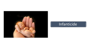
- 1. Infanticide
- 2. DEFINITION • Unlawful destruction of a child • Under one year of age /12 months of age • Does not include : • death of foetus during labour • When it is destroyed by craniotomy or decapitation
- 3. • Feticide- killing of a fetus anytime prior to its birth • Intrapartum death – death occurring during labor an delivery • Neonaticide – the act of killing an infant within the first 24 hours of life • Neonatal death – Death of a liveborn infant within first 28 days of life • Perinatal mortality- Stillbirths + early neonatal death (<7 days)
- 4. Filicide/ prolicide • Killing of a child older than 24 hrs by its own parents • Maternal filicide (commoner than paternal filicide) • MLI postpartum psychosis • Charged under a lesser offence in ENGLAND • (s1 infanticide act of England, 1938) • Defense is not available to anyone else
- 5. Legal Aspects • India tried under Section 302 IPC • Causing death of a living child in the womb Section 299 IPC
- 6. Stillbirth/ SADS (sudden antenatal death syndrome) • WHO defines • “One which is born after 28th week of pregnancy • And which did not breathe/show any other signs of life after being completely born” • UK (Still birth Act 1992), period is reduced to 24 weeks
- 8. Investigation of such a case • Examination of mother signs of recent delivery • her psychiatric condition • Examination of the child stillborn/deadborn • viable /not • whether the child was born alive?? • if born alive, how long did he live??? • what was the cause of death???
- 9. Causes • Anoxia • Birth trauma (MC: intracranial haemorrage due to excessive moulding ) • Congenital defects • Erythroblastosis fetalis • Placental abnormalities • Prematurity • Toxaemia of pregnancy
- 10. Deadborn child • One in which the foetus has died in-utero, • shows one of the following signs after it is completely born 1. Rigor mortis normal change of death 2. Maceration no air in the liquor 3. Putrefaction air ++ 4. Adipocere liquor amnii is retained for longer 5. Mummificationif all liquor amnii has been absoarbed /drained
- 11. Characteristics Still born fetus Dead born fetus Period of gestation > 28 weeks any Condition in uterus Was live before labour Was lying dead in uterus Predominance Illegitimate and immature male children in primiparae No such predominance Cardinal features Signs of prolonged labour Any signs- mummification/ rigor mortis/etc Cause Anoxia, birth trauma Congenital anamolies, ABO incompatibility
- 12. Maceration • (Latin macerare, to soften by soaking) • is degenerative change occurring in a fetus retained in utero after death. • It occurs due to the softening effect of soaking on solid tissues (pulpy fetus).
- 13. Salient features: (1) The earliest sign of maceration is skin slippage (12 hours after IUD) exposes a red, shiny, moist dermal surface, particularly noticeable over bony prominences. (2) 24 hours- fluid-filled bullae
- 14. (3) 48 hours (i)Sweetish, disagreeable odor (ii) hemolysis purplish discoloration of internal organs (iii) Dark, red-stained fluid accumulates in the serous cavities. (iv) deep dark red appearance (“tobacco juice”)- amniotic fluid or in the event of passage of meconium, a thick brown appearance. (v) laxity of joints - Bones are flexible and readily detached from soft parts (vi) Abdominal organs may show green discoloration due to leakage of bile pigments from the gallbladder
- 15. • (4) 3 to 4 days" Umbilical cord is red, soft and smooth • (5) 5th day"S/c edema up to 5 mm. • (6) >1 week" (i) Meconium released (ii) Protruding autolyzed liver mass - mistaken for omphalocele in USG (iii) fluid accumulation ~ hydrops fetalis. (iv) fluid accumulation at the nape of the neck ~cystic hygroma (v) distortion of skull occurs during vaginal delivery ~ hydrocephalus. (vi) from purple to brown discoloration. • (7) Several weeks – (i) yellow–gray color (ii) Dehydration results in shrinkage and compaction - formation of “fetus papyraceous
- 16. Lithopedion • .Very occasionally an extrauterine pregnancy may be retained for years forming so-called • (Greek lithos, stone, paidion, small child), a calcified fetus retained in the abdominal cavity.
- 17. Radiological Signs of IU Deaths 1. Robert’s sign (1944) • Earliest sign to appear. • 12 hours after death. • Air in aorta, pulmonary vessels or abdomen.
- 18. 2. Spalding’s sign (1922) Appears 2 days after death. Overlapping of fetal skull bones seen on x-ray (or ultrasound) examination. Due to shrinkage of cerebrum after intrauterine death. [D/d. May also be positive (i) during or just prior to labor (ii) scanty liquor amnii and diminished fetal vitality (iii) microcephaly (iv) craniostenosis]
- 20. 3. Deuel’s halo sign (1947) 3 days after death. Separation of the subcutaneous fat of the fetal scalp from the cranial bones. Seen as a halo on radiography and USG. First described by Deuel in 1947.
- 21. Live birth • [Explanation 3 of S.299, IPC]. • A child is “live born”, if any part of that child has been brought forth, even though the child may not have breathed or been completely born • The Registration of Births And Deaths Act, 1969 • complete expulsion of fetus from its mother, irrespective of the duration of pregnancy, and who after such expulsion breathes or shows any other evidence of life
- 22. Signs of Live birth •Changes in Lungs •Changes in stomach and intestines •Changes in middle ear •Other signs
- 23. Before respiration After respiration Shape of chest 1-2cm below umbilicus Drum shaped Position of diaphragm 4th/5th rib 6th/7th rib Lungs colour Uniformly reddish Mosaic/mottled Volume Smaller, not covering hear Larger, covers heart Surface smooth uneven margins sharp rounded consistency liverlike Soft, crepitant Blood within lungs less more Weight of lungs 35 g (1/70 of body wt) 70g (1/35 of body weight) Pleura loose stretched
- 24. Cut section - lungs Before respiration After respiration oozing Little frothless blood Abundant frothy blood oozes on cut section Bronchi and bronchioles empty Contained blood stained froth Alveoli Not inflated inflated MLI Still born/dead born Live born
- 25. Hydrostatic test (Raygat’s test) Principle • Upon breathing, both wt and vol of lungs are increased increased. • Wt due to inflow of blood and • Vol due to inflow of air. • vol >>>wt specific gravity of lungs is decreased.
- 26. Procedure • Stage 1 – • Whole thoracic pluck - both lungs and heart are placed in a bucket of water. If the pluck floats If the pluck sinks "indicates air in lungs“ move to second stage. ** liver - control (floats putrefaction)
- 27. Stage 2 • Each brochus is tied, and lungs severed above the ligature. • Each lung is then placed separately in water. • If either lung floats, it indicates that the infant may have born alive. • If either lung sinks, move to third stage.
- 28. Stage 3 • Cut each lung in 12-20 pieces (a) Roll a piece of lung b/w a finger and thumb crackling crepitant noise. (squeeze between thumb and finger bubbles) (c) observe if they float independently. If not next stage
- 29. Stage 4 • Each piece is now taken out of water, wrapped in a piece of cloth and squeezed by putting a weight. • remove the expiratory reserve volume air, and tidal air. • Residual air still remains within the alveoli, which can not be taken out by any means.
- 30. Note:-- • floating in earlier stages due to artefacts • (such as gases introduced by artificial respiration), • the pieces of lung would not continue to float till the last stage.
- 31. Fallacies (a) Child respired after birth yet lungs sink Causes (1) Absorption of air - Circulation continued after stoppage of respiration (2) Atelectasis (non expansion) of lungs. (3) Alveolar duct membrane - Causing obstruction to entry of air in alveoli (4) Diseases.
- 32. (b) Child did not respire after birth yet lungs float • Causes: (1) Artificial respiration - air may be found in stomach too. (2) Putrefaction - Putrefactive gases will make the lungs float. (3) Respiration within the womb [vagitus uterinus]- if membranes have ruptured, but may die from natural causes (4) Respiration within the Vagina [vagitus vaginalis] - Similar to above.
- 33. Hydrostatic test is not Necessary One is sure fetus was born dead – (i) Born before age of viability [28 wks] (ii) Macerated or mummified (iii)Monster [eg anencephalic] (iv)Bruising on lungs - indicating efforts at artificial respiration. One is sure fetus was born alive (i) Stomach - contains milk (i) Umbilical cord - has separated and a scar has formed.
- 34. Changes in Stomach and intestines • (1) Breslau’s second life test; Stomach and intestines are removed after tying double ligatures at each end, put in water. float if respiration had taken place; otherwise they sink Principle - Air is swallowed into the stomach and intestines during respiration, making them buoyant
- 35. (3) False +ve – (i) Resuscitation attempts (ii) Bacterial gas formation [putrefaction] (4) Survival period – can be calculated: (i) Immediate after birth – gas in stomach (upto 15 mins) (ii) 1-2 h – Gas reaches small intestines (iii) 5-6 h – colon (iv) 12 h - Rectum.
- 36. • 5) Drawbacks and Fallacies – • (i) Useless in putrefaction • (ii) Air may be swallowed by the child in attempting to free the air passages of fluid obstructions in cases of stillbirth.
- 37. Changes in the middle ear • (1) Wreden’s test – • Principle - During embryonic life, the middle ear contains gelatinous tissue. • During efforts of breathing, • the sphincter at the pharyngeal end of Eustachian tube relaxes and some air enters the middle ear.
- 38. • (i) Regular procedure – • Skull cap is removed and base of skull submerged in water. • Petrous part of temporal bone [which forms roof of the middle ear] is opened. • If a bubble of air escapes from middle ear, the child was born alive. • (ii) Simpler method – • Dip ears in water. Puncture tympanic membrane"Bubble of air escapes" Live birth.
- 39. Other signs a. Blood • (1) Nucleated RBC"disappear in 24 h • (2) Fetal Hb [synthesized mainly in liver]“ (i) Before birth"80-90% (ii) 3 rd month"7-8% (iii) 6 m"disappears completely. b. Meconium Completely excreted from the large intestine in 24-48 h after normal birth May be completely excreted before birth in (a) breech presentation (b) severe anoxia.
- 40. Caput succadeneum vs cephalheamatoma Caput succadeneum Cephalhaematoma Cause Edema b/w skin and galea Rupture of periosteal capillary Contents Serosanguinous fluid blood Suture lines Extends across suture lines Never extends across them Size larger Smaller Site bilateral unilateral Colour No discoloration Discolouration+
- 41. Caput succadeneum Cephalhaematoma Impulse on crying _ - Appearance Immediately after birth 24-48 hours Complications None Jaundice Calcification and ossification Disappearance After birth it starts disappearing Increases for 24-48 hours then recedes MLI Sign of live birth Regression~period of survival
- 45. • D. Changes in skin • E. Changes in Umbilical cord • F. Placenta • G. Circulation Blood vessel Time of closure Umbilical arteries 10hrs to 3 days Left umbilical vein 3-5 days Ductus venosus 3-5 days Ductus arteriousus 7-10 days Foramen Ovale 2-3 months
- 46. CAUSES OF DEATH Precipitate labour is the expulsion of fetus within less than 3 h of commencement of contractions Salient features • (1) All three stages of labor are merged in one • (2) Delivery occurs suddenly and rapidly even without the knowledge of mother • (3) Delivery may occur even unconsciously during like intoxication, anaesthasia
- 47. Risk factors are • (i) Multipara [extremely rare in primi] • (ii) Placental abruption • (iii) Roomy pelvis • (iv) Small premature baby.
- 48. Maternal complications i. cervical and grade 3 perineal tear ii. post-partum hemorrhage iii. retained placenta Child may die from (i) Suffocation (ii) Drowning (iii) Head injury – (iv) Hemorrhage - from torn end of the cord
- 49. PM findings (i) Placenta – is attached to newborn (ii) Umbilical cord – lacerated, torn (iii) Hair and injured scalp – show grass, gravel, mud, sand. (iv) Head – (a) Caput succedaneum and moulding of head - absent (b) fracture of skull. (c) IC hemorrhages (v) Air passages and lungs –depending on where the child fell or drowned.
- 50. Medico-legal Importance • (i) Infanticide - Plea • (ii) False allegation - Alternatively in a true precipitate labor case, mother may be accused of infanticide.
- 51. Head injury in precipitate labour and Blunt trauma Precipitate labour Blunt trauma Bruises on the vertex Bruises- anywhere Lacerations on the scalp- Present Absent Fractures- parietal bones- fissured fractures meeting sagittal suture at right angles Extensive comminuted and depressed fractures Brain- not injured Brain- contusions, haemorrhages
- 52. Criminal causes Act of commission Act of Omission Asphyxia Burns Trauma Poisons Abandoned child is called FONDLING Person in-charge of the infant [mother, father, guardian etc] can be charged u/s 302, IPC if they fail to take care of child and allow him to die. Failure to (i) Clear the air passages – these may be obstructed by amniotic fluid or mucus (ii) Tie the cord – after it is cut. May cause death by hemorrhage
- 53. Abandoning of children Child < 12years Section 317 IPC 7 years imprisonment
- 54. Concealment of birth by secret disposal of dead body before, after or during its birth, 2 y imprisonment or fine or both [Section.318, IPC].
- 55. • CHILD ABUSE Child abuse is the • physical, • sexual or • emotional mistreatment or neglect of a child.
- 56. Battered baby syndrome • Caffey’s syndrome, • Caffey-Kempe syndrome, • Maltreatment syndrome, • Non Accidental Injury of Childhood (NAIC), • parent-infant traumatic stress syndrome (PITS), • Tardieu’s syndrome
- 57. • clinical condition in which young children • usually under 3 years of age • are beaten repeatedly over the most trivial provocation
- 58. • Salient features: • (1) Lack of provocation • (2) Precipitating factors – Often child’s irritating actions eg crying, refusal to be quiet, persistent soiling of napkins etc • (3) Deprivation of nutrition, care and affection is present • (4) Delay - attention sought • (5) Cerebral palsy - 10-15% cases of cerebral palsy may be the result of the battered baby syndrome. • (6) History is incompatible with injuries
- 59. Injuries • Injuries are due to direct manual violence [commonest]. (1) Tear of the frenulum (2) Soft tissue Pinch marks – butterfly marks Slap marks – show as clear bruises [or as lines of petechial hgs] resembling fingers (3)Scalp injuries – very characteristic. Caused by vigorous pulling on scalp hair • Subgaleal hematoma • Traumatic alopecia – bald patches on scalp
- 63. • (4) Eyes (i) Black eye (ii) Hemorrhages [Retinal, Subconjunctival, Subhyaloid, Vitreous] (iii) Lens displacement (iv) Retinal separation. • (5) Skeletal injuries (i) Skull fractures, especially in occipito-parietal area (ii) Transverse and spiral fractures of long bones, (twisting of arms and legs etc )
- 64. (6) Traction lesions - jerking of shaking a child’s limbs Injuries of the periosteum of long bones are seen without fractures (i) Periosteal hematomas (ii) Periosteal shearing (iii) Epiphyseal separation – seen radiologically as small fragments separated from the main bone (iv) Avulsion of metaphysis or chipping of the edges of metaphysis. - corner fractures and bucket handle fractures.
- 65. • (7) Anteroposterior compression of chest Fractures of ribs in midaxillary line • (8) Side-to-side compression of chest – along posterior angles of ribs. After 1-2 weeks, callus forms. X-Rays these are seen as a “string of beads” [syn, rosary bead] paravertebral gutter [Nobbing fracture]
- 67. Visceral injuries (1) Brain and meninges (2) Lungs – Post-traumatic pulmonary pseudocysts [PTPPCs] (3) Liver and spleen - Bursting injuries (4) Hollow viscera – Ruptures Tearing of mesentery
- 69. Thank you