SlideShare une entreprise Scribd logo
1  sur  39
-DR. NIRAV MODI
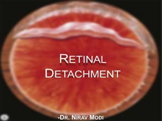
DEFINITION
 A Retinal Detachment (RD) describes the
separation of the neurosensory retina (NSR) from
the retinal pigment epithelium (RPE).
 This results in the accumulation of sub-retinal fluid
(SRF) in the potential space between the NSR and
RPE.
TYPES
 There are three types of retinal detachment:
1) Rhegmatogenous
2) Tractional
3) Exudative
RHEGMATOGENOUS
RETINAL DETACHMENT
=================================================
=================================================
 Rhegmatogenous retinal detachment occurs due to a
break in the retina (called a retinal tear)
 Retinal breaks are the predisposing lesions than can go
into RRD.
 These retinal breaks can be of 3 types
 Retinal Tear.
 Retinal Hole,
 Retinal Dialyses,
PATHOGENESIS
 Presence of retinal break in concert with Vitreo-retinal
traction allows accumulation of liquefied vitreous under
the Neuro-Sensory retina, separating it from RPE.
 Even though Retinal break is present, at least partially
liquefied vitreous and/or traction must be there.
 Vitreous degeneration, PVD, Lattice degeneration,
Snail-track degeneration, WWP, high myopic eyes &
vitreous loss during cataract or YAG capsulotomy are
predisposing.
Symptoms
 1) Photopsia is the subjective sensation of a flash of
light. In eyes with acute PVD it is probably caused by
traction at sites of vitreoretinal adhesion.
 The cessation of photopsia is the result of either
separation of the adhesion or complete tearing away of a
piece of retina (operculum).
 Patient will often describe it as an arc of golden or white
light induced by eye movements and is more noticeable
in dim illumination.
 2) Floaters are moving vitreous opacities which are perceived
when they cast shadows on the retina. Vitreous opacities in
eyes with acute PVD are of the following three types:
 Weiss ring is a solitary floater consisting of the detached
annular attachment of vitreous to the margin of the optic
disc.
 Vitreous Hemorrhage secondary to tearing of a peripheral
retinal blood vessel is indicated by sudden shower of
minute red-coloured or dark spots. Usually Vitreous
haemorrhage here is usually sparse due to the small calibre
of peripheral retinal vessels.
 3) Visual field defect is perceived as a ‘black curtain’. In
some patients it may not be present on waking in the morning,
due to spontaneous absorption of SRF while lying inactive
overnight, only to reappear later in the day.
 A lower field defect is usually appreciated more quickly by
the patient than an upper field defect.
 The quadrant of the visual field in which the field defect first
appears is useful in predicting the location of the primary
retinal break, which will be in the opposite quadrant.
LINCOFF’S RULES
 A shallow inferior RD in which the SRF is slightly higher on the
temporal side points to a primary break located inferiorly on that
side
 A primary break located at 6 o’clock will cause an inferior RD with
equal fluid levels
 In a bullous inferior RD the primary break usually lies above the
horizontal meridian
 If the primary break is located in the upper nasal quadrant the SRF
will revolve around the optic disc and then rise on the temporal side
until it is level with the primary break
 A subtotal RD with a superior wedge of attached retina points to a
primary break located in the periphery nearest its highest border
 When the SRF crosses the vertical midline above, the primary break
is near to 12 o’clock, the lower edge of the RD corresponding to the
side of the break
SIGNS
 1) Marcus Gunn pupil (relative afferent pupillary defect) is
present in an eye with an extensive RD irrespective of the
type.
 2) Intraocular pressure is usually lower by about 5 mmHg
compared with the normal eye. If the intraocular pressure is
extremely low, an associated choroidal detachment may be
present.
 3) Iritis is very common but usually mild.
 4) Tobacco dust consisting of pigment cells is seen in the
anterior vitreous.
 5) Retinal breaks are usually red because of the colour
contrast between the sensory retina and underlying choroid.
However, in eyes with hypo-pigmented choroid (as in high
myopia), the colour contrast is decreased and small breaks
may be missed.
FRESH RETINAL DETACHMENT
 1) The RD has a convex configuration and a slightly opaque
and corrugated appearance as a result of retinal oedema.
 There is loss of the underlying choroidal pattern and retinal
blood vessels appear darker than in flat retina.
 2) SRF extends up to the ora serrata, except in the rare cases
caused by a macular hole in which the SRF is initially
confined to the posterior pole. Because of the thinness of the
retina at the fovea, a pseudohole is frequently seen if the
posterior pole is detached. This should not be mistaken for a
true macular hole, which may give rise to RD in highly
myopic eyes or following blunt ocular trauma.
 3 B-scan ultrasonography shows good mobility of the retina
and vitreous.
(A)U-tear
(B) Superior
bullous RD
(C) Shallow
temporal RD
(D) B-scan-
totally detached
retina with
open funnel
LONG-STANDING RETINAL DETACHMENT
 1) Retinal thinning secondary to atrophy is a characteristic
finding which must not be mistaken for retinoschisis.
 2) Secondary intra-retinal cysts may develop if the RD has
been present for about 1 year; these tend to disappear after
retinal reattachment.
 3) Subretinal demarcation lines ‘‘high water marks’’ caused
by proliferation of RPE cells at the junction of flat and
detached retina are common and take about 3 months to
develop. They are initially pigmented but tend to lose this
with time. Demarcation lines are convex with respect to the
ora serrata and, although they represent sites of increased
adhesion, they do not invariably limit spread of SRF.
Long-standing RD
(A) Secondary retinal cyst.
(B) B-scan shows a retinal cyst
(C) ‘high water mark’ in an eye
with an inferior RD
PROLIFERATIVE VITREO-RETINOPATHY
 Proliferative vitreoretinopathy (PVR) is caused by epiretinal
and subretinal membrane formation. Cell-mediated
contraction of these membranes causes tangential retinal
traction and fixed retinal folds.
 Usually, PVR occurs following surgery for rhegmatogenous
RD or penetrating injury. However, it may also occur in eyes
with rhegmatogenous RD that have not had previous
vitreoretinal surgery.
 The main features are retinal folds and rigidity so that retinal
mobility induced by eye movements or scleral indentation is
decreased.
ADVANCED PVR WITH FUNNEL LIKE RD
EXUDATIVE
RETINAL DETACHMENT
===========================================
===========================================
 Exudative, Serous or Secondary
 Occurs due to inflammation, injury or vascular
abnormalities.
 Fluid accumulating underneath the retina
without the presence of a hole, tear, or break.
 Rare
PATHOGENESIS
 Exudative RD occurs in a variety of vascular, inflammatory
and neoplastic diseases involving the NSR, RPE and choroid
in which fluid leaks outside the vessels and accumulates under
the retina.
 As long as the RPE is able to compensate by pumping the
leaking fluid into the choroidal circulation, no fluid
accumulates in the sub-retinal space and RD does not occur.
 But if the normal RPE pump is overwhelmed or if the RPE
activity is decreased, then fluid starts to accumulate in the
sub-retinal space
 The main causes are the following:
1) Choroidal tumours such as melanomas, haemangiomas and
metastases; it is therefore very important to consider that
exudative RD is caused by an intraocular tumour until proved
otherwise.
2) Inflammation such as Harada disease (Part of VKH syndrome)
and posterior scleritis.
3) Bullous central serous chorioretinopathy is a rare cause.
4) Iatrogenic causes include retinal detachment surgery and
panretinal photocoagulation.
5) Subretinal neovascularization which may leak and give rise to
extensive subretinal accumulation of fluid at the posterior pole.
6) Hypertensive choroidopathy, as may occur in toxaemia of
pregnancy, is a very rare cause.
7) Idiopathic such as the uveal effusion syndrome.
Symptoms:
 Photopsia is absent because there is no vitreoretinal traction,
although floaters may be present if there is associated vitritis.
 The visual field defect may develop suddenly and progress
rapidly.
 Depending on the cause both eyes may be involved
simultaneously (e.g. Harada disease).
Signs :
 The RD has a convex configuration, just like a
rhegmatogenous RD, but its surface is smooth and not
corrugated.
 The detached retina is very mobile and exhibits the
phenomenon of ‘shifting fluid’ in which SRF responds to the
force of gravity and detaches the area of retina under which it
accumulates.
 For example, in the upright position the SRF collects under
the inferior retina, but on assuming the supine position for
several minutes, the inferior retina flattens and the SRF shifts
posteriorly detaching the superior retina.
 The cause of the RD, such as a choroidal tumour, may be
apparent when the fundus is examined, or the patient may
have an associated systemic disease responsible for the RD
(e.g. Harada disease, toxaemia of pregnancy).
 ‘Leopard spots’ consisting of scattered areas of sub-retinal
pigment clumping may be seen after the detachment has
flattened.
Exudative RD showing shifting fluid.
(A)Inferior collection of sub-retinal
fluid with the patient sitting;
(B) the subretinal fluid shifts upwards
when the patient assumes the supine.
Exudative RD caused by Choroidal melanoma
TRACTIONAL
RETINAL DETACHMENT
============================================
============================================
 Tractional retinal detachment
occurs when fibrous or fibro-vascular tissue,
pulls the sensory retina from the retinal pigment
epithelium.
Main causes of tractional RD are
(a) proliferative retinopathy such as diabetic
and retinopathy of prematurity,
(b) penetrating posterior segment trauma
PATHOGENESIS
 1) Tractional RD is caused by progressive contraction of
fibrovascular membranes over large areas of vitreoretinal
adhesion.
 It is thought to be caused by leakage of plasma constituents
into the vitreous gel from a fibrovascular network adherent to
the posterior vitreous surface.
 Owing to the strong adhesions of the cortical vitreous to areas
of fibrovascular proliferation, PVD is usually incomplete.
 In the very rare event of a subsequent complete PVD,the new
blood vessels are avulsed and RD does not develop
 2) Static vitreo-retinal traction of the following three types
is recognized.
 A) Tangential traction is caused by the contraction of
epiretinal fibrovascular membranes with puckering of the
retina and distortion of retinal blood vessels.
 B) Anteroposterior traction is caused by the contraction of
fibrovascular membranes extending from the retina
posteriorly, usually in association with the major arcades,
to the vitreous base anteriorly.
 C) Bridging (trampoline) traction is the result of
contraction of fibrovascular membranes which stretch from
one part of the posterior retina to another or between the
vascular arcades, tending to pull the two involved points
together.
Tractional RD associated with Antero-posterior and bridging traction
Symptoms: Photopsia and floaters are usually absent because
vitreoretinal traction develops insidiously and is not associated
with acute PVD. The visual field defect usually progresses
slowly and may become stationary for months or even years.
Signs :
o The RD has a concave configuration and breaks are absent.
o Retinal mobility is severely reduced and shifting fluid is
absent.
o The SRF is shallower than in a rhegmatogenous RD and
seldom extends to the ora serrata.
o The highest elevation of the retina occurs at sites of
vitreoretinal traction.
o If a tractional RD develops a break it assumes the
characteristics of a rhegmatogenous RD and progresses more
quickly (combined tractional-rhegmatogenous RD).
(A) Tractional RD in severe
proliferative diabetic retinopathy
(B) B-scan- Incomplete posterior
vitreous detachment and a
shallow tractional RD
 B-scan ultrasonography shows incomplete posterior vitreous
detachment and a relatively immobile retina.
Retinal detachment presentation

Contenu connexe

Tendances

Vitreous substitutes
Vitreous substitutesVitreous substitutes
Vitreous substitutesSSSIHMS-PG
 
The vitreous for undergraduate
The vitreous for undergraduateThe vitreous for undergraduate
The vitreous for undergraduateAbdelmonem Hamed
 
Cryotherapy in Ophthalmology
Cryotherapy in OphthalmologyCryotherapy in Ophthalmology
Cryotherapy in OphthalmologyDevanshu Arora
 
Corneal degenerations
Corneal degenerationsCorneal degenerations
Corneal degenerationsdrkvasantha
 
Tear film Dr Ferdous
Tear film Dr Ferdous  Tear film Dr Ferdous
Tear film Dr Ferdous Ferdous101531
 
Retinal detachment
Retinal detachment Retinal detachment
Retinal detachment jyotigontia
 
Treatment of retinal detachment
Treatment of retinal detachmentTreatment of retinal detachment
Treatment of retinal detachmentslidenka
 
Retinal vascular occlusions
Retinal vascular occlusions Retinal vascular occlusions
Retinal vascular occlusions Pooja Kandula
 
Ectropion and entropion
Ectropion and entropionEctropion and entropion
Ectropion and entropionchethanadr
 
FUNDUS FLUORESCEIN ANGIOGRAPHY
FUNDUS FLUORESCEIN ANGIOGRAPHYFUNDUS FLUORESCEIN ANGIOGRAPHY
FUNDUS FLUORESCEIN ANGIOGRAPHYAnuraag Singh
 
Dissociated vertical deviation
Dissociated vertical deviationDissociated vertical deviation
Dissociated vertical deviationdoc_angie Shah
 
Uveitis classification & clinical features 1
Uveitis classification & clinical features 1Uveitis classification & clinical features 1
Uveitis classification & clinical features 1lijulk
 
CENTRAL SEROUS CHORIO RETINOPATHY
CENTRAL SEROUS CHORIO RETINOPATHYCENTRAL SEROUS CHORIO RETINOPATHY
CENTRAL SEROUS CHORIO RETINOPATHYSSSIHMS-PG
 
Lenses in ophthalmology
Lenses in ophthalmologyLenses in ophthalmology
Lenses in ophthalmologyReshma Peter
 

Tendances (20)

Vitreous substitutes
Vitreous substitutesVitreous substitutes
Vitreous substitutes
 
The vitreous for undergraduate
The vitreous for undergraduateThe vitreous for undergraduate
The vitreous for undergraduate
 
Cryotherapy in Ophthalmology
Cryotherapy in OphthalmologyCryotherapy in Ophthalmology
Cryotherapy in Ophthalmology
 
Ectropion
EctropionEctropion
Ectropion
 
Complications of cataract surgery
Complications of cataract surgeryComplications of cataract surgery
Complications of cataract surgery
 
Corneal degenerations
Corneal degenerationsCorneal degenerations
Corneal degenerations
 
Tear film Dr Ferdous
Tear film Dr Ferdous  Tear film Dr Ferdous
Tear film Dr Ferdous
 
Retinal detachment
Retinal detachment Retinal detachment
Retinal detachment
 
Treatment of retinal detachment
Treatment of retinal detachmentTreatment of retinal detachment
Treatment of retinal detachment
 
Retinal vascular occlusions
Retinal vascular occlusions Retinal vascular occlusions
Retinal vascular occlusions
 
RHEGMATOGENOUS Retinal detachment
 RHEGMATOGENOUS Retinal detachment RHEGMATOGENOUS Retinal detachment
RHEGMATOGENOUS Retinal detachment
 
Retinal Vein Occlusion
Retinal Vein OcclusionRetinal Vein Occlusion
Retinal Vein Occlusion
 
Ectropion and entropion
Ectropion and entropionEctropion and entropion
Ectropion and entropion
 
Macular hole
Macular holeMacular hole
Macular hole
 
FUNDUS FLUORESCEIN ANGIOGRAPHY
FUNDUS FLUORESCEIN ANGIOGRAPHYFUNDUS FLUORESCEIN ANGIOGRAPHY
FUNDUS FLUORESCEIN ANGIOGRAPHY
 
Dissociated vertical deviation
Dissociated vertical deviationDissociated vertical deviation
Dissociated vertical deviation
 
Uveitis classification & clinical features 1
Uveitis classification & clinical features 1Uveitis classification & clinical features 1
Uveitis classification & clinical features 1
 
CENTRAL SEROUS CHORIO RETINOPATHY
CENTRAL SEROUS CHORIO RETINOPATHYCENTRAL SEROUS CHORIO RETINOPATHY
CENTRAL SEROUS CHORIO RETINOPATHY
 
Hypermetropia
HypermetropiaHypermetropia
Hypermetropia
 
Lenses in ophthalmology
Lenses in ophthalmologyLenses in ophthalmology
Lenses in ophthalmology
 

Similaire à Retinal detachment presentation

Retina Detachment.pptx
Retina Detachment.pptxRetina Detachment.pptx
Retina Detachment.pptxSHAYRI PILLAI
 
RETINAL DETACHMENT
RETINAL DETACHMENTRETINAL DETACHMENT
RETINAL DETACHMENTp K
 
Rhegmatogenous retinal detachment
Rhegmatogenous retinal detachmentRhegmatogenous retinal detachment
Rhegmatogenous retinal detachmentSamuel Ponraj
 
Tractional RD
Tractional RD Tractional RD
Tractional RD Nikhil Rp
 
retinal_detachment 4.pptx
retinal_detachment 4.pptxretinal_detachment 4.pptx
retinal_detachment 4.pptxHarshika Malik
 
Retinal Detachment_Pradeep Bastola.pptx
Retinal Detachment_Pradeep Bastola.pptxRetinal Detachment_Pradeep Bastola.pptx
Retinal Detachment_Pradeep Bastola.pptxDr. Pradeep Bastola
 
Rhegmatogenous retinal detachment (rrd)
Rhegmatogenous retinal detachment (rrd)Rhegmatogenous retinal detachment (rrd)
Rhegmatogenous retinal detachment (rrd)Pavan Mahajan
 
3 mirror, retinal break.pptx
3 mirror, retinal break.pptx3 mirror, retinal break.pptx
3 mirror, retinal break.pptxTimothyLiew3
 
Gonioscopy and optic nerve head evaluation
Gonioscopy and optic nerve head evaluationGonioscopy and optic nerve head evaluation
Gonioscopy and optic nerve head evaluationAhmedfaik
 
5_sudden_loss_of_vision_disorders.pdf
5_sudden_loss_of_vision_disorders.pdf5_sudden_loss_of_vision_disorders.pdf
5_sudden_loss_of_vision_disorders.pdfMohamadAbusaad
 
Choroidal neovascularization
Choroidal neovascularizationChoroidal neovascularization
Choroidal neovascularizationdrkvasantha
 
role of oct in diagnosis and ttt of AMD
role of oct in diagnosis and ttt of AMDrole of oct in diagnosis and ttt of AMD
role of oct in diagnosis and ttt of AMDDoaa Mahmoud
 
18. Retinal detachment.pptx
18. Retinal detachment.pptx18. Retinal detachment.pptx
18. Retinal detachment.pptxannieamjad1
 

Similaire à Retinal detachment presentation (20)

Retina Detachment.pptx
Retina Detachment.pptxRetina Detachment.pptx
Retina Detachment.pptx
 
RETINAL DETACHMENT
RETINAL DETACHMENTRETINAL DETACHMENT
RETINAL DETACHMENT
 
Rhegmatogenous retinal detachment
Rhegmatogenous retinal detachmentRhegmatogenous retinal detachment
Rhegmatogenous retinal detachment
 
Retinal detachment
Retinal detachment Retinal detachment
Retinal detachment
 
Tractional RD
Tractional RD Tractional RD
Tractional RD
 
Retinal detachment
Retinal detachment  Retinal detachment
Retinal detachment
 
retinal_detachment 4.pptx
retinal_detachment 4.pptxretinal_detachment 4.pptx
retinal_detachment 4.pptx
 
Retinal Detachment_Pradeep Bastola.pptx
Retinal Detachment_Pradeep Bastola.pptxRetinal Detachment_Pradeep Bastola.pptx
Retinal Detachment_Pradeep Bastola.pptx
 
Rhegmatogenous retinal detachment (rrd)
Rhegmatogenous retinal detachment (rrd)Rhegmatogenous retinal detachment (rrd)
Rhegmatogenous retinal detachment (rrd)
 
Retinal detachment 2016
Retinal detachment 2016Retinal detachment 2016
Retinal detachment 2016
 
3 mirror, retinal break.pptx
3 mirror, retinal break.pptx3 mirror, retinal break.pptx
3 mirror, retinal break.pptx
 
Retinoschisis
RetinoschisisRetinoschisis
Retinoschisis
 
Nw2013 RetinalDetachment
Nw2013 RetinalDetachmentNw2013 RetinalDetachment
Nw2013 RetinalDetachment
 
Gonioscopy and optic nerve head evaluation
Gonioscopy and optic nerve head evaluationGonioscopy and optic nerve head evaluation
Gonioscopy and optic nerve head evaluation
 
5_sudden_loss_of_vision_disorders.pdf
5_sudden_loss_of_vision_disorders.pdf5_sudden_loss_of_vision_disorders.pdf
5_sudden_loss_of_vision_disorders.pdf
 
Retinal detachment
Retinal detachmentRetinal detachment
Retinal detachment
 
Choroidal neovascularization
Choroidal neovascularizationChoroidal neovascularization
Choroidal neovascularization
 
role of oct in diagnosis and ttt of AMD
role of oct in diagnosis and ttt of AMDrole of oct in diagnosis and ttt of AMD
role of oct in diagnosis and ttt of AMD
 
Rrd
RrdRrd
Rrd
 
18. Retinal detachment.pptx
18. Retinal detachment.pptx18. Retinal detachment.pptx
18. Retinal detachment.pptx
 

Dernier

🚺LEELA JOSHI WhatsApp Number +91-9930245274 ✔ Unsatisfied Bhabhi Call Girls T...
🚺LEELA JOSHI WhatsApp Number +91-9930245274 ✔ Unsatisfied Bhabhi Call Girls T...🚺LEELA JOSHI WhatsApp Number +91-9930245274 ✔ Unsatisfied Bhabhi Call Girls T...
🚺LEELA JOSHI WhatsApp Number +91-9930245274 ✔ Unsatisfied Bhabhi Call Girls T...soniya pandit
 
💚Chandigarh Call Girls Service 💯Piya 📲🔝8868886958🔝Call Girls In Chandigarh No...
💚Chandigarh Call Girls Service 💯Piya 📲🔝8868886958🔝Call Girls In Chandigarh No...💚Chandigarh Call Girls Service 💯Piya 📲🔝8868886958🔝Call Girls In Chandigarh No...
💚Chandigarh Call Girls Service 💯Piya 📲🔝8868886958🔝Call Girls In Chandigarh No...Sheetaleventcompany
 
Circulatory Shock, types and stages, compensatory mechanisms
Circulatory Shock, types and stages, compensatory mechanismsCirculatory Shock, types and stages, compensatory mechanisms
Circulatory Shock, types and stages, compensatory mechanismsMedicoseAcademics
 
Kolkata Call Girls Naktala 💯Call Us 🔝 8005736733 🔝 💃 Top Class Call Girl Se...
Kolkata Call Girls Naktala  💯Call Us 🔝 8005736733 🔝 💃  Top Class Call Girl Se...Kolkata Call Girls Naktala  💯Call Us 🔝 8005736733 🔝 💃  Top Class Call Girl Se...
Kolkata Call Girls Naktala 💯Call Us 🔝 8005736733 🔝 💃 Top Class Call Girl Se...Namrata Singh
 
Dehradun Call Girls Service {8854095900} ❤️VVIP ROCKY Call Girl in Dehradun U...
Dehradun Call Girls Service {8854095900} ❤️VVIP ROCKY Call Girl in Dehradun U...Dehradun Call Girls Service {8854095900} ❤️VVIP ROCKY Call Girl in Dehradun U...
Dehradun Call Girls Service {8854095900} ❤️VVIP ROCKY Call Girl in Dehradun U...Sheetaleventcompany
 
Control of Local Blood Flow: acute and chronic
Control of Local Blood Flow: acute and chronicControl of Local Blood Flow: acute and chronic
Control of Local Blood Flow: acute and chronicMedicoseAcademics
 
Genuine Call Girls Hyderabad 9630942363 Book High Profile Call Girl in Hydera...
Genuine Call Girls Hyderabad 9630942363 Book High Profile Call Girl in Hydera...Genuine Call Girls Hyderabad 9630942363 Book High Profile Call Girl in Hydera...
Genuine Call Girls Hyderabad 9630942363 Book High Profile Call Girl in Hydera...GENUINE ESCORT AGENCY
 
💰Call Girl In Bangalore☎️63788-78445💰 Call Girl service in Bangalore☎️Bangalo...
💰Call Girl In Bangalore☎️63788-78445💰 Call Girl service in Bangalore☎️Bangalo...💰Call Girl In Bangalore☎️63788-78445💰 Call Girl service in Bangalore☎️Bangalo...
💰Call Girl In Bangalore☎️63788-78445💰 Call Girl service in Bangalore☎️Bangalo...gragneelam30
 
Jaipur Call Girl Service 📞9xx000xx09📞Just Call Divya📲 Call Girl In Jaipur No💰...
Jaipur Call Girl Service 📞9xx000xx09📞Just Call Divya📲 Call Girl In Jaipur No💰...Jaipur Call Girl Service 📞9xx000xx09📞Just Call Divya📲 Call Girl In Jaipur No💰...
Jaipur Call Girl Service 📞9xx000xx09📞Just Call Divya📲 Call Girl In Jaipur No💰...Sheetaleventcompany
 
tongue disease lecture Dr Assadawy legacy
tongue disease lecture Dr Assadawy legacytongue disease lecture Dr Assadawy legacy
tongue disease lecture Dr Assadawy legacyDrMohamed Assadawy
 
Goa Call Girl Service 📞9xx000xx09📞Just Call Divya📲 Call Girl In Goa No💰Advanc...
Goa Call Girl Service 📞9xx000xx09📞Just Call Divya📲 Call Girl In Goa No💰Advanc...Goa Call Girl Service 📞9xx000xx09📞Just Call Divya📲 Call Girl In Goa No💰Advanc...
Goa Call Girl Service 📞9xx000xx09📞Just Call Divya📲 Call Girl In Goa No💰Advanc...Sheetaleventcompany
 
💚Chandigarh Call Girls 💯Riya 📲🔝8868886958🔝Call Girls In Chandigarh No💰Advance...
💚Chandigarh Call Girls 💯Riya 📲🔝8868886958🔝Call Girls In Chandigarh No💰Advance...💚Chandigarh Call Girls 💯Riya 📲🔝8868886958🔝Call Girls In Chandigarh No💰Advance...
💚Chandigarh Call Girls 💯Riya 📲🔝8868886958🔝Call Girls In Chandigarh No💰Advance...Sheetaleventcompany
 
Call 8250092165 Patna Call Girls ₹4.5k Cash Payment With Room Delivery
Call 8250092165 Patna Call Girls ₹4.5k Cash Payment With Room DeliveryCall 8250092165 Patna Call Girls ₹4.5k Cash Payment With Room Delivery
Call 8250092165 Patna Call Girls ₹4.5k Cash Payment With Room DeliveryJyoti singh
 
Whitefield { Call Girl in Bangalore ₹7.5k Pick Up & Drop With Cash Payment 63...
Whitefield { Call Girl in Bangalore ₹7.5k Pick Up & Drop With Cash Payment 63...Whitefield { Call Girl in Bangalore ₹7.5k Pick Up & Drop With Cash Payment 63...
Whitefield { Call Girl in Bangalore ₹7.5k Pick Up & Drop With Cash Payment 63...dishamehta3332
 
Chandigarh Call Girls Service ❤️🍑 9809698092 👄🫦Independent Escort Service Cha...
Chandigarh Call Girls Service ❤️🍑 9809698092 👄🫦Independent Escort Service Cha...Chandigarh Call Girls Service ❤️🍑 9809698092 👄🫦Independent Escort Service Cha...
Chandigarh Call Girls Service ❤️🍑 9809698092 👄🫦Independent Escort Service Cha...Sheetaleventcompany
 
Most Beautiful Call Girl in Chennai 7427069034 Contact on WhatsApp
Most Beautiful Call Girl in Chennai 7427069034 Contact on WhatsAppMost Beautiful Call Girl in Chennai 7427069034 Contact on WhatsApp
Most Beautiful Call Girl in Chennai 7427069034 Contact on WhatsAppjimmihoslasi
 
ANATOMY AND PHYSIOLOGY OF RESPIRATORY SYSTEM.pptx
ANATOMY AND PHYSIOLOGY OF RESPIRATORY SYSTEM.pptxANATOMY AND PHYSIOLOGY OF RESPIRATORY SYSTEM.pptx
ANATOMY AND PHYSIOLOGY OF RESPIRATORY SYSTEM.pptxSwetaba Besh
 
Kolkata Call Girls Shobhabazar 💯Call Us 🔝 8005736733 🔝 💃 Top Class Call Gir...
Kolkata Call Girls Shobhabazar  💯Call Us 🔝 8005736733 🔝 💃  Top Class Call Gir...Kolkata Call Girls Shobhabazar  💯Call Us 🔝 8005736733 🔝 💃  Top Class Call Gir...
Kolkata Call Girls Shobhabazar 💯Call Us 🔝 8005736733 🔝 💃 Top Class Call Gir...Namrata Singh
 
VIP Hyderabad Call Girls KPHB 7877925207 ₹5000 To 25K With AC Room 💚😋
VIP Hyderabad Call Girls KPHB 7877925207 ₹5000 To 25K With AC Room 💚😋VIP Hyderabad Call Girls KPHB 7877925207 ₹5000 To 25K With AC Room 💚😋
VIP Hyderabad Call Girls KPHB 7877925207 ₹5000 To 25K With AC Room 💚😋mahima pandey
 
Bhawanipatna Call Girls 📞9332606886 Call Girls in Bhawanipatna Escorts servic...
Bhawanipatna Call Girls 📞9332606886 Call Girls in Bhawanipatna Escorts servic...Bhawanipatna Call Girls 📞9332606886 Call Girls in Bhawanipatna Escorts servic...
Bhawanipatna Call Girls 📞9332606886 Call Girls in Bhawanipatna Escorts servic...Dipal Arora
 

Dernier (20)

🚺LEELA JOSHI WhatsApp Number +91-9930245274 ✔ Unsatisfied Bhabhi Call Girls T...
🚺LEELA JOSHI WhatsApp Number +91-9930245274 ✔ Unsatisfied Bhabhi Call Girls T...🚺LEELA JOSHI WhatsApp Number +91-9930245274 ✔ Unsatisfied Bhabhi Call Girls T...
🚺LEELA JOSHI WhatsApp Number +91-9930245274 ✔ Unsatisfied Bhabhi Call Girls T...
 
💚Chandigarh Call Girls Service 💯Piya 📲🔝8868886958🔝Call Girls In Chandigarh No...
💚Chandigarh Call Girls Service 💯Piya 📲🔝8868886958🔝Call Girls In Chandigarh No...💚Chandigarh Call Girls Service 💯Piya 📲🔝8868886958🔝Call Girls In Chandigarh No...
💚Chandigarh Call Girls Service 💯Piya 📲🔝8868886958🔝Call Girls In Chandigarh No...
 
Circulatory Shock, types and stages, compensatory mechanisms
Circulatory Shock, types and stages, compensatory mechanismsCirculatory Shock, types and stages, compensatory mechanisms
Circulatory Shock, types and stages, compensatory mechanisms
 
Kolkata Call Girls Naktala 💯Call Us 🔝 8005736733 🔝 💃 Top Class Call Girl Se...
Kolkata Call Girls Naktala  💯Call Us 🔝 8005736733 🔝 💃  Top Class Call Girl Se...Kolkata Call Girls Naktala  💯Call Us 🔝 8005736733 🔝 💃  Top Class Call Girl Se...
Kolkata Call Girls Naktala 💯Call Us 🔝 8005736733 🔝 💃 Top Class Call Girl Se...
 
Dehradun Call Girls Service {8854095900} ❤️VVIP ROCKY Call Girl in Dehradun U...
Dehradun Call Girls Service {8854095900} ❤️VVIP ROCKY Call Girl in Dehradun U...Dehradun Call Girls Service {8854095900} ❤️VVIP ROCKY Call Girl in Dehradun U...
Dehradun Call Girls Service {8854095900} ❤️VVIP ROCKY Call Girl in Dehradun U...
 
Control of Local Blood Flow: acute and chronic
Control of Local Blood Flow: acute and chronicControl of Local Blood Flow: acute and chronic
Control of Local Blood Flow: acute and chronic
 
Genuine Call Girls Hyderabad 9630942363 Book High Profile Call Girl in Hydera...
Genuine Call Girls Hyderabad 9630942363 Book High Profile Call Girl in Hydera...Genuine Call Girls Hyderabad 9630942363 Book High Profile Call Girl in Hydera...
Genuine Call Girls Hyderabad 9630942363 Book High Profile Call Girl in Hydera...
 
💰Call Girl In Bangalore☎️63788-78445💰 Call Girl service in Bangalore☎️Bangalo...
💰Call Girl In Bangalore☎️63788-78445💰 Call Girl service in Bangalore☎️Bangalo...💰Call Girl In Bangalore☎️63788-78445💰 Call Girl service in Bangalore☎️Bangalo...
💰Call Girl In Bangalore☎️63788-78445💰 Call Girl service in Bangalore☎️Bangalo...
 
Jaipur Call Girl Service 📞9xx000xx09📞Just Call Divya📲 Call Girl In Jaipur No💰...
Jaipur Call Girl Service 📞9xx000xx09📞Just Call Divya📲 Call Girl In Jaipur No💰...Jaipur Call Girl Service 📞9xx000xx09📞Just Call Divya📲 Call Girl In Jaipur No💰...
Jaipur Call Girl Service 📞9xx000xx09📞Just Call Divya📲 Call Girl In Jaipur No💰...
 
tongue disease lecture Dr Assadawy legacy
tongue disease lecture Dr Assadawy legacytongue disease lecture Dr Assadawy legacy
tongue disease lecture Dr Assadawy legacy
 
Goa Call Girl Service 📞9xx000xx09📞Just Call Divya📲 Call Girl In Goa No💰Advanc...
Goa Call Girl Service 📞9xx000xx09📞Just Call Divya📲 Call Girl In Goa No💰Advanc...Goa Call Girl Service 📞9xx000xx09📞Just Call Divya📲 Call Girl In Goa No💰Advanc...
Goa Call Girl Service 📞9xx000xx09📞Just Call Divya📲 Call Girl In Goa No💰Advanc...
 
💚Chandigarh Call Girls 💯Riya 📲🔝8868886958🔝Call Girls In Chandigarh No💰Advance...
💚Chandigarh Call Girls 💯Riya 📲🔝8868886958🔝Call Girls In Chandigarh No💰Advance...💚Chandigarh Call Girls 💯Riya 📲🔝8868886958🔝Call Girls In Chandigarh No💰Advance...
💚Chandigarh Call Girls 💯Riya 📲🔝8868886958🔝Call Girls In Chandigarh No💰Advance...
 
Call 8250092165 Patna Call Girls ₹4.5k Cash Payment With Room Delivery
Call 8250092165 Patna Call Girls ₹4.5k Cash Payment With Room DeliveryCall 8250092165 Patna Call Girls ₹4.5k Cash Payment With Room Delivery
Call 8250092165 Patna Call Girls ₹4.5k Cash Payment With Room Delivery
 
Whitefield { Call Girl in Bangalore ₹7.5k Pick Up & Drop With Cash Payment 63...
Whitefield { Call Girl in Bangalore ₹7.5k Pick Up & Drop With Cash Payment 63...Whitefield { Call Girl in Bangalore ₹7.5k Pick Up & Drop With Cash Payment 63...
Whitefield { Call Girl in Bangalore ₹7.5k Pick Up & Drop With Cash Payment 63...
 
Chandigarh Call Girls Service ❤️🍑 9809698092 👄🫦Independent Escort Service Cha...
Chandigarh Call Girls Service ❤️🍑 9809698092 👄🫦Independent Escort Service Cha...Chandigarh Call Girls Service ❤️🍑 9809698092 👄🫦Independent Escort Service Cha...
Chandigarh Call Girls Service ❤️🍑 9809698092 👄🫦Independent Escort Service Cha...
 
Most Beautiful Call Girl in Chennai 7427069034 Contact on WhatsApp
Most Beautiful Call Girl in Chennai 7427069034 Contact on WhatsAppMost Beautiful Call Girl in Chennai 7427069034 Contact on WhatsApp
Most Beautiful Call Girl in Chennai 7427069034 Contact on WhatsApp
 
ANATOMY AND PHYSIOLOGY OF RESPIRATORY SYSTEM.pptx
ANATOMY AND PHYSIOLOGY OF RESPIRATORY SYSTEM.pptxANATOMY AND PHYSIOLOGY OF RESPIRATORY SYSTEM.pptx
ANATOMY AND PHYSIOLOGY OF RESPIRATORY SYSTEM.pptx
 
Kolkata Call Girls Shobhabazar 💯Call Us 🔝 8005736733 🔝 💃 Top Class Call Gir...
Kolkata Call Girls Shobhabazar  💯Call Us 🔝 8005736733 🔝 💃  Top Class Call Gir...Kolkata Call Girls Shobhabazar  💯Call Us 🔝 8005736733 🔝 💃  Top Class Call Gir...
Kolkata Call Girls Shobhabazar 💯Call Us 🔝 8005736733 🔝 💃 Top Class Call Gir...
 
VIP Hyderabad Call Girls KPHB 7877925207 ₹5000 To 25K With AC Room 💚😋
VIP Hyderabad Call Girls KPHB 7877925207 ₹5000 To 25K With AC Room 💚😋VIP Hyderabad Call Girls KPHB 7877925207 ₹5000 To 25K With AC Room 💚😋
VIP Hyderabad Call Girls KPHB 7877925207 ₹5000 To 25K With AC Room 💚😋
 
Bhawanipatna Call Girls 📞9332606886 Call Girls in Bhawanipatna Escorts servic...
Bhawanipatna Call Girls 📞9332606886 Call Girls in Bhawanipatna Escorts servic...Bhawanipatna Call Girls 📞9332606886 Call Girls in Bhawanipatna Escorts servic...
Bhawanipatna Call Girls 📞9332606886 Call Girls in Bhawanipatna Escorts servic...
 

Retinal detachment presentation

  • 2. DEFINITION  A Retinal Detachment (RD) describes the separation of the neurosensory retina (NSR) from the retinal pigment epithelium (RPE).  This results in the accumulation of sub-retinal fluid (SRF) in the potential space between the NSR and RPE.
  • 3. TYPES  There are three types of retinal detachment: 1) Rhegmatogenous 2) Tractional 3) Exudative
  • 5.  Rhegmatogenous retinal detachment occurs due to a break in the retina (called a retinal tear)  Retinal breaks are the predisposing lesions than can go into RRD.  These retinal breaks can be of 3 types  Retinal Tear.  Retinal Hole,  Retinal Dialyses,
  • 6. PATHOGENESIS  Presence of retinal break in concert with Vitreo-retinal traction allows accumulation of liquefied vitreous under the Neuro-Sensory retina, separating it from RPE.  Even though Retinal break is present, at least partially liquefied vitreous and/or traction must be there.  Vitreous degeneration, PVD, Lattice degeneration, Snail-track degeneration, WWP, high myopic eyes & vitreous loss during cataract or YAG capsulotomy are predisposing.
  • 7. Symptoms  1) Photopsia is the subjective sensation of a flash of light. In eyes with acute PVD it is probably caused by traction at sites of vitreoretinal adhesion.  The cessation of photopsia is the result of either separation of the adhesion or complete tearing away of a piece of retina (operculum).  Patient will often describe it as an arc of golden or white light induced by eye movements and is more noticeable in dim illumination.
  • 8.  2) Floaters are moving vitreous opacities which are perceived when they cast shadows on the retina. Vitreous opacities in eyes with acute PVD are of the following three types:  Weiss ring is a solitary floater consisting of the detached annular attachment of vitreous to the margin of the optic disc.  Vitreous Hemorrhage secondary to tearing of a peripheral retinal blood vessel is indicated by sudden shower of minute red-coloured or dark spots. Usually Vitreous haemorrhage here is usually sparse due to the small calibre of peripheral retinal vessels.
  • 9.  3) Visual field defect is perceived as a ‘black curtain’. In some patients it may not be present on waking in the morning, due to spontaneous absorption of SRF while lying inactive overnight, only to reappear later in the day.  A lower field defect is usually appreciated more quickly by the patient than an upper field defect.  The quadrant of the visual field in which the field defect first appears is useful in predicting the location of the primary retinal break, which will be in the opposite quadrant.
  • 10. LINCOFF’S RULES  A shallow inferior RD in which the SRF is slightly higher on the temporal side points to a primary break located inferiorly on that side  A primary break located at 6 o’clock will cause an inferior RD with equal fluid levels  In a bullous inferior RD the primary break usually lies above the horizontal meridian  If the primary break is located in the upper nasal quadrant the SRF will revolve around the optic disc and then rise on the temporal side until it is level with the primary break  A subtotal RD with a superior wedge of attached retina points to a primary break located in the periphery nearest its highest border  When the SRF crosses the vertical midline above, the primary break is near to 12 o’clock, the lower edge of the RD corresponding to the side of the break
  • 11.
  • 12. SIGNS  1) Marcus Gunn pupil (relative afferent pupillary defect) is present in an eye with an extensive RD irrespective of the type.  2) Intraocular pressure is usually lower by about 5 mmHg compared with the normal eye. If the intraocular pressure is extremely low, an associated choroidal detachment may be present.  3) Iritis is very common but usually mild.  4) Tobacco dust consisting of pigment cells is seen in the anterior vitreous.
  • 13.  5) Retinal breaks are usually red because of the colour contrast between the sensory retina and underlying choroid. However, in eyes with hypo-pigmented choroid (as in high myopia), the colour contrast is decreased and small breaks may be missed.
  • 14. FRESH RETINAL DETACHMENT  1) The RD has a convex configuration and a slightly opaque and corrugated appearance as a result of retinal oedema.  There is loss of the underlying choroidal pattern and retinal blood vessels appear darker than in flat retina.  2) SRF extends up to the ora serrata, except in the rare cases caused by a macular hole in which the SRF is initially confined to the posterior pole. Because of the thinness of the retina at the fovea, a pseudohole is frequently seen if the posterior pole is detached. This should not be mistaken for a true macular hole, which may give rise to RD in highly myopic eyes or following blunt ocular trauma.  3 B-scan ultrasonography shows good mobility of the retina and vitreous.
  • 15. (A)U-tear (B) Superior bullous RD (C) Shallow temporal RD (D) B-scan- totally detached retina with open funnel
  • 16. LONG-STANDING RETINAL DETACHMENT  1) Retinal thinning secondary to atrophy is a characteristic finding which must not be mistaken for retinoschisis.  2) Secondary intra-retinal cysts may develop if the RD has been present for about 1 year; these tend to disappear after retinal reattachment.  3) Subretinal demarcation lines ‘‘high water marks’’ caused by proliferation of RPE cells at the junction of flat and detached retina are common and take about 3 months to develop. They are initially pigmented but tend to lose this with time. Demarcation lines are convex with respect to the ora serrata and, although they represent sites of increased adhesion, they do not invariably limit spread of SRF.
  • 17. Long-standing RD (A) Secondary retinal cyst. (B) B-scan shows a retinal cyst (C) ‘high water mark’ in an eye with an inferior RD
  • 18. PROLIFERATIVE VITREO-RETINOPATHY  Proliferative vitreoretinopathy (PVR) is caused by epiretinal and subretinal membrane formation. Cell-mediated contraction of these membranes causes tangential retinal traction and fixed retinal folds.  Usually, PVR occurs following surgery for rhegmatogenous RD or penetrating injury. However, it may also occur in eyes with rhegmatogenous RD that have not had previous vitreoretinal surgery.  The main features are retinal folds and rigidity so that retinal mobility induced by eye movements or scleral indentation is decreased.
  • 19. ADVANCED PVR WITH FUNNEL LIKE RD
  • 21.  Exudative, Serous or Secondary  Occurs due to inflammation, injury or vascular abnormalities.  Fluid accumulating underneath the retina without the presence of a hole, tear, or break.  Rare
  • 22. PATHOGENESIS  Exudative RD occurs in a variety of vascular, inflammatory and neoplastic diseases involving the NSR, RPE and choroid in which fluid leaks outside the vessels and accumulates under the retina.  As long as the RPE is able to compensate by pumping the leaking fluid into the choroidal circulation, no fluid accumulates in the sub-retinal space and RD does not occur.  But if the normal RPE pump is overwhelmed or if the RPE activity is decreased, then fluid starts to accumulate in the sub-retinal space
  • 23.  The main causes are the following: 1) Choroidal tumours such as melanomas, haemangiomas and metastases; it is therefore very important to consider that exudative RD is caused by an intraocular tumour until proved otherwise. 2) Inflammation such as Harada disease (Part of VKH syndrome) and posterior scleritis. 3) Bullous central serous chorioretinopathy is a rare cause. 4) Iatrogenic causes include retinal detachment surgery and panretinal photocoagulation. 5) Subretinal neovascularization which may leak and give rise to extensive subretinal accumulation of fluid at the posterior pole. 6) Hypertensive choroidopathy, as may occur in toxaemia of pregnancy, is a very rare cause. 7) Idiopathic such as the uveal effusion syndrome.
  • 24. Symptoms:  Photopsia is absent because there is no vitreoretinal traction, although floaters may be present if there is associated vitritis.  The visual field defect may develop suddenly and progress rapidly.  Depending on the cause both eyes may be involved simultaneously (e.g. Harada disease).
  • 25. Signs :  The RD has a convex configuration, just like a rhegmatogenous RD, but its surface is smooth and not corrugated.  The detached retina is very mobile and exhibits the phenomenon of ‘shifting fluid’ in which SRF responds to the force of gravity and detaches the area of retina under which it accumulates.  For example, in the upright position the SRF collects under the inferior retina, but on assuming the supine position for several minutes, the inferior retina flattens and the SRF shifts posteriorly detaching the superior retina.
  • 26.  The cause of the RD, such as a choroidal tumour, may be apparent when the fundus is examined, or the patient may have an associated systemic disease responsible for the RD (e.g. Harada disease, toxaemia of pregnancy).  ‘Leopard spots’ consisting of scattered areas of sub-retinal pigment clumping may be seen after the detachment has flattened.
  • 27. Exudative RD showing shifting fluid. (A)Inferior collection of sub-retinal fluid with the patient sitting; (B) the subretinal fluid shifts upwards when the patient assumes the supine.
  • 28. Exudative RD caused by Choroidal melanoma
  • 30.  Tractional retinal detachment occurs when fibrous or fibro-vascular tissue, pulls the sensory retina from the retinal pigment epithelium.
  • 31. Main causes of tractional RD are (a) proliferative retinopathy such as diabetic and retinopathy of prematurity, (b) penetrating posterior segment trauma
  • 32. PATHOGENESIS  1) Tractional RD is caused by progressive contraction of fibrovascular membranes over large areas of vitreoretinal adhesion.  It is thought to be caused by leakage of plasma constituents into the vitreous gel from a fibrovascular network adherent to the posterior vitreous surface.  Owing to the strong adhesions of the cortical vitreous to areas of fibrovascular proliferation, PVD is usually incomplete.  In the very rare event of a subsequent complete PVD,the new blood vessels are avulsed and RD does not develop
  • 33.  2) Static vitreo-retinal traction of the following three types is recognized.  A) Tangential traction is caused by the contraction of epiretinal fibrovascular membranes with puckering of the retina and distortion of retinal blood vessels.  B) Anteroposterior traction is caused by the contraction of fibrovascular membranes extending from the retina posteriorly, usually in association with the major arcades, to the vitreous base anteriorly.  C) Bridging (trampoline) traction is the result of contraction of fibrovascular membranes which stretch from one part of the posterior retina to another or between the vascular arcades, tending to pull the two involved points together.
  • 34. Tractional RD associated with Antero-posterior and bridging traction
  • 35. Symptoms: Photopsia and floaters are usually absent because vitreoretinal traction develops insidiously and is not associated with acute PVD. The visual field defect usually progresses slowly and may become stationary for months or even years.
  • 36. Signs : o The RD has a concave configuration and breaks are absent. o Retinal mobility is severely reduced and shifting fluid is absent. o The SRF is shallower than in a rhegmatogenous RD and seldom extends to the ora serrata. o The highest elevation of the retina occurs at sites of vitreoretinal traction. o If a tractional RD develops a break it assumes the characteristics of a rhegmatogenous RD and progresses more quickly (combined tractional-rhegmatogenous RD).
  • 37. (A) Tractional RD in severe proliferative diabetic retinopathy (B) B-scan- Incomplete posterior vitreous detachment and a shallow tractional RD
  • 38.  B-scan ultrasonography shows incomplete posterior vitreous detachment and a relatively immobile retina.

Notes de l'éditeur

  1. Dialyses- circumferential tear along ora serrata
  2. PVD- seperation of cortical vit, along wid delineating post hyaloid memb,from NSR towards vit base. Latt- discontinuity of ILM wid atrophy of underlying NSR and liq.vitreous Snail- precursor to lattice WWP- strong adhesion of vit gel to retina Myop- holes+