SlideShare une entreprise Scribd logo
1  sur  152
Télécharger pour lire hors ligne
DR NIYATI N PATEL
MPT IN NEUROLOGICAL CONDITIONS
 Neuron or nerve cell is defined as the
structural and functional unit of nervous
system.
 It is different from other cells by two ways:
 1.Neuron has branches or processes called axon
and dendrites
 2. Neuron does not have centrosome. So, it
cannot undergo division.
 Neurons are classified by three different
methods.
 A. Depending upon the number of poles
 B. Depending upon the function
 C. Depending upon the length of axon.
DEPENDING UPON THE NUMBER OF POLES
1. Unipolar Neurons
 Unipolar neurons are the neurons that have only one pole. From a single
pole, both axon and dendrite arise
 This type of nerve cells is present only in embryonic stage in human beings.
2. Bipolar Neurons
 Neurons with two poles are known as bipolar neurons.
 Axon arises from one pole and dendrites arise from the other pole.
3. Multipolar Neurons
 Multipolar neurons are the neurons which have many poles.
 One of the poles gives rise to axon and all other poles give rise to
dendrites.
DEPENDING UPON THE FUNCTION
 On the basis of function, nerve cells are classified into two
types:
 1. Motor or efferent neurons
 2. Sensory or afferent neurons.
 1. Motor or Efferent Neurons
 Motor or efferent neurons are the neurons which carry the
motor impulses from central nervous system to peripheral
effector organs like muscles, glands, blood vessels, etc.
 Generally, each motor neuron has a long axon and short
dendrites.
 2. Sensory or Afferent Neurons
 Sensory or afferent neurons are the neurons which carry the
sensory impulses from periphery to central nervous system.
 Generally, each sensory neuron has a short axon and long
dendrites.
DEPENDING UPON THE LENGTH OF AXON
 Depending upon the length of axon, neurons are divided into two
types:
 1. Golgi type I neurons
 2. Golgi type II neurons.
 1. Golgi Type I Neurons
 Golgi type I neurons have long axons.
 Cell body of these neurons is in different parts of central
nervous system and their axons reach the remote peripheral
organs.
 2. Golgi Type II Neurons
 Neurons of this type have short axons.
 These neurons are present in cerebral cortex and spinal cord.
 Neuron is made up of three
parts:
 1. Nerve cell body
 2. Dendrite
 3. Axon
 Dendrite and axon form the
processes of neuron
 Dendrites are short processes
and the axons are long
processes.
 Dendrites and axons are
usually called nerve fibers.
 NERVE CELL BODY
 Nucleus
 Nissl Bodies (Ribosomes)
 Neurofibrils
 Mitochondria
 Golgi Apparatus
 DENDRITE (Branched process)
 AXON (Longer process)
 Non myelinated nerve fiber
 Myelinated nerve fiber
1. Depending upon structure
2. Depending upon distribution
3. Depending upon origin
4. Depending upon function
5. Depending upon secretion of
neurotransmitter
6. Depending upon diameter and conduction of
impulse (ErlangerGasser classification)
 DEPENDING UPON STRUCTURE
 Myelinated Nerve Fibers
 Myelinated nerve fibers are the nerve fibers that are
covered by myelin sheath.
 Non-myelinated Nerve Fibers
 Nonmyelinated nerve fibers are the nerve fibers which
are not covered by myelin sheath.
 MYELIN SHEATH
 Myelin sheath is a thick lipoprotein sheath that
insulates the myelinated nerve fiber.
 Myelin sheath is not a continuous sheath.
 It is absent at regular intervals.
 The area where myelin sheath is absent is called
node of Ranvier.
 Segment of the nerve fiber between two nodes
is called internode.
 Myelin sheath is responsible for white color of
nerve fibers.
 DEPENDING UPON DISTRIBUTION
 Somatic Nerve Fibers
 Somatic nerve fibers supply the skeletal muscles of
the body.
 Visceral or Autonomic Nerve Fibers
 Autonomic nerve fibers supply the various internal
organs of the body.
 DEPENDING UPON ORIGIN
 Cranial Nerve Fibers
 Nerve fibers arising from brain are called cranial
nerve fibers.
 Spinal Nerve Fibers
 Nerve fibers arising from spinal cord are called spinal
nerve fibers.
 DEPENDING UPON FUNCTION
 Sensory Nerve Fibers (afferent nerve Fibers)
 Sensory nerve fibers carry sensory impulses from
different parts of the body to the central nervous
system
 Motor Nerve Fibers (efferent nerve fibers)
 Motor nerve fibers carry motor impulses from central
nervous system to different parts of the body.
 DEPENDING UPON SECRETION OF
NEUROTRANSMITTER
 Adrenergic Nerve Fibers
 Adrenergic nerve fibers secrete nor adrenaline.
 Cholinergic Nerve Fibers
 Cholinergic nerve fibers secrete acetylcholine.
 DEPENDING UPON DIAMETER AND
CONDUCTION OF IMPULSE (ERLANGER-
GASSER CLASSIFICATION)
Type Diameter (u) Velocity of
conduction
(m/sec)
A Alpha 12 To 24 70 to 120
A Beta 6 to 12 30 to 70
A Gamma 5 to 6 15 to 30
A Delta 2 to 5 12 to 15
B 1 to 2 3 to 10
C < 1.5 0.5 to 2
 Excitability
 Conductivity
 Refractory Period
 Summation
 Adaptation
 Infatigability
 All Or None Law
 Defined as the physiochemical change that
occurs in a tissue when stimulus is applied
 Response Due to Stimulation of Nerve
Fiber
 Action potential or nerve impulse (Propagated)
 Electrotonic potential or local potential (Non
Propagated)
ACTION POTENTIAL
 Action potential is defined as a series of electrical
changes that occur in the membrane potential when
the muscle or nerve is stimulated.
 Action potential occurs in three phases:
 1. Latent Period - Latent period is the period when no
change occurs in the electrical potential immediately
after applying the stimulus. It is a very short period with
duration of 0.5 to 1 millisecond.
 2. Depolarization - action potential in which inside of
the muscle becomes positive and outside becomes
negative.
 3. Repolarization - after depolarization the inside of
muscle becomes negative and outside becomes
positive. So, the polarized state of the muscle is re
established.
Action potential in
a skeletal muscle
A = Opening of few
Na+ channels
B = Opening of many
Na+ channels
C = Closure of Na+
channels and
opening
of K+ channels
D = Closure of K+
channels
 Types of action potential
 Monophasic Action potential
 Biphasic Action potential
 Compound Action potential
Properties of action potential and graded
potential
Action potential Graded
potential/Electronic
potential
•Propagative
•Long distance signal
•Both depolarization &
repolarization
•Obeys all or none law
•Summation is not possible
•Has refractory period
•Non propagative
•Short distance signal
•Only depolarization/
Hyperpolarization
•Does not obey all or none law
•Summation is possible
•No refractory period
 Conductivity is the
ability of nerve
fibers to transmit
the impulse from
the area of
stimulation to the
other areas.
 Refractory period is the period at which the
nerve does not give any response to a
stimulus.
 TWO TYPES OF REFRACTORY PERIODS
1. Absolute Refractory Period
1. Absolute refractory period is the period during which
the nerve does not show any response at all,
whatever may be the strength of stimulus.
2. 2. Relative Refractory Period
1. It is the period, during which the nerve fiber shows
response, if the strength of stimulus is increased to
maximum.
 When one subliminal stimulus is applied, it
does not produce any response in the nerve
fiber because, the subliminal stimulus is very
weak.
 However, if two or more subliminal stimuli
are applied within a short interval of about
0.5 millisecond, the response is produced.
 It is because the subliminal stimuli are
summed up together to become strong
enough to produce the response.
 This phenomenon is known as summation.
 While stimulating a nerve fiber continuously,
the excitability of the nerve fiber is greater in
the beginning.
 Later the response decreases slowly and finally
the nerve fiber does not show any response at
all.
 This phenomenon is known as adaptation or
accommodation.
 Cause for Adaptation
 When a nerve fiber is stimulated continuously,
depolarization occurs continuously.
 Continuous de polarization inactivates the sodium
pump and increases the efflux of potassium ions.
 Nerve fiber cannot be fatigued, even if it is
stimulated continuously for a long time.
 The reason is that nerve fiber can conduct
only one action potential at a time.
 At that time, it is completely refractory and
does not conduct another action potential.
 All-or-none law states that when a nerve is
stimulated by a stimulus it gives maximum
response or does not give response at all.
 When a nerve fiber is injured, various changes occur
in the nerve fiber and nerve cell body. All these
changes are together called the degenerative
changes.
 The term regeneration refers to regrowth of lost or
destroyed part of a tissue. The injured and
degenerated nerve fiber can regenerate. It starts as
early as 4th day after injury, but becomes more
effective only after 30 days and is completed in
about 80 days.
 Causes for Injury
 1. Obstruction of blood flow
 2. Local injection of toxic substances
 3. Crushing of nerve fiber
 4. Transection of nerve fiber.
 Neuroglia or glia (glia = glue) is the
supporting cell of the nervous system.
 Neuroglial cells are non-excitable.
CENTRAL NEUROGLIAL CELLS PERIPHERAL NEUROGLIAL CELLS
ASTROCYTES (STAR SHAPE &
PRESENT IN BRAIN)
„MICROGLIA (PHAGOCYTIC CELL &
MACROPHAGES OF CNS)
„OLIGODENDROCYTES (MYELIN
SHEATH IN CNS)
SCHWANN CELLS (MYELIN
SHEATH IN PNS)
„SATELLITE CELLS (EXTERIOR
SURFACE OF PNS)
 Receptors are sensory (afferent) nerve
endings that terminate in periphery as bare
unmyelinated endings or in the form of
specialized capsulated structures.
 Classification of receptors
Exteroceptors (outside of
the body)
Interoceptors (within
body)
Mechanoreceptor/cutenous
receptors (skin area)
Chemoreceptors (chemical
stimuli)
Telereceptors (distance
receptor)
Visceroceptors (viscera)
Proprioceptors (joint
position)
Cutaneous
receptors
 PROPERTIES OF RECEPTORS
 SPECIFICITY OF RESPONSE –MÜLLER LAW
 ADAPTATION – SENSORY ADAPTATION
 RESPONSE TO INCREASE IN STRENGTH OF
STIMULUS – WEBERFECHNER LAW
 SENSORY TRANSDUCTION
 RECEPTOR POTENTIAL
 LAW OF PROJECTION
 Synapse is the junction between two
neurons.
 It is not an anatomical continuation But, it is
only a physiological continuity between two
nerve cells.
 CLASSIFICATION OF SYNAPSE
Anatomical classification
(depends on ending of
axon)
Physiological classification
(depends on mode of
impulse transmission)
Axoaxonic synapse
Axodendritic synapse
Axosomatic synapse
Electrical synapse
Chemical synapse
Anatomical Physiological
 Presynaptic
 Mitochondria, which help in the synthesis of
neurotransmitter substance
 Synaptic vesicles, which store neurotransmitter
substance.
 Synaptic cleft & Post synaptic
 Postsynaptic neuron is called postsynaptic
membrane.
 It contains some receptor proteins.
 Small space in between the presynaptic
membrane and the postsynaptic membrane is
called synaptic cleft.
 FUNCTIONS OF SYNAPSE
 Main function of the synapse is to transmit
the impulses, i.e. action potential from one
neuron to another
Excitatorypotential
Inhibitorypotential
 PROPERTIES OF SYNAPSE
 ONE WAY CONDUCTION – BELL-MAGENDIE
LAW
 According to BellMagendie law, the impulses are
transmitted only in one direction in synapse,
i.e. from presynaptic neuron to postsynaptic
neuron.
 SYNAPTIC DELAY
 Release of neurotransmitter
 Passage of neurotransmitter from axon terminal
to postsynaptic membrane
 Action of the neurotransmitter to open the ionic
channels in postsynaptic membrane.
 FATIGUE
 Fatigue at synapse is due to the depletion of
neurotransmitter substance, acetylcholine.
 Depletion of acetylcholine occurs because of two
factors:
 i. Soon after the action, acetylcholine is destroyed by
acetylcholinesterase
 ii. Due to continuous action, new acetylcholine is not
synthesize
 SUMMATION
 Summation is the fusion of effects or progressive
increase when single presynaptic terminal is
stimulated repeatedly.
CONVERGENCE
Convergence is the process by which many presynaptic neurons
terminate on a single postsynaptic neuron
„DIVERGENCE
Divergence is the process by which one presynaptic neuron
terminates on many postsynaptic neurons.
 Reflex activity is the response to a peripheral
nervous stimulation that occurs without our
consciousness.
 It is a type of protective mechanism and it
protects the body from irreparable
damages.
 REFLEX ARC
 Reflex arc is the anatomical nervous pathway
for a reflex action. A simple reflex arc
includes five components
 CLASSIFICATION OF REFLEXES
1. Depending upon whether inborn or acquired
2. Depending upon situation – anatomical
classification
 Cortical, Cerebellum, Midbrain, Bulbar/Medullary &
Spinal Reflexes
3. Depending upon purpose – physiological
Classification
 Protective/flexor & antigravity/extensor
4. Depending upon number of synapse
 Monosynaptic & polysynaptic
5. Depending upon whether visceral or somatic
6. Depending upon clinical basis
 Deep reflex & superficial reflex
Deep tendon
reflex
Superficial mucous
membrane reflexes
Superficial
cutaneous
reflexes
Pathological
reflexes
Jaw jerk Corneal reflex Scapular reflex Babinski sign
Biceps jerk Conjunctival reflex Upper abdominal
reflex
Clonus
Triceps jerk Nasal reflex
(sneezing reflex)
Lower abdominal
reflex
Pendular
movements
Supinator jerk or
radial periosteal
reflex
Pharyngeal
reflex
Cremasteric
reflex
Wrist tendon or
finger flexion reflex
Uvular reflex Gluteal reflex
Knee jerk or patellar
tendon reflex
Plantar reflex
Ankle jerk or
Achilles tendon
reflex
Bulbocavernous
reflex
Anal reflex
 REFLEXES IN MOTOR NEURON LESION
1. UPPER MOTOR NEURON LESION
 During upper motor neuron lesion, all the
superficial reflexes are lost. Deep reflexes
are exaggerated and the Babinski sign is
positive.
2. LOWER MOTOR NEURON LESION
 During lower motor lesion, all the superficial
and deep reflexes are lost.
 Situation and Extent
 Vertebral canal
 It extends from foramen magnum where
it is continuous with medulla oblongata,
above and up to the lower border of
first lumbar vertebra
 Coverings
 Spinal cord is covered by sheaths called
meninges, which are membranous in
nature.
 Meninges are pia mater, arachnoid
mater & dura mater.
 These coverings continue as coverings of
brain.
 Meninges are responsible for protection
and nourishment of the nervous tissues.
 Shape and Length
 Shape- cylindrical
 Length - 45 cm in males and about 43 cm in females.
 Enlargements
 Cervical enlargements – upper extremities
 lumbar enlargements - lower extremities
 Conus Medullaris and Filum Terminale
 Below the lumbar enlargement, spinal cord rapidly
narrows to a cone-shaped termination called conus
medullaris.
 A slender non-nervous filament called filum terminale
extends from conus medullaris downward to the
fundus of the dural sac at the level of second
sacralvertebra.
1. Cervical segments/Cervical
spinal nerves = 8
2. Thoracic segments/Thoracic
spinal nerves = 12
3. Lumbar segments/Lumbar
spinal nerves = 5
4. Sacral segments/Sacral
spinal nerves = 5
5. Coccygeal
segment/Coccygeal spinal
nerves = 1
 Total = 31
 Neurons in Grey matter
 Golgi type 1 (anterior
aspect, long axons-tracts)
 Golgi type 2 (posterior
aspect, short axons-
interneurons)
 Contents of grey matter
 Nuclie
 Alpha motor neurons
 Gamma motor neurons
 Renshaw cells
 Lamina
 White matter
 Anterior or Ventral White
Column
 Lateral White Column
 posterior or dorsal White
Column
 Tracts
 Shorts tracts
 Long tracts
 Ascending tracts
 Descending tracts
 ASCENDING TRACTS OF SPINAL CORD
 First Order Neurons
 Receptors to sensory neurons present in the
posterior gray horn of spinal cord
 Nerve cell bodies - located in the posterior
nerve root ganglion.
 Second Order Neurons
 Posterior gray horn to ascending tracts of
spinal cord.
 These fibers carry sensory impulses from
spinal cord to different brain areas below
cerebral cortex (subcortical areas) such as
thalamus.
 Third Order Neurons
 Subcortical areas
 Fibers of these neurons carry the sensory
impulses from subcortical areas to cerebral
cortex.
 Ascending tracts
White column Tract
Anterior white column 1.Anterior spinothalamic tract
Lateral white column 1. Lateral spinothalamic tract
2. Ventral spinocerebellar tract
3. Dorsal spinocerebellar tract
4. Spinotectal tract
5. Fasiculus dorsolateralis
6. Spinoreticular tract
7. Spino-olivary tract
8. Spinovestibular tract
Posterior white column 1. Fasciculus gracilis
2. Fasciculus cuneatus
 Tract : Anterior
spinothalamic tract
 Origin: Chief sensory
nucleus
 Course: Crossing in
spinal cord Forms
spinal lemniscus
 Termination: Ventral
posterolateral
nucleus of thalamus
 Function: Crude
touch sensation
 Tract : Lateral
spinothalamic tract
 Origin: Substantia
gelatinosa
 Course: Crossing in
spinal cord Forms
spinal lemniscus
 Termination: Ventral
posterolateral nucleus
of thalamus
 Function: Pain and
temperature sensation
Ant&latspinothalamictract
 Tract : Ventral
spinocerebellar tract
 Origin: Marginal
nucleus
 Course: Crossing in
spinal cord
 Termination: Anterior
lobe of cerebellum
 Function:
Subconscious
kinesthetic sensations
Tract : Dorsal
spinocerebellar tract
Origin: Clarke nucleus
 Course: Uncrossed
fibers
 Termination: Anterior
lobe of cerebellum
 Function:
Subconscious
kinesthetic sensations
 Tract : Spinotectal
tract
 Origin: Chief sensory
nucleus
 Course: Crossing in
spinal cord
 Termination:
Superior colliculus
 Function:
Spinovisual reflex
Tract : Fasiculus
dorsolateralis
 Origin: Posterior nerve
root ganglion
 Course: Component of
lateral spinothalamic
tract
 Termination:
Substantia gelatinosa
 Function: Pain and
temperature sensations
 Tract :Spinoreticular
tract
 Origin: Inter
mediolateral cells
 Course: Crossed and
uncrossed fibers
 Termination:
Reticular formation
of brainstem
 Function:Consciousn
ess andawareness
 Tract : Spino-olivary
tract
 Origin: Non-specific
 Course: Uncrossed
fibers
 Termination: Olivary
nucleus
 Function:Proprioce
ption
 Tract :
Spinovestibular tract
 Origin:
 Course: Non-specific
Crossed and
uncrossed fibers
 Termination:
Lateral vestibular
nucleus
 Function:
Proprioception
 Tract : Fasciculus cuneatus
 Origin: Posterior nerve root
ganglia
 Course: Uncrossed fibers No
synapse in spinal cord
 Termination: Nucleus
cuneatus in medulla
 Function:
Tactile sensation
Tactile localization
Tactile discrimination
Vibratory sensation
Conscious kinesthetic
sensation
Stereognosis
 Tract : Fasciculus gracilis
 Origin: Posterior nerve
root Ganglia
 Course: Uncrossed fibers
No synapse in spinal cord
 Termination: Nucleus
gracilis in medulla
 Function:
Tactile sensation
Tactile localization
Tactile discrimination
Vibratory sensation
Conscious kinesthetic
sensation
Stereognosis
 Descending tracts
Types Tracts
Pyramidal tracts 1.Anterior corticospinal tract
2.Lateral corticospinal tract
Extrapyramidal tracts 1. Medial longitudinal fasciculus
2. Anterior vestibulospinal tract
3. Lateral vestibulospinal tract
4. Reticulospinal tract
5. Tectospinal tract
6. Rubrospinal tract
7. Olivospinal tract
 Tract : Anterior
corticospinal tract
 Situation: Anterior
white column
 Origin: Betz cells and
other cells of motor
area
 Course: Uncrossed
fibers
 Function:
i. Control of voluntary
movements
ii. Form upper motor
neurons
 Tract : lateral
corticospinal tract
 Situation : Lateral
white column
 Origin: Betz cells and
other cells of motor
area
 Course: Crossed
fibers
 Function:
i. Control of voluntary
movements
ii. Form upper motor
neurons
 Tract : Anterior
vestibulospinal tract
 Situation: Anterior
white column
 Origin: Medial vestibular
nucleus
 Course: Uncrossed
fibers Extend up to
upper thoracic segments
 Function:
i. Maintenance of muscle
tone and posture
ii. Maintenance of position
of head and body during
acceleration
Tract : Lateral
vestibulospinal tract
 Situation: Lateral
white column
 Origin: Lateral
vestibular nucleus
 Course: Mostly
uncrossed Extend to
all segments
 Function:
i. Maintenance of muscle
tone and posture
ii. Maintenance of position
of head and body during
acceleration
 Tract : Reticulospinal tract
 Situation: Lateral white
fasciculus
 Origin: Reticular formation
of pons and medulla
 Course: Mostly uncrossed
Extend up to thoracic
segments
 Function:
 i. Coordination of
voluntary and reflex
movements
 ii. Control of muscle tone
 iii.Control of respiration
and diameter of blood
vessels
 Tract : Tectospinal tract
 Situation: Anterior
white column
 Origin: Superior
colliculus
 Course: Crossed fibers
Extend up to lower
cervical segments
 Function:
Control of
movement of head
in response to
visual and auditory
impulses
 Tract : Rubrospinal
tract
 Situation: Lateral
white column
 Origin: Red nucleus
 Course: Crossed
fibers Extend up to
thoracic segments
 Function:
Facilitatory influence
on flexor muscle
tone
 Tract : Olivospinal
tract
 Situation: Lateral
white column
 Origin: Inferior olivary
nucleus
 Course: Mostly
crossed Extent – not
clear
 Function: Control of
movements due to
proprioception
COMPLETE TRANSECTION OF SPINAL CORD
 Complete transection of spinal cord occurs
due to:
 1. Bullet injury, which causes dislocation of
spinal cord
 2. Accidents, which cause dislocation of spinal
cord or occlusion of blood vessels.
 Complete transection causes immediate loss
of sensation and voluntary movement
below the level of lesion.
 Effects occur in three stages:
 1. Stage of spinal shock
 2. Stage of reflex activity
 3. Stage of reflex failure
 1. Stage of Spinal Shock
 Stage of spinal shock is the first stage of effects
that occurs immediately after injury. It is also
called stage of flaccidity.
1. Paralysis of limbs
1. Cervical injury – quadriplegia/tetraplegia
2. Thoracic/lumbar injury- paraplegia
2. Flaccid paralysis
3. Loss of reflexes
4. Loss of sensations
5. Effect on visceral organs
6. Heart rate
7. Venous return
8. Effect on blood pressure
2. Stage of Reflex Activity
 Stage of reflex activity is also called stage of recovery.
After 3 weeks period, depending largely upon the general
health of the patient
 functional activities return to smooth muscles
 Next, the sympathetic tone to blood vessels returns.
 Lastly, after another 3 months, the tone in skeletal muscle
returns
 Flexor reflexes appear first. To elicit the flexor reflex, a
painful stimulus is required. First reflex, which usually
appears, is the Babinski reflex.
 After a variable period of 1 to 5 weeks of reappearance of
flexor reflexes, the extensor reflexes return.
 This reaction constitutes the spasm in flexor muscles of
both the lower limbs, evacuation of urinary bladder and
profuse sweating. This is known as the mass reflex.
 Stage of Reflex Failure
 Though the reflex movements return,
muscles below the level of injury have less
power and less resistance.
 deterioration of condition with infection
 Brainstem is the part of
brain formed by
medulla
oblongata,pons and
midbrain.
 Brainstem contains
ascending and
descending tracts
between brain and
spinal cord.
 It also contains many
centers for regulation
of vital functions in the
body.
 Medulla oblongata or medulla
is the lowermost part of
brain.
 It is situated below pons and
is continued downwards as
spinal cord.
 Medulla forms the main
pathway for ascending and
descending tracts of the
spinal cord.
 1. Respiratory Centers
 Dorsal and ventral group of neurons form the
medullary respiratory centers, which maintain
normal rhythmic respiration
 2. Vasomotor Center
 Vasomotor center controls blood pressure and
heart rate.
 3. Deglutition Center
 Deglutition center regulates the pharyngeal and
esophageal stages of deglutition
 4. Vomiting Center
 Vomiting center induces vomiting during
irritation or inflammation of gastrointestinal
(GI) tract
 5. Superior and Inferior Salivatory Nuclei
 Salivatory nuclei control the secretion of saliva
 6. Cranial Nerve Nuclei
 Nuclei of 10th, 11th, 12th and some nuclei of 5th
and 8th cranial nerves are located in the medulla
oblongata
 7. Vestibular Nuclei
 Vestibular nuclei contain the second order
neurons of vestibular nerve
 Superior, medial, lateral and inferior
vestibular nuclei.
 Medial and inferior vestibular nuclei extend
into medulla.
 Pons forms a bridge between medulla and
midbrain
 Axons of pontine nuclei join to the middle
cerebellar peduncle & pons pathway that
connects cerebellum with cerebral cortex.
 Pyramidal tracts pass through the pons
 Medial lemniscus is joined by the fibers of
5th, 7th, 9th and 10th cranial nerves in
pons
 Nuclei of 5th, 6th, 7th and 8th cranial
nerves are located in pons
 Pons contains the pneumotaxic and
apneustic centers for regulation of
respiration
 Midbrain lies between pons and diencephalon.
Midbrain
Tectum Cerebral peduncles
Superior colliculus :
1. Small structure & imp center for reflex
2. Tectospinal tract, superior colliculus
controls the movements of the eyes,
head, trunk and limbs, in response to
visual impulses
Basis pedunculus:
Laterally temperopontine fibres
Medially frontopontine fibers
Middle pyramidal tract fibers
Inferior colliculus:
The center for auditory reflexes ,
produces reflex vocalization
Substantia nigra:
Situated below the red nucleus.
Tegmentum:
Dorsal to substantia nigra, upward to the
reticular formation
Superior cerebellar peduncle,
Crossing of rubrospinal tracts
Efferent fibers of 3rd, 4th and 6th
Cranial nerves.
 Red nucleus is a large oval or round
mass of gray matter, extending
between the superior colliculus and
hypothalamus.
 Two parts
 Nucleus magnocellularis (larger)
 This fibers form the rubrospinal and
rubrobulbar tracts
 Nucleus parvocellularis (smaller)
 This fibers form the rubroreticular tract
 Afferent connection
 Corticorubral fibers
 Pallidorubral fibers
 Cerebellorubral tracts
 Efferent connections
 Rubrospinal tract to spinal cord
 Rubrobulbar tract to medulla
 Rubroreticular fibers to reticular formation
 Rubrothalamic tract to lateral ventral nucleus of
thalamus
 Rubroolivary tract to inferior olivary nucleus
 Fibers to nuclei of 3rd, 4th and 6th cranial nerves.
 Control of muscle tone
 Control of complex muscular movements
 Control of righting reflexes
 Control of movements of eyeball
 Control of skilled movements
 Cerebellum consists of a narrow, worm-like
central body called vermis and two lateral
lobes, the right and left cerebellar hemispheres
 VERMIS :
Superior vermis
 1. Lingula
 2. Central lobe
 3. Culmen
 4. Lobulus simplex
 5. Declive
Inferior vermis
 6. Tuber
 7. Pyramid
 8. Uvula
 9. Nodulus + flocculi =
floculonodular lobe
 CEREBELLAR HEMISPHERES
 1. Lobulus ansiformis or ansiform lobe -
larger portion of cerebellar hemisphere
 2. Lobulus paramedianus or paramedian
lobe - smaller portion of cerebellar
hemisphere.
 A. Anatomical divisions
 B. Phylogenetic divisions
 C. Physiological or functional divisions
 ANATOMICAL DIVISIONS
 1. Anterior Lobe
 Anterior lobe includes lingula, central lobe and
culmen..
 2. Posterior Lobe
 Posterior lobe consists of lobulus simplex,
declive, tuber, pyramid, uvula, paraflocculi and
the two portions of hemispheres, viz. ansiform
lobe and paramedian lobe.
 3. Flocculonodular Lobe
 Flocculonodular lobe includes nodulus and the
lateral extension on either side called flocculus.
 PHYLOGENETIC DIVISIONS
 1. Paleocerebellum:
 i. Archicerebellum, which includes
flocculonodular lobe
 ii. Paleocerebellum proper, which includes
lingula, central lobe, culmen, lobulus simplex,
pyramid, uvula and paraflocculi.
 2. Neocerebellum
 It includes declive, tuber and the two portions
of cerebellar hemispheres, viz. lobulus ansiformis
and lobulus paramedianus.
 PHYSIOLOGICAL OR FUNCTIONAL DIVISIONS
 1. Vestibulocerebellum
 Vestibulocerebellum includes flocculonodular
lobe that forms the archicerebellum.
 2. Spinocerebellum
 Spinocerebellum includes lingula, central lobe,
culmen, lobulus simplex, declive, tuber, pyramid,
uvula and paraflocculi and medial portions of
lobulus ansiformis and lobulus paramedianus.
 3. Corticocerebellum
 Corticocerebellum includes lateral portions of
lobulus ansiformis and lobulus paramedianus.
 GRAY MATTER
 1. Outer molecular (Parallel, climbing, Dendrites
of Purkinje cells and Golgi cells)
 2. Intermediate Purkinje layer
 3. Inner granular layer (granule cells and Golgi
cells)
 CEREBELLAR NUCLEI
 Fastigial Nucleus-
midline on the roof of IV
ventricle.
 Globosus Nucleus-
lateral to nucleus fastigi
 Emboliform Nucleus-
below the nucleus
fastigi and nucleus
globosus
 Dentate Nucleus-
lateral to all the other
nuclei
 WHITE MATTER OF
CEREBELLUM
 Superior cerebellar
peduncles between
cerebellum and midbrain
 Middle cerebellar peduncles
between cerebellum and
pons
 Inferior cerebellar peduncles
between cerebellum and
medulla oblongata
 VESTIBULOCEREBELLUM
 Vestibulocerebellum is connected with the
vestibular apparatus and so it is known as
vestibulocerebellum
 STRUCTURES
 Includes the flocculonodular lobe that is formed
by the nodulus of vermis and its lateral
extensions called flocculi
 FUNCTIONS
 Tone, posture and equilibrium
 SPINOCEREBELLUM
 Spinocerebellum is connected with spinal cord
 COMPONENTS
 Medial Portions Of Cerebellar Hemisphere,
Paraflocculi And The Parts Of Vermis, Viz.
Lingula, Central Lobe, Culmen, Lobulus Simplex,
Declive, Tuber, Pyramid And Uvula
 FUNCTIONS
 Tone, posture and equilibrium
 Localization of tactile
 CORTICOCEREBELLUM
 Corticocerebellum is the largest part of
cerebellum, Because of its connection with
cerebral cortex
 COMPONENTS
 Corticocerebellum includes the lateral portions
of cerebellar hemispheres
 „FUNCTIONS
 Integration and regulation of well-coordinated
muscular activities
 Basal ganglia are the
scattered masses of gray
matter submerged in
subcortical substance of
cerebral hemisphere
 Basal ganglia form the
part of extra pyramidal
system, which is
concerned with motor
activities.
 1. Corpus striatum
 2. Substantia nigra
 3. Subthalamic
nucleus
 1. Corpus striatum
 Mass of gray matter situated at the base of cerebral
hemispheres in close relation to thalamus
Caudate Nucleus : Caudate nucleus is an
elongated arched gray mass, lying medial to
internal capsule.
 Caudate nucleus has a head portion and a tail portion
Lenticular Nucleus: Lenticular nucleus is a
wedgeshaped gray mass, situated lateral to
internal capsule. A vertical plate of white
matter called external medullary lamina
 Outer putamen
 Inner globus pallidus
 SUBSTANTIA NIGRA
 Substantia nigra is situated below red nucleus.
 It is made up of large pigmented and small
nonpigmented cells.
 The pigment contains high quantity of iron.
 SUBTHALAMIC NUCLEUS OF LUYS
 Subthalamic nucleus is situated lateral to red
nucleus and dorsal to substantia nigra.
 1. Putamen to globus pallidus
 2. Caudate nucleus to globus pallidus
 3. Caudate nucleus to putamen.
Component Afferent connections
from
Efferent
connections to
Corpus striatum 1. Thalamic nuclei to caudate
nucleus and
putamen
2. Cerebral cortex to caudate
nucleus and
putamen
3. Substantia nigra to putamen
4. Subthalamic nucleus to
globus pallidus
1. Thalamic nuclei
2. Subthalamic nucleus
3. Red nucleus
4. Substantia nigra
5. Hypothalamus
6. Reticular formation
(Most of the
Substantia nigra 1. Putamen
2. Frontal lobe of cerebral
cortex
3. Superior colliculus
4. Mamillary body of
hypothalamus
5. Medial and
Putamen
Subthalamic
nucleus
Globus pallidus 1. Globus pallidus
2. Red nucleus
Afferent connections
of corpus striatum
Efferent connections
of corpus striatum
 1. CONTROL OF MUSCLE TONE
 Gamma motor neurons of spinal cord are
responsible for development of tone in the
muscles
 Decrease the muscle tone by inhibiting gamma
motor neurons through descending inhibitory
reticular system in brainstem
 During the lesion of basal ganglia, muscle tone
increases leading to rigidity
 2. CONTROL OF MOTOR ACTIVITY
 Regulation of Voluntary Movements
 a. Premotor area
 b. Primary motor area
 c. Supplementary motor area
 Regulation of Conscious Movements
 Fibers between cerebral cortex and caudate
nucleus are concerned with regulation of
conscious movements (cognitive control)
 Regulation of Subconscious Movements
 Cortical fibers reaching putamen  regulation of
some subconscious movement
 3. CONTROL OF REFLEX MUSCULAR
ACTIVITY
 Visual and labyrinthine reflexes are important
in maintaining the posture
 4. CONTROL OF AUTOMATIC ASSOCIATED
MOVEMENTS
 Automatic associated movements are the
movements in the body (motor activities)
 5. ROLE IN AROUSAL MECHANISM
 Globus pallidus and red nucleus are involved in
arousal mechanism because of their connections
with reticular formation
 6. ROLE OF NEUROTRANSMITTERS IN THE
FUNCTIONS OF BASAL GANGLIA
 Dopamine released by dopaminergic fibers from
substantia nigra to corpus striatum
 Gamma aminobutyric acid (GABA) secreted by
intrinsic fibers of substantia nigra to corpus striatum
 Acetylcholine released by fibers from cerebral
cortex to caudate nucleus and putamen
 Substance P & Enkephalins released by fibers from
globus pallidus reaching substantia nigra
 Noradrenaline secreted by fibers between basal
ganglia and reticular formation
 Glutamic acid secreted by fibers from subthalamic
nucleus to globus pallidus and substantia nigra
 APPLIED PHYSIOLOGY – DISORDERS OF
BASAL GANGLIA
 PARKINSON DISEASE
 WILSON DISEASE
 CHOREA
 ATHETOSIS
 CHOREOATHETOSIS
 HUNTINGTON CHOREA
 HEMIBALLISMUS
 KERNICTERUS
 Cerebral cortex is also called pallidum and
it consists of two hemispheres
 Both the cerebral hemispheres are
separated by a deep vertical fissure
 Separation is complete anteriorly and posteriorly
 Middle portion, the fissure extends only up to
corpusm callosum (connecting the two
hemispheres)
 Surface of the cerebral cortex is
characterized by
 Sulci - Slight Depression
 Gyri - Raised Ridge
 HISTOLOGY OF CEREBRAL CORTEX
 LAYERS OF CEREBRAL CORTEX
 Gray matter
 White matter
 Molecular or Plexiform Layer - dendrites or axons
from cells of deeper layers
 External Granular Layer – Dendrites & Axons end
(axons enter white substance of the hemisphere)
 Outer Pyramidal Layer
 Medium pyramidal cells - outer portion
 larger pyramidal cells - deeper portion
 Internal Granular Layer – horizontal fibers, which
appear as a white strip known as outer strip
 Ganglionic Layer or Internal Pyramidal Layer -
Pyramidal cells in this region are otherwise known as
Betz cells or giant cells
 Fusiform Cell Layer - white matter of cerebral
hemisphere
 LOBES OF CEREBRAL CORTEX
 Four lobes
 1. Frontal lobe
 2. Parietal lobe
 3. Occipital lobe
 4. Temporal lobe
 Four main fissures and sulci
 Central sulcus (frontal and parietal lobes)
 Parieto-occipital sulcus (parietal and occipital lobe)
 lateral sulcus (parietal and temporal lobes)
 Callosomarginal fissure (temporal lobe and limbic
area)
 CEREBRAL DOMINANCE
 Cerebral dominance is defined as the
dominance of one cerebral hemisphere
over the other in the control of cerebral
functions
 Right handed – left hemisphere
(representational hemisphere)
 Left handed – right hemisphere
(representational hemisphere)
 Frontal lobe of cerebral cortex is divided into
two parts:
 A.Precentral cortex, which is situated posteriorly
 B.Prefrontal cortex, which is situated anteriorly
 PRECENTRAL CORTEX
 Includes
 Central sulcus
 Precentral gyrus
 Posterior portions of superior, middle and inferior
frontal gyri
 Precentral cortex is further divided
into three functional areas:
 1. Primary motor area
 2. Premotor area
 3. Supplementary motor area.
 Primary Motor Area (giant pyramidal cells)
 Functions of primary motor area - initiation
of voluntary movements and speech
 Area 4
 It activates both α-motor neurons and γ-motor
neurons simultaneously by the process called
coactivation
 Effect of lesion of area 4
 The ability to walk is not affected
 Hemiplegia with spastic paralysis.
 (Hemiplegia means the paralysis in one half of
the body)
 Premotor Area (6, 8, 44 and 45)
 Area 6 - Posterior portions of superior, middle
and inferior frontal gyri
 Functions of area 6 - coordination of movements &
cortical center for extrapyramidal system
 Area 8 - frontal eye field
 Function of area 8 - Frontal eye field is concerned
with conjugate movement of eyeballs
 Area 44 & 45 (Broca area) - motor area for
speech- dominant hemisphere
 Function of Broca area - movements of tongue, lips
and larynx, which are involved in speech
 Supplementary Motor Area
 Medial surface of frontal lobe rostral to primary
motor area
 Motor movements are elicited by electrical
stimulation of this area like raising the
contralateral arm, turning the head and eye
and movements of synergistic muscles of
trunk and legs
 Function of supplementary motor area -
coordinated skilled movements
 Prefrontal cortex is the anterior part of
frontal lobe of cerebral cortex
 Areas present in prefrontal cortex are 9, 10,
11, 12, 13, 14, 23, 24, 29 and 32.
 Medial Surface Areas -12, 13, 14, 23, 24, 29 & 32
 Lateral Surface Areas - 9, 10 & 11
 Functions of Prefrontal Cortex
 The higher functions like emotion, learning,
memory , social behavior, short-term memories,
intelligence & personality
 APPLIED PHYSIOLOGY – FRONTAL LOBE
SYNDROME
 Emotional instability
 Lack of concentration
 Impairment of recent memory
 Apart from mental defects, there are some
functional abnormalities also:
 i. Hyperphagia (increased food intake)
 ii. Loss of control over sphincter of the urinary
bladder or rectum
 iii. Disturbances in orientation
 iv. Slight tremor
 Parietal lobe is divided into three functional
areas:
 A. Somesthetic area I
 B. Somesthetic area II
 C. Somesthetic association area
 SOMESTHETIC AREA I (SOMATOSENSORY
AREA I OR PRIMARY SOMESTHETIC OR
PRIMARY SENSORY AREA)
 Areas of Somesthetic Area I (3,1 & 2)-
Anterior part – 3, Posterior part – 1 & 2
 Functions of Somesthetic Area I
 Responsible for perception and integration of
cutaneous and kinesthetic sensations
 Sensory feedback to the premotor area
 The movements of head and eyeballs
 SOMESTHETIC AREA II
 Functions of Somesthetic Area II -
perception of sensation
 SOMESTHETIC ASSOCIATION AREA (area 5 &
7)
 Functions of Somesthetic Association Area
- sensations like stereognosis
 APPLIED PHYSIOLOGY – PARIETAL LOBE
 1. Contralateral disturbance of cutaneous
sensations
 2. Disturbances in kinesthetic sensations
 3. Loss of tactile localization and
discrimination
 Temporal lobe of cerebral cortex includes
three functional areas
 A. Primary auditory area
 B. Secondary auditory area or auditopsychic area
 C. Area for equilibrium
 PRIMARY AUDITORY AREA (area 41,42 &
wernicke area)
 Functions of Primary Auditory Area
 Primary auditory area is concerned with
perception of auditory impulses, analysis of pitch
and determination of intensity and source of
sound
 SECONDARY AUDITORY AREA
 Secondary auditory area occupies the superior
temporal gyrus. It is also called or auditopsychic
area or auditory association area.
 It includes area 22
 AREA FOR EQUILIBRIUM
 Area for equilibrium is in the posterior part of
superior temporal gyrus
 It is concerned with the maintenance of
equilibrium of the body
 Stimulation of this area causes dizziness,
swaying, falling and feeling of rotation
 APPLIED PHYSIOLOGY – TEMPORAL LOBE
SYNDROME
 Kluver- Bucy syndrome
 Aphasia
 Auditory disturbances s/a frequent attacks
of tinnitus, auditory hallucinations with
sounds like buzzing, ringing or humming
 Disturbances in smell and taste sensations
 Visual hallucinations
 Occipital lobe is called the visual cortex
 AREAS OF VISUAL CORTEX
 1. Primary visual area (area 17)
 2. Secondary visual area or visuopsychic area (area
18)
 3. Occipital eye field (area 19)
 Functions of Occipital Lobe
 Primary visual area (area 17) is concerned with
perception of visual sensation
 Secondary visual area (area 18) is concerned with
interpretation of visual sensation
 Occipital eye field (area 19) is concerned with reflex
movement of eyeballs
 APPLIED PHYSIOLOGY - Occipital Lobe
 Lesion in the upper or lower part of visual
cortex results in hemianopia
 Bilateral lesion leads to total blindness
 Limbic system is
a complex system
of cortical and
subcortical
structures that
form a ring
around the hilus
of cerebral
hemisphere
 FUNCTIONS OF LIMBIC SYSTEM
 „1. OLFACTION
 Piriform cortex and amygdaloid nucleus form the
olfactory centers.
 „2. REGULATION OF ENDOCRINE GLANDS
 Hypothalamus plays an important role in regulation of
endocrine secretion
 „3. REGULATION OF AUTONOMIC FUNCTIONS
 Hypothalamus plays an important role in regulating
the autonomic functions such as:
 i. Heart rate
 ii. Blood pressure
 iii. Water balance
 iv. Body temperature
 4. REGULATION OF FOOD INTAKE
 Along with amygdaloid complex, the feeding
center and satiety center present in
hypothalamus regulate food intake
 5. CONTROL OF CIRCADIAN RHYTHM
 Hypothalamus is taking major role in the
circadian fluctuations of various physiological
activities
 6. REGULATION OF SEXUAL FUNCTIONS
 Hypothalamus is responsible for maintaining
sexual functions in both man and animals
 7. ROLE IN EMOTIONAL STATE
 Emotional state of human beings is maintained
by hippocampus along with hypothalamus
 8. ROLE IN MEMORY
 Hippocampus and Papez circuit play an important
role in memory
 9. ROLE IN MOTIVATION
 Reward and punishment centers present in
hypothalamus and other structures of limbic
system are responsible for motivation and the
behavior pattern of human beings
 Proprioceptors are the receptors, which
detect and give response to movement and
change in position of different parts of the
body
 These receptors are also called kinesthetic
receptors
 Situation of proprioceptors
 Muscle spindle - Skeletal muscles
 Golgi tendon organ - Tendons
 Pacinian corpuscle
 Skin
 Fascia over muscles
 Tendons
 Tissues around joint
 Joint capsule
 Free nerve ending
 Skin
 Skeletal muscles
 Tendons
 Fascia over muscles
 Joints
 Labyrinthine proprioceptors -Labyrinth

Contenu connexe

Tendances (20)

Classification of nerve fibers, Nervous System Physiology
Classification of nerve fibers, Nervous System PhysiologyClassification of nerve fibers, Nervous System Physiology
Classification of nerve fibers, Nervous System Physiology
 
The synapse
The synapseThe synapse
The synapse
 
motor cortex
motor cortexmotor cortex
motor cortex
 
Neuron
NeuronNeuron
Neuron
 
LIMBIC SYSTEM
LIMBIC SYSTEMLIMBIC SYSTEM
LIMBIC SYSTEM
 
NEUROMUSCULAR JUNCTION.pptx
NEUROMUSCULAR JUNCTION.pptxNEUROMUSCULAR JUNCTION.pptx
NEUROMUSCULAR JUNCTION.pptx
 
Organization of the nervous system
Organization of the nervous systemOrganization of the nervous system
Organization of the nervous system
 
Posture and equilibrium
Posture and equilibriumPosture and equilibrium
Posture and equilibrium
 
Action potential
Action potential Action potential
Action potential
 
Properties of nerve fiber by Pandian M, Dept Physiology DYPMCKOP, this ppt fo...
Properties of nerve fiber by Pandian M, Dept Physiology DYPMCKOP, this ppt fo...Properties of nerve fiber by Pandian M, Dept Physiology DYPMCKOP, this ppt fo...
Properties of nerve fiber by Pandian M, Dept Physiology DYPMCKOP, this ppt fo...
 
NERVE PHYSIOLOGY- CLASSIFICATION & PROPERTIES OF NERVE FIBERS
NERVE PHYSIOLOGY- CLASSIFICATION & PROPERTIES OF NERVE FIBERSNERVE PHYSIOLOGY- CLASSIFICATION & PROPERTIES OF NERVE FIBERS
NERVE PHYSIOLOGY- CLASSIFICATION & PROPERTIES OF NERVE FIBERS
 
Cerebellum Physiology
Cerebellum  PhysiologyCerebellum  Physiology
Cerebellum Physiology
 
General organization of nervous system
General organization of nervous systemGeneral organization of nervous system
General organization of nervous system
 
Central nervous system
Central nervous systemCentral nervous system
Central nervous system
 
Synaptic transmission
Synaptic transmissionSynaptic transmission
Synaptic transmission
 
Sensory Receptors 2023 (2).pdf
Sensory Receptors 2023 (2).pdfSensory Receptors 2023 (2).pdf
Sensory Receptors 2023 (2).pdf
 
Neuromuscular junction
Neuromuscular junctionNeuromuscular junction
Neuromuscular junction
 
Action potential
Action potentialAction potential
Action potential
 
Neurophysiology
NeurophysiologyNeurophysiology
Neurophysiology
 
Sensory Physiology
Sensory PhysiologySensory Physiology
Sensory Physiology
 

Similaire à Cns physiology

CENTRAL NERVOUS SYSTEM.pptx
CENTRAL NERVOUS SYSTEM.pptxCENTRAL NERVOUS SYSTEM.pptx
CENTRAL NERVOUS SYSTEM.pptxSanthi Dasari
 
2-Motor Examination reflexes practical.pptx
2-Motor Examination reflexes practical.pptx2-Motor Examination reflexes practical.pptx
2-Motor Examination reflexes practical.pptxomarmoo7787
 
Nervous system.pdf
Nervous system.pdfNervous system.pdf
Nervous system.pdfWdEaAlBoNy
 
Lect 1 introduction to nerve
Lect 1 introduction to nerveLect 1 introduction to nerve
Lect 1 introduction to nerveAyub Abdi
 
Organization and nerve cells
Organization and nerve cellsOrganization and nerve cells
Organization and nerve cellsSado Anatomist
 
RDP_UPDATED_HAP-II_NERVOUS SYSTEM_ BRAIN.pdf
RDP_UPDATED_HAP-II_NERVOUS SYSTEM_ BRAIN.pdfRDP_UPDATED_HAP-II_NERVOUS SYSTEM_ BRAIN.pdf
RDP_UPDATED_HAP-II_NERVOUS SYSTEM_ BRAIN.pdfrishi2789
 
CNS Physiology.pptx
CNS Physiology.pptxCNS Physiology.pptx
CNS Physiology.pptxFarazaJaved
 
basic nervous system-CNS-PNS -cell bodie- axon-dendron-grye matter- white mat...
basic nervous system-CNS-PNS -cell bodie- axon-dendron-grye matter- white mat...basic nervous system-CNS-PNS -cell bodie- axon-dendron-grye matter- white mat...
basic nervous system-CNS-PNS -cell bodie- axon-dendron-grye matter- white mat...shailesh sangle
 
Unit 11 nervous system
Unit 11 nervous systemUnit 11 nervous system
Unit 11 nervous systemCHANDAN260
 
Excitable tissues nerve
Excitable tissues nerveExcitable tissues nerve
Excitable tissues nerveDanielZulu8
 
Neuron structure and functions by Dr. Pandian M, Dept of Physiology, DYPMCK...
Neuron   structure and functions by Dr. Pandian M, Dept of Physiology, DYPMCK...Neuron   structure and functions by Dr. Pandian M, Dept of Physiology, DYPMCK...
Neuron structure and functions by Dr. Pandian M, Dept of Physiology, DYPMCK...Pandian M
 
UNIT VII. THE NERVOUS SYSTEM ANATOMY.pptx
UNIT VII. THE NERVOUS SYSTEM ANATOMY.pptxUNIT VII. THE NERVOUS SYSTEM ANATOMY.pptx
UNIT VII. THE NERVOUS SYSTEM ANATOMY.pptxJRRolfNeuqelet
 

Similaire à Cns physiology (20)

Nervous System .pdf
Nervous System .pdfNervous System .pdf
Nervous System .pdf
 
CENTRAL NERVOUS SYSTEM.pptx
CENTRAL NERVOUS SYSTEM.pptxCENTRAL NERVOUS SYSTEM.pptx
CENTRAL NERVOUS SYSTEM.pptx
 
Nerve physiology
Nerve physiologyNerve physiology
Nerve physiology
 
nervous system
 nervous system nervous system
nervous system
 
Neurophysiology of pain
Neurophysiology of pain Neurophysiology of pain
Neurophysiology of pain
 
2-Motor Examination reflexes practical.pptx
2-Motor Examination reflexes practical.pptx2-Motor Examination reflexes practical.pptx
2-Motor Examination reflexes practical.pptx
 
Nervous system.pdf
Nervous system.pdfNervous system.pdf
Nervous system.pdf
 
Lect 1 introduction to nerve
Lect 1 introduction to nerveLect 1 introduction to nerve
Lect 1 introduction to nerve
 
Organization and nerve cells
Organization and nerve cellsOrganization and nerve cells
Organization and nerve cells
 
RDP_UPDATED_HAP-II_NERVOUS SYSTEM_ BRAIN.pdf
RDP_UPDATED_HAP-II_NERVOUS SYSTEM_ BRAIN.pdfRDP_UPDATED_HAP-II_NERVOUS SYSTEM_ BRAIN.pdf
RDP_UPDATED_HAP-II_NERVOUS SYSTEM_ BRAIN.pdf
 
CNS Physiology.pptx
CNS Physiology.pptxCNS Physiology.pptx
CNS Physiology.pptx
 
basic nervous system-CNS-PNS -cell bodie- axon-dendron-grye matter- white mat...
basic nervous system-CNS-PNS -cell bodie- axon-dendron-grye matter- white mat...basic nervous system-CNS-PNS -cell bodie- axon-dendron-grye matter- white mat...
basic nervous system-CNS-PNS -cell bodie- axon-dendron-grye matter- white mat...
 
Nervous system
Nervous systemNervous system
Nervous system
 
Unit 11 nervous system
Unit 11 nervous systemUnit 11 nervous system
Unit 11 nervous system
 
Excitable tissues nerve
Excitable tissues nerveExcitable tissues nerve
Excitable tissues nerve
 
Human Nervous system
Human Nervous systemHuman Nervous system
Human Nervous system
 
Neuron structure and functions by Dr. Pandian M, Dept of Physiology, DYPMCK...
Neuron   structure and functions by Dr. Pandian M, Dept of Physiology, DYPMCK...Neuron   structure and functions by Dr. Pandian M, Dept of Physiology, DYPMCK...
Neuron structure and functions by Dr. Pandian M, Dept of Physiology, DYPMCK...
 
Nervous system.pptx
Nervous system.pptxNervous system.pptx
Nervous system.pptx
 
UNIT VII. THE NERVOUS SYSTEM ANATOMY.pptx
UNIT VII. THE NERVOUS SYSTEM ANATOMY.pptxUNIT VII. THE NERVOUS SYSTEM ANATOMY.pptx
UNIT VII. THE NERVOUS SYSTEM ANATOMY.pptx
 
Coordination 1
Coordination 1Coordination 1
Coordination 1
 

Plus de DR NIYATI PATEL

9.X-RAY (BIOMEDICAL PHYSICS).pdf
9.X-RAY (BIOMEDICAL PHYSICS).pdf9.X-RAY (BIOMEDICAL PHYSICS).pdf
9.X-RAY (BIOMEDICAL PHYSICS).pdfDR NIYATI PATEL
 
8. ELECTRO MAGNETIC SPECTRUM (Biomedical Physics).pdf
8. ELECTRO MAGNETIC SPECTRUM  (Biomedical Physics).pdf8. ELECTRO MAGNETIC SPECTRUM  (Biomedical Physics).pdf
8. ELECTRO MAGNETIC SPECTRUM (Biomedical Physics).pdfDR NIYATI PATEL
 
7.Sound (Biomedical Physics).pdf
7.Sound (Biomedical Physics).pdf7.Sound (Biomedical Physics).pdf
7.Sound (Biomedical Physics).pdfDR NIYATI PATEL
 
1. GROWTH & DEVELOPMENT.pdf
1. GROWTH & DEVELOPMENT.pdf1. GROWTH & DEVELOPMENT.pdf
1. GROWTH & DEVELOPMENT.pdfDR NIYATI PATEL
 
26.IMMUNIZATION PROGRAM.pdf
26.IMMUNIZATION PROGRAM.pdf26.IMMUNIZATION PROGRAM.pdf
26.IMMUNIZATION PROGRAM.pdfDR NIYATI PATEL
 
25.VITAMIN DEFICIENCY.pdf
25.VITAMIN DEFICIENCY.pdf25.VITAMIN DEFICIENCY.pdf
25.VITAMIN DEFICIENCY.pdfDR NIYATI PATEL
 
14.MALNUTRITION DISEASES.pdf
14.MALNUTRITION DISEASES.pdf14.MALNUTRITION DISEASES.pdf
14.MALNUTRITION DISEASES.pdfDR NIYATI PATEL
 
8. COMMON PERONEAL NERVE.pdf
8. COMMON PERONEAL NERVE.pdf8. COMMON PERONEAL NERVE.pdf
8. COMMON PERONEAL NERVE.pdfDR NIYATI PATEL
 
1.Force (Biomedical Physics) (2).pdf
1.Force (Biomedical Physics) (2).pdf1.Force (Biomedical Physics) (2).pdf
1.Force (Biomedical Physics) (2).pdfDR NIYATI PATEL
 

Plus de DR NIYATI PATEL (20)

9.X-RAY (BIOMEDICAL PHYSICS).pdf
9.X-RAY (BIOMEDICAL PHYSICS).pdf9.X-RAY (BIOMEDICAL PHYSICS).pdf
9.X-RAY (BIOMEDICAL PHYSICS).pdf
 
8. ELECTRO MAGNETIC SPECTRUM (Biomedical Physics).pdf
8. ELECTRO MAGNETIC SPECTRUM  (Biomedical Physics).pdf8. ELECTRO MAGNETIC SPECTRUM  (Biomedical Physics).pdf
8. ELECTRO MAGNETIC SPECTRUM (Biomedical Physics).pdf
 
7.Sound (Biomedical Physics).pdf
7.Sound (Biomedical Physics).pdf7.Sound (Biomedical Physics).pdf
7.Sound (Biomedical Physics).pdf
 
Malaria.pdf
Malaria.pdfMalaria.pdf
Malaria.pdf
 
MENTAL RETARDATION.pdf
MENTAL RETARDATION.pdfMENTAL RETARDATION.pdf
MENTAL RETARDATION.pdf
 
1. GROWTH & DEVELOPMENT.pdf
1. GROWTH & DEVELOPMENT.pdf1. GROWTH & DEVELOPMENT.pdf
1. GROWTH & DEVELOPMENT.pdf
 
17.BREAST FEEDING.pdf
17.BREAST FEEDING.pdf17.BREAST FEEDING.pdf
17.BREAST FEEDING.pdf
 
20.TETANUS.pdf
20.TETANUS.pdf20.TETANUS.pdf
20.TETANUS.pdf
 
23.MEASLES.pdf
23.MEASLES.pdf23.MEASLES.pdf
23.MEASLES.pdf
 
22.CHICKEN POX.pdf
22.CHICKEN POX.pdf22.CHICKEN POX.pdf
22.CHICKEN POX.pdf
 
21.DIPTHERIA.pdf
21.DIPTHERIA.pdf21.DIPTHERIA.pdf
21.DIPTHERIA.pdf
 
26.IMMUNIZATION PROGRAM.pdf
26.IMMUNIZATION PROGRAM.pdf26.IMMUNIZATION PROGRAM.pdf
26.IMMUNIZATION PROGRAM.pdf
 
25.VITAMIN DEFICIENCY.pdf
25.VITAMIN DEFICIENCY.pdf25.VITAMIN DEFICIENCY.pdf
25.VITAMIN DEFICIENCY.pdf
 
14.MALNUTRITION DISEASES.pdf
14.MALNUTRITION DISEASES.pdf14.MALNUTRITION DISEASES.pdf
14.MALNUTRITION DISEASES.pdf
 
5.FEMORAL NERVE.pdf
5.FEMORAL NERVE.pdf5.FEMORAL NERVE.pdf
5.FEMORAL NERVE.pdf
 
9. TIBIAL NERVE.pdf
9. TIBIAL NERVE.pdf9. TIBIAL NERVE.pdf
9. TIBIAL NERVE.pdf
 
4.OBTURATOR NERVE.pdf
4.OBTURATOR NERVE.pdf4.OBTURATOR NERVE.pdf
4.OBTURATOR NERVE.pdf
 
8. COMMON PERONEAL NERVE.pdf
8. COMMON PERONEAL NERVE.pdf8. COMMON PERONEAL NERVE.pdf
8. COMMON PERONEAL NERVE.pdf
 
7.SCIATIC NERVE.pdf
7.SCIATIC NERVE.pdf7.SCIATIC NERVE.pdf
7.SCIATIC NERVE.pdf
 
1.Force (Biomedical Physics) (2).pdf
1.Force (Biomedical Physics) (2).pdf1.Force (Biomedical Physics) (2).pdf
1.Force (Biomedical Physics) (2).pdf
 

Dernier

Thrissur Call Girls 👙 6297143586 👙 Genuine WhatsApp Number for Real Meet
Thrissur Call Girls 👙 6297143586 👙 Genuine WhatsApp Number for Real MeetThrissur Call Girls 👙 6297143586 👙 Genuine WhatsApp Number for Real Meet
Thrissur Call Girls 👙 6297143586 👙 Genuine WhatsApp Number for Real MeetCall Girls Service
 
kochi Call Girls 👙 6297143586 👙 Genuine WhatsApp Number for Real Meet
kochi Call Girls 👙 6297143586 👙 Genuine WhatsApp Number for Real Meetkochi Call Girls 👙 6297143586 👙 Genuine WhatsApp Number for Real Meet
kochi Call Girls 👙 6297143586 👙 Genuine WhatsApp Number for Real MeetCall Girls Service
 
Jalna Call Girls 👙 6297143586 👙 Genuine WhatsApp Number for Real Meet
Jalna Call Girls 👙 6297143586 👙 Genuine WhatsApp Number for Real MeetJalna Call Girls 👙 6297143586 👙 Genuine WhatsApp Number for Real Meet
Jalna Call Girls 👙 6297143586 👙 Genuine WhatsApp Number for Real MeetCall Girls Service
 
Mangalore Call Girls 👙 6297143586 👙 Genuine WhatsApp Number for Real Meet
Mangalore Call Girls 👙 6297143586 👙 Genuine WhatsApp Number for Real MeetMangalore Call Girls 👙 6297143586 👙 Genuine WhatsApp Number for Real Meet
Mangalore Call Girls 👙 6297143586 👙 Genuine WhatsApp Number for Real MeetCall Girls Service
 
Sangli Call Girls 👙 6297143586 👙 Genuine WhatsApp Number for Real Meet
Sangli Call Girls 👙 6297143586 👙 Genuine WhatsApp Number for Real MeetSangli Call Girls 👙 6297143586 👙 Genuine WhatsApp Number for Real Meet
Sangli Call Girls 👙 6297143586 👙 Genuine WhatsApp Number for Real MeetCall Girls Service
 
Vip Call Girls Makarba 👙 6367187148 👙 Genuine WhatsApp Number for Real Meet
Vip Call Girls Makarba 👙 6367187148 👙 Genuine WhatsApp Number for Real MeetVip Call Girls Makarba 👙 6367187148 👙 Genuine WhatsApp Number for Real Meet
Vip Call Girls Makarba 👙 6367187148 👙 Genuine WhatsApp Number for Real MeetAhmedabad Call Girls
 
Premium Call Girls Bangalore {7304373326} ❤️VVIP POOJA Call Girls in Bangalor...
Premium Call Girls Bangalore {7304373326} ❤️VVIP POOJA Call Girls in Bangalor...Premium Call Girls Bangalore {7304373326} ❤️VVIP POOJA Call Girls in Bangalor...
Premium Call Girls Bangalore {7304373326} ❤️VVIP POOJA Call Girls in Bangalor...Sheetaleventcompany
 
Bihar Sharif Call Girls 👙 6297143586 👙 Genuine WhatsApp Number for Real Meet
Bihar Sharif Call Girls 👙 6297143586 👙 Genuine WhatsApp Number for Real MeetBihar Sharif Call Girls 👙 6297143586 👙 Genuine WhatsApp Number for Real Meet
Bihar Sharif Call Girls 👙 6297143586 👙 Genuine WhatsApp Number for Real MeetCall Girls Service
 
bhubaneswar Call Girls 👙 6297143586 👙 Genuine WhatsApp Number for Real Meet
bhubaneswar Call Girls 👙 6297143586 👙 Genuine WhatsApp Number for Real Meetbhubaneswar Call Girls 👙 6297143586 👙 Genuine WhatsApp Number for Real Meet
bhubaneswar Call Girls 👙 6297143586 👙 Genuine WhatsApp Number for Real MeetCall Girls Service
 
(Deeksha) 💓 9920725232 💓High Profile Call Girls Navi Mumbai You Can Get The S...
(Deeksha) 💓 9920725232 💓High Profile Call Girls Navi Mumbai You Can Get The S...(Deeksha) 💓 9920725232 💓High Profile Call Girls Navi Mumbai You Can Get The S...
(Deeksha) 💓 9920725232 💓High Profile Call Girls Navi Mumbai You Can Get The S...Ahmedabad Call Girls
 
Tirupati Call Girls 👙 6297143586 👙 Genuine WhatsApp Number for Real Meet
Tirupati Call Girls 👙 6297143586 👙 Genuine WhatsApp Number for Real MeetTirupati Call Girls 👙 6297143586 👙 Genuine WhatsApp Number for Real Meet
Tirupati Call Girls 👙 6297143586 👙 Genuine WhatsApp Number for Real MeetCall Girls Service
 
Patna Call Girls 👙 6297143586 👙 Genuine WhatsApp Number for Real Meet
Patna Call Girls 👙 6297143586 👙 Genuine WhatsApp Number for Real MeetPatna Call Girls 👙 6297143586 👙 Genuine WhatsApp Number for Real Meet
Patna Call Girls 👙 6297143586 👙 Genuine WhatsApp Number for Real MeetCall Girls Service
 
Rajkot Call Girls 👙 6297143586 👙 Genuine WhatsApp Number for Real Meet
Rajkot Call Girls 👙 6297143586 👙 Genuine WhatsApp Number for Real MeetRajkot Call Girls 👙 6297143586 👙 Genuine WhatsApp Number for Real Meet
Rajkot Call Girls 👙 6297143586 👙 Genuine WhatsApp Number for Real MeetCall Girls Service
 
Muzaffarpur Call Girls 👙 6297143586 👙 Genuine WhatsApp Number for Real Meet
Muzaffarpur Call Girls 👙 6297143586 👙 Genuine WhatsApp Number for Real MeetMuzaffarpur Call Girls 👙 6297143586 👙 Genuine WhatsApp Number for Real Meet
Muzaffarpur Call Girls 👙 6297143586 👙 Genuine WhatsApp Number for Real MeetCall Girls Service
 
Kochi call girls Mallu escort girls available 7877702510
Kochi call girls Mallu escort girls available 7877702510Kochi call girls Mallu escort girls available 7877702510
Kochi call girls Mallu escort girls available 7877702510Vipesco
 
Sambalpur Call Girls 👙 6297143586 👙 Genuine WhatsApp Number for Real Meet
Sambalpur Call Girls 👙 6297143586 👙 Genuine WhatsApp Number for Real MeetSambalpur Call Girls 👙 6297143586 👙 Genuine WhatsApp Number for Real Meet
Sambalpur Call Girls 👙 6297143586 👙 Genuine WhatsApp Number for Real MeetCall Girls Service
 
dhanbad Call Girls 👙 6297143586 👙 Genuine WhatsApp Number for Real Meet
dhanbad Call Girls 👙 6297143586 👙 Genuine WhatsApp Number for Real Meetdhanbad Call Girls 👙 6297143586 👙 Genuine WhatsApp Number for Real Meet
dhanbad Call Girls 👙 6297143586 👙 Genuine WhatsApp Number for Real MeetCall Girls Service
 
Nanded Call Girls 👙 6297143586 👙 Genuine WhatsApp Number for Real Meet
Nanded Call Girls 👙 6297143586 👙 Genuine WhatsApp Number for Real MeetNanded Call Girls 👙 6297143586 👙 Genuine WhatsApp Number for Real Meet
Nanded Call Girls 👙 6297143586 👙 Genuine WhatsApp Number for Real MeetCall Girls Service
 
kozhikode Call Girls 👙 6297143586 👙 Genuine WhatsApp Number for Real Meet
kozhikode Call Girls 👙 6297143586 👙 Genuine WhatsApp Number for Real Meetkozhikode Call Girls 👙 6297143586 👙 Genuine WhatsApp Number for Real Meet
kozhikode Call Girls 👙 6297143586 👙 Genuine WhatsApp Number for Real MeetCall Girls Service
 

Dernier (20)

Thrissur Call Girls 👙 6297143586 👙 Genuine WhatsApp Number for Real Meet
Thrissur Call Girls 👙 6297143586 👙 Genuine WhatsApp Number for Real MeetThrissur Call Girls 👙 6297143586 👙 Genuine WhatsApp Number for Real Meet
Thrissur Call Girls 👙 6297143586 👙 Genuine WhatsApp Number for Real Meet
 
kochi Call Girls 👙 6297143586 👙 Genuine WhatsApp Number for Real Meet
kochi Call Girls 👙 6297143586 👙 Genuine WhatsApp Number for Real Meetkochi Call Girls 👙 6297143586 👙 Genuine WhatsApp Number for Real Meet
kochi Call Girls 👙 6297143586 👙 Genuine WhatsApp Number for Real Meet
 
Jalna Call Girls 👙 6297143586 👙 Genuine WhatsApp Number for Real Meet
Jalna Call Girls 👙 6297143586 👙 Genuine WhatsApp Number for Real MeetJalna Call Girls 👙 6297143586 👙 Genuine WhatsApp Number for Real Meet
Jalna Call Girls 👙 6297143586 👙 Genuine WhatsApp Number for Real Meet
 
Mangalore Call Girls 👙 6297143586 👙 Genuine WhatsApp Number for Real Meet
Mangalore Call Girls 👙 6297143586 👙 Genuine WhatsApp Number for Real MeetMangalore Call Girls 👙 6297143586 👙 Genuine WhatsApp Number for Real Meet
Mangalore Call Girls 👙 6297143586 👙 Genuine WhatsApp Number for Real Meet
 
Sangli Call Girls 👙 6297143586 👙 Genuine WhatsApp Number for Real Meet
Sangli Call Girls 👙 6297143586 👙 Genuine WhatsApp Number for Real MeetSangli Call Girls 👙 6297143586 👙 Genuine WhatsApp Number for Real Meet
Sangli Call Girls 👙 6297143586 👙 Genuine WhatsApp Number for Real Meet
 
Vip Call Girls Makarba 👙 6367187148 👙 Genuine WhatsApp Number for Real Meet
Vip Call Girls Makarba 👙 6367187148 👙 Genuine WhatsApp Number for Real MeetVip Call Girls Makarba 👙 6367187148 👙 Genuine WhatsApp Number for Real Meet
Vip Call Girls Makarba 👙 6367187148 👙 Genuine WhatsApp Number for Real Meet
 
Premium Call Girls Bangalore {7304373326} ❤️VVIP POOJA Call Girls in Bangalor...
Premium Call Girls Bangalore {7304373326} ❤️VVIP POOJA Call Girls in Bangalor...Premium Call Girls Bangalore {7304373326} ❤️VVIP POOJA Call Girls in Bangalor...
Premium Call Girls Bangalore {7304373326} ❤️VVIP POOJA Call Girls in Bangalor...
 
Bihar Sharif Call Girls 👙 6297143586 👙 Genuine WhatsApp Number for Real Meet
Bihar Sharif Call Girls 👙 6297143586 👙 Genuine WhatsApp Number for Real MeetBihar Sharif Call Girls 👙 6297143586 👙 Genuine WhatsApp Number for Real Meet
Bihar Sharif Call Girls 👙 6297143586 👙 Genuine WhatsApp Number for Real Meet
 
bhubaneswar Call Girls 👙 6297143586 👙 Genuine WhatsApp Number for Real Meet
bhubaneswar Call Girls 👙 6297143586 👙 Genuine WhatsApp Number for Real Meetbhubaneswar Call Girls 👙 6297143586 👙 Genuine WhatsApp Number for Real Meet
bhubaneswar Call Girls 👙 6297143586 👙 Genuine WhatsApp Number for Real Meet
 
(Deeksha) 💓 9920725232 💓High Profile Call Girls Navi Mumbai You Can Get The S...
(Deeksha) 💓 9920725232 💓High Profile Call Girls Navi Mumbai You Can Get The S...(Deeksha) 💓 9920725232 💓High Profile Call Girls Navi Mumbai You Can Get The S...
(Deeksha) 💓 9920725232 💓High Profile Call Girls Navi Mumbai You Can Get The S...
 
Tirupati Call Girls 👙 6297143586 👙 Genuine WhatsApp Number for Real Meet
Tirupati Call Girls 👙 6297143586 👙 Genuine WhatsApp Number for Real MeetTirupati Call Girls 👙 6297143586 👙 Genuine WhatsApp Number for Real Meet
Tirupati Call Girls 👙 6297143586 👙 Genuine WhatsApp Number for Real Meet
 
Patna Call Girls 👙 6297143586 👙 Genuine WhatsApp Number for Real Meet
Patna Call Girls 👙 6297143586 👙 Genuine WhatsApp Number for Real MeetPatna Call Girls 👙 6297143586 👙 Genuine WhatsApp Number for Real Meet
Patna Call Girls 👙 6297143586 👙 Genuine WhatsApp Number for Real Meet
 
Rajkot Call Girls 👙 6297143586 👙 Genuine WhatsApp Number for Real Meet
Rajkot Call Girls 👙 6297143586 👙 Genuine WhatsApp Number for Real MeetRajkot Call Girls 👙 6297143586 👙 Genuine WhatsApp Number for Real Meet
Rajkot Call Girls 👙 6297143586 👙 Genuine WhatsApp Number for Real Meet
 
Muzaffarpur Call Girls 👙 6297143586 👙 Genuine WhatsApp Number for Real Meet
Muzaffarpur Call Girls 👙 6297143586 👙 Genuine WhatsApp Number for Real MeetMuzaffarpur Call Girls 👙 6297143586 👙 Genuine WhatsApp Number for Real Meet
Muzaffarpur Call Girls 👙 6297143586 👙 Genuine WhatsApp Number for Real Meet
 
Punjab Call Girls Contact Number +919053,900,678 Punjab Call Girls
Punjab Call Girls Contact Number +919053,900,678 Punjab Call GirlsPunjab Call Girls Contact Number +919053,900,678 Punjab Call Girls
Punjab Call Girls Contact Number +919053,900,678 Punjab Call Girls
 
Kochi call girls Mallu escort girls available 7877702510
Kochi call girls Mallu escort girls available 7877702510Kochi call girls Mallu escort girls available 7877702510
Kochi call girls Mallu escort girls available 7877702510
 
Sambalpur Call Girls 👙 6297143586 👙 Genuine WhatsApp Number for Real Meet
Sambalpur Call Girls 👙 6297143586 👙 Genuine WhatsApp Number for Real MeetSambalpur Call Girls 👙 6297143586 👙 Genuine WhatsApp Number for Real Meet
Sambalpur Call Girls 👙 6297143586 👙 Genuine WhatsApp Number for Real Meet
 
dhanbad Call Girls 👙 6297143586 👙 Genuine WhatsApp Number for Real Meet
dhanbad Call Girls 👙 6297143586 👙 Genuine WhatsApp Number for Real Meetdhanbad Call Girls 👙 6297143586 👙 Genuine WhatsApp Number for Real Meet
dhanbad Call Girls 👙 6297143586 👙 Genuine WhatsApp Number for Real Meet
 
Nanded Call Girls 👙 6297143586 👙 Genuine WhatsApp Number for Real Meet
Nanded Call Girls 👙 6297143586 👙 Genuine WhatsApp Number for Real MeetNanded Call Girls 👙 6297143586 👙 Genuine WhatsApp Number for Real Meet
Nanded Call Girls 👙 6297143586 👙 Genuine WhatsApp Number for Real Meet
 
kozhikode Call Girls 👙 6297143586 👙 Genuine WhatsApp Number for Real Meet
kozhikode Call Girls 👙 6297143586 👙 Genuine WhatsApp Number for Real Meetkozhikode Call Girls 👙 6297143586 👙 Genuine WhatsApp Number for Real Meet
kozhikode Call Girls 👙 6297143586 👙 Genuine WhatsApp Number for Real Meet
 

Cns physiology

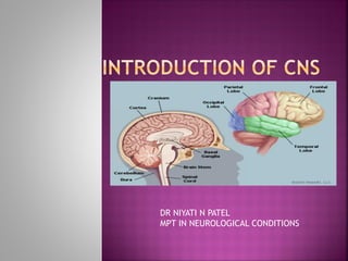
  • 1. DR NIYATI N PATEL MPT IN NEUROLOGICAL CONDITIONS
  • 2.
  • 3.
  • 4.  Neuron or nerve cell is defined as the structural and functional unit of nervous system.  It is different from other cells by two ways:  1.Neuron has branches or processes called axon and dendrites  2. Neuron does not have centrosome. So, it cannot undergo division.
  • 5.  Neurons are classified by three different methods.  A. Depending upon the number of poles  B. Depending upon the function  C. Depending upon the length of axon.
  • 6. DEPENDING UPON THE NUMBER OF POLES 1. Unipolar Neurons  Unipolar neurons are the neurons that have only one pole. From a single pole, both axon and dendrite arise  This type of nerve cells is present only in embryonic stage in human beings. 2. Bipolar Neurons  Neurons with two poles are known as bipolar neurons.  Axon arises from one pole and dendrites arise from the other pole. 3. Multipolar Neurons  Multipolar neurons are the neurons which have many poles.  One of the poles gives rise to axon and all other poles give rise to dendrites.
  • 7.
  • 8. DEPENDING UPON THE FUNCTION  On the basis of function, nerve cells are classified into two types:  1. Motor or efferent neurons  2. Sensory or afferent neurons.  1. Motor or Efferent Neurons  Motor or efferent neurons are the neurons which carry the motor impulses from central nervous system to peripheral effector organs like muscles, glands, blood vessels, etc.  Generally, each motor neuron has a long axon and short dendrites.  2. Sensory or Afferent Neurons  Sensory or afferent neurons are the neurons which carry the sensory impulses from periphery to central nervous system.  Generally, each sensory neuron has a short axon and long dendrites.
  • 9. DEPENDING UPON THE LENGTH OF AXON  Depending upon the length of axon, neurons are divided into two types:  1. Golgi type I neurons  2. Golgi type II neurons.  1. Golgi Type I Neurons  Golgi type I neurons have long axons.  Cell body of these neurons is in different parts of central nervous system and their axons reach the remote peripheral organs.  2. Golgi Type II Neurons  Neurons of this type have short axons.  These neurons are present in cerebral cortex and spinal cord.
  • 10.  Neuron is made up of three parts:  1. Nerve cell body  2. Dendrite  3. Axon  Dendrite and axon form the processes of neuron  Dendrites are short processes and the axons are long processes.  Dendrites and axons are usually called nerve fibers.
  • 11.  NERVE CELL BODY  Nucleus  Nissl Bodies (Ribosomes)  Neurofibrils  Mitochondria  Golgi Apparatus  DENDRITE (Branched process)  AXON (Longer process)  Non myelinated nerve fiber  Myelinated nerve fiber
  • 12. 1. Depending upon structure 2. Depending upon distribution 3. Depending upon origin 4. Depending upon function 5. Depending upon secretion of neurotransmitter 6. Depending upon diameter and conduction of impulse (ErlangerGasser classification)
  • 13.  DEPENDING UPON STRUCTURE  Myelinated Nerve Fibers  Myelinated nerve fibers are the nerve fibers that are covered by myelin sheath.  Non-myelinated Nerve Fibers  Nonmyelinated nerve fibers are the nerve fibers which are not covered by myelin sheath.
  • 14.  MYELIN SHEATH  Myelin sheath is a thick lipoprotein sheath that insulates the myelinated nerve fiber.  Myelin sheath is not a continuous sheath.  It is absent at regular intervals.  The area where myelin sheath is absent is called node of Ranvier.  Segment of the nerve fiber between two nodes is called internode.  Myelin sheath is responsible for white color of nerve fibers.
  • 15.  DEPENDING UPON DISTRIBUTION  Somatic Nerve Fibers  Somatic nerve fibers supply the skeletal muscles of the body.  Visceral or Autonomic Nerve Fibers  Autonomic nerve fibers supply the various internal organs of the body.
  • 16.  DEPENDING UPON ORIGIN  Cranial Nerve Fibers  Nerve fibers arising from brain are called cranial nerve fibers.  Spinal Nerve Fibers  Nerve fibers arising from spinal cord are called spinal nerve fibers.
  • 17.  DEPENDING UPON FUNCTION  Sensory Nerve Fibers (afferent nerve Fibers)  Sensory nerve fibers carry sensory impulses from different parts of the body to the central nervous system  Motor Nerve Fibers (efferent nerve fibers)  Motor nerve fibers carry motor impulses from central nervous system to different parts of the body.
  • 18.  DEPENDING UPON SECRETION OF NEUROTRANSMITTER  Adrenergic Nerve Fibers  Adrenergic nerve fibers secrete nor adrenaline.  Cholinergic Nerve Fibers  Cholinergic nerve fibers secrete acetylcholine.
  • 19.  DEPENDING UPON DIAMETER AND CONDUCTION OF IMPULSE (ERLANGER- GASSER CLASSIFICATION) Type Diameter (u) Velocity of conduction (m/sec) A Alpha 12 To 24 70 to 120 A Beta 6 to 12 30 to 70 A Gamma 5 to 6 15 to 30 A Delta 2 to 5 12 to 15 B 1 to 2 3 to 10 C < 1.5 0.5 to 2
  • 20.  Excitability  Conductivity  Refractory Period  Summation  Adaptation  Infatigability  All Or None Law
  • 21.  Defined as the physiochemical change that occurs in a tissue when stimulus is applied  Response Due to Stimulation of Nerve Fiber  Action potential or nerve impulse (Propagated)  Electrotonic potential or local potential (Non Propagated)
  • 22. ACTION POTENTIAL  Action potential is defined as a series of electrical changes that occur in the membrane potential when the muscle or nerve is stimulated.  Action potential occurs in three phases:  1. Latent Period - Latent period is the period when no change occurs in the electrical potential immediately after applying the stimulus. It is a very short period with duration of 0.5 to 1 millisecond.  2. Depolarization - action potential in which inside of the muscle becomes positive and outside becomes negative.  3. Repolarization - after depolarization the inside of muscle becomes negative and outside becomes positive. So, the polarized state of the muscle is re established.
  • 23. Action potential in a skeletal muscle A = Opening of few Na+ channels B = Opening of many Na+ channels C = Closure of Na+ channels and opening of K+ channels D = Closure of K+ channels
  • 24.  Types of action potential  Monophasic Action potential  Biphasic Action potential  Compound Action potential
  • 25. Properties of action potential and graded potential Action potential Graded potential/Electronic potential •Propagative •Long distance signal •Both depolarization & repolarization •Obeys all or none law •Summation is not possible •Has refractory period •Non propagative •Short distance signal •Only depolarization/ Hyperpolarization •Does not obey all or none law •Summation is possible •No refractory period
  • 26.  Conductivity is the ability of nerve fibers to transmit the impulse from the area of stimulation to the other areas.
  • 27.  Refractory period is the period at which the nerve does not give any response to a stimulus.  TWO TYPES OF REFRACTORY PERIODS 1. Absolute Refractory Period 1. Absolute refractory period is the period during which the nerve does not show any response at all, whatever may be the strength of stimulus. 2. 2. Relative Refractory Period 1. It is the period, during which the nerve fiber shows response, if the strength of stimulus is increased to maximum.
  • 28.  When one subliminal stimulus is applied, it does not produce any response in the nerve fiber because, the subliminal stimulus is very weak.  However, if two or more subliminal stimuli are applied within a short interval of about 0.5 millisecond, the response is produced.  It is because the subliminal stimuli are summed up together to become strong enough to produce the response.  This phenomenon is known as summation.
  • 29.  While stimulating a nerve fiber continuously, the excitability of the nerve fiber is greater in the beginning.  Later the response decreases slowly and finally the nerve fiber does not show any response at all.  This phenomenon is known as adaptation or accommodation.  Cause for Adaptation  When a nerve fiber is stimulated continuously, depolarization occurs continuously.  Continuous de polarization inactivates the sodium pump and increases the efflux of potassium ions.
  • 30.  Nerve fiber cannot be fatigued, even if it is stimulated continuously for a long time.  The reason is that nerve fiber can conduct only one action potential at a time.  At that time, it is completely refractory and does not conduct another action potential.
  • 31.  All-or-none law states that when a nerve is stimulated by a stimulus it gives maximum response or does not give response at all.
  • 32.  When a nerve fiber is injured, various changes occur in the nerve fiber and nerve cell body. All these changes are together called the degenerative changes.  The term regeneration refers to regrowth of lost or destroyed part of a tissue. The injured and degenerated nerve fiber can regenerate. It starts as early as 4th day after injury, but becomes more effective only after 30 days and is completed in about 80 days.  Causes for Injury  1. Obstruction of blood flow  2. Local injection of toxic substances  3. Crushing of nerve fiber  4. Transection of nerve fiber.
  • 33.
  • 34.  Neuroglia or glia (glia = glue) is the supporting cell of the nervous system.  Neuroglial cells are non-excitable. CENTRAL NEUROGLIAL CELLS PERIPHERAL NEUROGLIAL CELLS ASTROCYTES (STAR SHAPE & PRESENT IN BRAIN) „MICROGLIA (PHAGOCYTIC CELL & MACROPHAGES OF CNS) „OLIGODENDROCYTES (MYELIN SHEATH IN CNS) SCHWANN CELLS (MYELIN SHEATH IN PNS) „SATELLITE CELLS (EXTERIOR SURFACE OF PNS)
  • 35.  Receptors are sensory (afferent) nerve endings that terminate in periphery as bare unmyelinated endings or in the form of specialized capsulated structures.  Classification of receptors Exteroceptors (outside of the body) Interoceptors (within body) Mechanoreceptor/cutenous receptors (skin area) Chemoreceptors (chemical stimuli) Telereceptors (distance receptor) Visceroceptors (viscera) Proprioceptors (joint position)
  • 36.
  • 38.
  • 39.  PROPERTIES OF RECEPTORS  SPECIFICITY OF RESPONSE –MÜLLER LAW  ADAPTATION – SENSORY ADAPTATION  RESPONSE TO INCREASE IN STRENGTH OF STIMULUS – WEBERFECHNER LAW  SENSORY TRANSDUCTION  RECEPTOR POTENTIAL  LAW OF PROJECTION
  • 40.  Synapse is the junction between two neurons.  It is not an anatomical continuation But, it is only a physiological continuity between two nerve cells.  CLASSIFICATION OF SYNAPSE Anatomical classification (depends on ending of axon) Physiological classification (depends on mode of impulse transmission) Axoaxonic synapse Axodendritic synapse Axosomatic synapse Electrical synapse Chemical synapse
  • 42.
  • 43.  Presynaptic  Mitochondria, which help in the synthesis of neurotransmitter substance  Synaptic vesicles, which store neurotransmitter substance.  Synaptic cleft & Post synaptic  Postsynaptic neuron is called postsynaptic membrane.  It contains some receptor proteins.  Small space in between the presynaptic membrane and the postsynaptic membrane is called synaptic cleft.
  • 44.  FUNCTIONS OF SYNAPSE  Main function of the synapse is to transmit the impulses, i.e. action potential from one neuron to another
  • 47.  PROPERTIES OF SYNAPSE  ONE WAY CONDUCTION – BELL-MAGENDIE LAW  According to BellMagendie law, the impulses are transmitted only in one direction in synapse, i.e. from presynaptic neuron to postsynaptic neuron.  SYNAPTIC DELAY  Release of neurotransmitter  Passage of neurotransmitter from axon terminal to postsynaptic membrane  Action of the neurotransmitter to open the ionic channels in postsynaptic membrane.
  • 48.  FATIGUE  Fatigue at synapse is due to the depletion of neurotransmitter substance, acetylcholine.  Depletion of acetylcholine occurs because of two factors:  i. Soon after the action, acetylcholine is destroyed by acetylcholinesterase  ii. Due to continuous action, new acetylcholine is not synthesize
  • 49.  SUMMATION  Summation is the fusion of effects or progressive increase when single presynaptic terminal is stimulated repeatedly.
  • 50. CONVERGENCE Convergence is the process by which many presynaptic neurons terminate on a single postsynaptic neuron „DIVERGENCE Divergence is the process by which one presynaptic neuron terminates on many postsynaptic neurons.
  • 51.  Reflex activity is the response to a peripheral nervous stimulation that occurs without our consciousness.  It is a type of protective mechanism and it protects the body from irreparable damages.
  • 52.  REFLEX ARC  Reflex arc is the anatomical nervous pathway for a reflex action. A simple reflex arc includes five components
  • 53.  CLASSIFICATION OF REFLEXES 1. Depending upon whether inborn or acquired 2. Depending upon situation – anatomical classification  Cortical, Cerebellum, Midbrain, Bulbar/Medullary & Spinal Reflexes 3. Depending upon purpose – physiological Classification  Protective/flexor & antigravity/extensor 4. Depending upon number of synapse  Monosynaptic & polysynaptic 5. Depending upon whether visceral or somatic 6. Depending upon clinical basis  Deep reflex & superficial reflex
  • 54. Deep tendon reflex Superficial mucous membrane reflexes Superficial cutaneous reflexes Pathological reflexes Jaw jerk Corneal reflex Scapular reflex Babinski sign Biceps jerk Conjunctival reflex Upper abdominal reflex Clonus Triceps jerk Nasal reflex (sneezing reflex) Lower abdominal reflex Pendular movements Supinator jerk or radial periosteal reflex Pharyngeal reflex Cremasteric reflex Wrist tendon or finger flexion reflex Uvular reflex Gluteal reflex Knee jerk or patellar tendon reflex Plantar reflex Ankle jerk or Achilles tendon reflex Bulbocavernous reflex Anal reflex
  • 55.  REFLEXES IN MOTOR NEURON LESION 1. UPPER MOTOR NEURON LESION  During upper motor neuron lesion, all the superficial reflexes are lost. Deep reflexes are exaggerated and the Babinski sign is positive. 2. LOWER MOTOR NEURON LESION  During lower motor lesion, all the superficial and deep reflexes are lost.
  • 56.
  • 57.  Situation and Extent  Vertebral canal  It extends from foramen magnum where it is continuous with medulla oblongata, above and up to the lower border of first lumbar vertebra  Coverings  Spinal cord is covered by sheaths called meninges, which are membranous in nature.  Meninges are pia mater, arachnoid mater & dura mater.  These coverings continue as coverings of brain.  Meninges are responsible for protection and nourishment of the nervous tissues.
  • 58.  Shape and Length  Shape- cylindrical  Length - 45 cm in males and about 43 cm in females.  Enlargements  Cervical enlargements – upper extremities  lumbar enlargements - lower extremities  Conus Medullaris and Filum Terminale  Below the lumbar enlargement, spinal cord rapidly narrows to a cone-shaped termination called conus medullaris.  A slender non-nervous filament called filum terminale extends from conus medullaris downward to the fundus of the dural sac at the level of second sacralvertebra.
  • 59. 1. Cervical segments/Cervical spinal nerves = 8 2. Thoracic segments/Thoracic spinal nerves = 12 3. Lumbar segments/Lumbar spinal nerves = 5 4. Sacral segments/Sacral spinal nerves = 5 5. Coccygeal segment/Coccygeal spinal nerves = 1  Total = 31
  • 60.
  • 61.  Neurons in Grey matter  Golgi type 1 (anterior aspect, long axons-tracts)  Golgi type 2 (posterior aspect, short axons- interneurons)  Contents of grey matter  Nuclie  Alpha motor neurons  Gamma motor neurons  Renshaw cells  Lamina
  • 62.  White matter  Anterior or Ventral White Column  Lateral White Column  posterior or dorsal White Column  Tracts  Shorts tracts  Long tracts  Ascending tracts  Descending tracts
  • 63.  ASCENDING TRACTS OF SPINAL CORD  First Order Neurons  Receptors to sensory neurons present in the posterior gray horn of spinal cord  Nerve cell bodies - located in the posterior nerve root ganglion.  Second Order Neurons  Posterior gray horn to ascending tracts of spinal cord.  These fibers carry sensory impulses from spinal cord to different brain areas below cerebral cortex (subcortical areas) such as thalamus.  Third Order Neurons  Subcortical areas  Fibers of these neurons carry the sensory impulses from subcortical areas to cerebral cortex.
  • 64.  Ascending tracts White column Tract Anterior white column 1.Anterior spinothalamic tract Lateral white column 1. Lateral spinothalamic tract 2. Ventral spinocerebellar tract 3. Dorsal spinocerebellar tract 4. Spinotectal tract 5. Fasiculus dorsolateralis 6. Spinoreticular tract 7. Spino-olivary tract 8. Spinovestibular tract Posterior white column 1. Fasciculus gracilis 2. Fasciculus cuneatus
  • 65.  Tract : Anterior spinothalamic tract  Origin: Chief sensory nucleus  Course: Crossing in spinal cord Forms spinal lemniscus  Termination: Ventral posterolateral nucleus of thalamus  Function: Crude touch sensation  Tract : Lateral spinothalamic tract  Origin: Substantia gelatinosa  Course: Crossing in spinal cord Forms spinal lemniscus  Termination: Ventral posterolateral nucleus of thalamus  Function: Pain and temperature sensation
  • 67.  Tract : Ventral spinocerebellar tract  Origin: Marginal nucleus  Course: Crossing in spinal cord  Termination: Anterior lobe of cerebellum  Function: Subconscious kinesthetic sensations Tract : Dorsal spinocerebellar tract Origin: Clarke nucleus  Course: Uncrossed fibers  Termination: Anterior lobe of cerebellum  Function: Subconscious kinesthetic sensations
  • 68.
  • 69.  Tract : Spinotectal tract  Origin: Chief sensory nucleus  Course: Crossing in spinal cord  Termination: Superior colliculus  Function: Spinovisual reflex Tract : Fasiculus dorsolateralis  Origin: Posterior nerve root ganglion  Course: Component of lateral spinothalamic tract  Termination: Substantia gelatinosa  Function: Pain and temperature sensations
  • 70.  Tract :Spinoreticular tract  Origin: Inter mediolateral cells  Course: Crossed and uncrossed fibers  Termination: Reticular formation of brainstem  Function:Consciousn ess andawareness  Tract : Spino-olivary tract  Origin: Non-specific  Course: Uncrossed fibers  Termination: Olivary nucleus  Function:Proprioce ption
  • 71.  Tract : Spinovestibular tract  Origin:  Course: Non-specific Crossed and uncrossed fibers  Termination: Lateral vestibular nucleus  Function: Proprioception
  • 72.  Tract : Fasciculus cuneatus  Origin: Posterior nerve root ganglia  Course: Uncrossed fibers No synapse in spinal cord  Termination: Nucleus cuneatus in medulla  Function: Tactile sensation Tactile localization Tactile discrimination Vibratory sensation Conscious kinesthetic sensation Stereognosis  Tract : Fasciculus gracilis  Origin: Posterior nerve root Ganglia  Course: Uncrossed fibers No synapse in spinal cord  Termination: Nucleus gracilis in medulla  Function: Tactile sensation Tactile localization Tactile discrimination Vibratory sensation Conscious kinesthetic sensation Stereognosis
  • 73.
  • 74.  Descending tracts Types Tracts Pyramidal tracts 1.Anterior corticospinal tract 2.Lateral corticospinal tract Extrapyramidal tracts 1. Medial longitudinal fasciculus 2. Anterior vestibulospinal tract 3. Lateral vestibulospinal tract 4. Reticulospinal tract 5. Tectospinal tract 6. Rubrospinal tract 7. Olivospinal tract
  • 75.  Tract : Anterior corticospinal tract  Situation: Anterior white column  Origin: Betz cells and other cells of motor area  Course: Uncrossed fibers  Function: i. Control of voluntary movements ii. Form upper motor neurons  Tract : lateral corticospinal tract  Situation : Lateral white column  Origin: Betz cells and other cells of motor area  Course: Crossed fibers  Function: i. Control of voluntary movements ii. Form upper motor neurons
  • 76.
  • 77.  Tract : Anterior vestibulospinal tract  Situation: Anterior white column  Origin: Medial vestibular nucleus  Course: Uncrossed fibers Extend up to upper thoracic segments  Function: i. Maintenance of muscle tone and posture ii. Maintenance of position of head and body during acceleration Tract : Lateral vestibulospinal tract  Situation: Lateral white column  Origin: Lateral vestibular nucleus  Course: Mostly uncrossed Extend to all segments  Function: i. Maintenance of muscle tone and posture ii. Maintenance of position of head and body during acceleration
  • 78.  Tract : Reticulospinal tract  Situation: Lateral white fasciculus  Origin: Reticular formation of pons and medulla  Course: Mostly uncrossed Extend up to thoracic segments  Function:  i. Coordination of voluntary and reflex movements  ii. Control of muscle tone  iii.Control of respiration and diameter of blood vessels  Tract : Tectospinal tract  Situation: Anterior white column  Origin: Superior colliculus  Course: Crossed fibers Extend up to lower cervical segments  Function: Control of movement of head in response to visual and auditory impulses
  • 79.  Tract : Rubrospinal tract  Situation: Lateral white column  Origin: Red nucleus  Course: Crossed fibers Extend up to thoracic segments  Function: Facilitatory influence on flexor muscle tone  Tract : Olivospinal tract  Situation: Lateral white column  Origin: Inferior olivary nucleus  Course: Mostly crossed Extent – not clear  Function: Control of movements due to proprioception
  • 80. COMPLETE TRANSECTION OF SPINAL CORD  Complete transection of spinal cord occurs due to:  1. Bullet injury, which causes dislocation of spinal cord  2. Accidents, which cause dislocation of spinal cord or occlusion of blood vessels.  Complete transection causes immediate loss of sensation and voluntary movement below the level of lesion.
  • 81.  Effects occur in three stages:  1. Stage of spinal shock  2. Stage of reflex activity  3. Stage of reflex failure
  • 82.  1. Stage of Spinal Shock  Stage of spinal shock is the first stage of effects that occurs immediately after injury. It is also called stage of flaccidity. 1. Paralysis of limbs 1. Cervical injury – quadriplegia/tetraplegia 2. Thoracic/lumbar injury- paraplegia 2. Flaccid paralysis 3. Loss of reflexes 4. Loss of sensations 5. Effect on visceral organs 6. Heart rate 7. Venous return 8. Effect on blood pressure
  • 83. 2. Stage of Reflex Activity  Stage of reflex activity is also called stage of recovery. After 3 weeks period, depending largely upon the general health of the patient  functional activities return to smooth muscles  Next, the sympathetic tone to blood vessels returns.  Lastly, after another 3 months, the tone in skeletal muscle returns  Flexor reflexes appear first. To elicit the flexor reflex, a painful stimulus is required. First reflex, which usually appears, is the Babinski reflex.  After a variable period of 1 to 5 weeks of reappearance of flexor reflexes, the extensor reflexes return.  This reaction constitutes the spasm in flexor muscles of both the lower limbs, evacuation of urinary bladder and profuse sweating. This is known as the mass reflex.
  • 84.  Stage of Reflex Failure  Though the reflex movements return, muscles below the level of injury have less power and less resistance.  deterioration of condition with infection
  • 85.  Brainstem is the part of brain formed by medulla oblongata,pons and midbrain.  Brainstem contains ascending and descending tracts between brain and spinal cord.  It also contains many centers for regulation of vital functions in the body.
  • 86.  Medulla oblongata or medulla is the lowermost part of brain.  It is situated below pons and is continued downwards as spinal cord.  Medulla forms the main pathway for ascending and descending tracts of the spinal cord.
  • 87.  1. Respiratory Centers  Dorsal and ventral group of neurons form the medullary respiratory centers, which maintain normal rhythmic respiration  2. Vasomotor Center  Vasomotor center controls blood pressure and heart rate.  3. Deglutition Center  Deglutition center regulates the pharyngeal and esophageal stages of deglutition
  • 88.  4. Vomiting Center  Vomiting center induces vomiting during irritation or inflammation of gastrointestinal (GI) tract  5. Superior and Inferior Salivatory Nuclei  Salivatory nuclei control the secretion of saliva  6. Cranial Nerve Nuclei  Nuclei of 10th, 11th, 12th and some nuclei of 5th and 8th cranial nerves are located in the medulla oblongata
  • 89.  7. Vestibular Nuclei  Vestibular nuclei contain the second order neurons of vestibular nerve  Superior, medial, lateral and inferior vestibular nuclei.  Medial and inferior vestibular nuclei extend into medulla.
  • 90.  Pons forms a bridge between medulla and midbrain
  • 91.  Axons of pontine nuclei join to the middle cerebellar peduncle & pons pathway that connects cerebellum with cerebral cortex.  Pyramidal tracts pass through the pons  Medial lemniscus is joined by the fibers of 5th, 7th, 9th and 10th cranial nerves in pons  Nuclei of 5th, 6th, 7th and 8th cranial nerves are located in pons  Pons contains the pneumotaxic and apneustic centers for regulation of respiration
  • 92.  Midbrain lies between pons and diencephalon. Midbrain Tectum Cerebral peduncles Superior colliculus : 1. Small structure & imp center for reflex 2. Tectospinal tract, superior colliculus controls the movements of the eyes, head, trunk and limbs, in response to visual impulses Basis pedunculus: Laterally temperopontine fibres Medially frontopontine fibers Middle pyramidal tract fibers Inferior colliculus: The center for auditory reflexes , produces reflex vocalization Substantia nigra: Situated below the red nucleus. Tegmentum: Dorsal to substantia nigra, upward to the reticular formation Superior cerebellar peduncle, Crossing of rubrospinal tracts Efferent fibers of 3rd, 4th and 6th Cranial nerves.
  • 93.  Red nucleus is a large oval or round mass of gray matter, extending between the superior colliculus and hypothalamus.  Two parts  Nucleus magnocellularis (larger)  This fibers form the rubrospinal and rubrobulbar tracts  Nucleus parvocellularis (smaller)  This fibers form the rubroreticular tract
  • 94.  Afferent connection  Corticorubral fibers  Pallidorubral fibers  Cerebellorubral tracts  Efferent connections  Rubrospinal tract to spinal cord  Rubrobulbar tract to medulla  Rubroreticular fibers to reticular formation  Rubrothalamic tract to lateral ventral nucleus of thalamus  Rubroolivary tract to inferior olivary nucleus  Fibers to nuclei of 3rd, 4th and 6th cranial nerves.
  • 95.  Control of muscle tone  Control of complex muscular movements  Control of righting reflexes  Control of movements of eyeball  Control of skilled movements
  • 96.  Cerebellum consists of a narrow, worm-like central body called vermis and two lateral lobes, the right and left cerebellar hemispheres
  • 97.  VERMIS : Superior vermis  1. Lingula  2. Central lobe  3. Culmen  4. Lobulus simplex  5. Declive Inferior vermis  6. Tuber  7. Pyramid  8. Uvula  9. Nodulus + flocculi = floculonodular lobe
  • 98.  CEREBELLAR HEMISPHERES  1. Lobulus ansiformis or ansiform lobe - larger portion of cerebellar hemisphere  2. Lobulus paramedianus or paramedian lobe - smaller portion of cerebellar hemisphere.
  • 99.  A. Anatomical divisions  B. Phylogenetic divisions  C. Physiological or functional divisions
  • 100.  ANATOMICAL DIVISIONS  1. Anterior Lobe  Anterior lobe includes lingula, central lobe and culmen..  2. Posterior Lobe  Posterior lobe consists of lobulus simplex, declive, tuber, pyramid, uvula, paraflocculi and the two portions of hemispheres, viz. ansiform lobe and paramedian lobe.  3. Flocculonodular Lobe  Flocculonodular lobe includes nodulus and the lateral extension on either side called flocculus.
  • 101.  PHYLOGENETIC DIVISIONS  1. Paleocerebellum:  i. Archicerebellum, which includes flocculonodular lobe  ii. Paleocerebellum proper, which includes lingula, central lobe, culmen, lobulus simplex, pyramid, uvula and paraflocculi.  2. Neocerebellum  It includes declive, tuber and the two portions of cerebellar hemispheres, viz. lobulus ansiformis and lobulus paramedianus.
  • 102.  PHYSIOLOGICAL OR FUNCTIONAL DIVISIONS  1. Vestibulocerebellum  Vestibulocerebellum includes flocculonodular lobe that forms the archicerebellum.  2. Spinocerebellum  Spinocerebellum includes lingula, central lobe, culmen, lobulus simplex, declive, tuber, pyramid, uvula and paraflocculi and medial portions of lobulus ansiformis and lobulus paramedianus.  3. Corticocerebellum  Corticocerebellum includes lateral portions of lobulus ansiformis and lobulus paramedianus.
  • 103.  GRAY MATTER  1. Outer molecular (Parallel, climbing, Dendrites of Purkinje cells and Golgi cells)  2. Intermediate Purkinje layer  3. Inner granular layer (granule cells and Golgi cells)
  • 104.  CEREBELLAR NUCLEI  Fastigial Nucleus- midline on the roof of IV ventricle.  Globosus Nucleus- lateral to nucleus fastigi  Emboliform Nucleus- below the nucleus fastigi and nucleus globosus  Dentate Nucleus- lateral to all the other nuclei
  • 105.  WHITE MATTER OF CEREBELLUM  Superior cerebellar peduncles between cerebellum and midbrain  Middle cerebellar peduncles between cerebellum and pons  Inferior cerebellar peduncles between cerebellum and medulla oblongata
  • 106.  VESTIBULOCEREBELLUM  Vestibulocerebellum is connected with the vestibular apparatus and so it is known as vestibulocerebellum  STRUCTURES  Includes the flocculonodular lobe that is formed by the nodulus of vermis and its lateral extensions called flocculi  FUNCTIONS  Tone, posture and equilibrium
  • 107.  SPINOCEREBELLUM  Spinocerebellum is connected with spinal cord  COMPONENTS  Medial Portions Of Cerebellar Hemisphere, Paraflocculi And The Parts Of Vermis, Viz. Lingula, Central Lobe, Culmen, Lobulus Simplex, Declive, Tuber, Pyramid And Uvula  FUNCTIONS  Tone, posture and equilibrium  Localization of tactile
  • 108.  CORTICOCEREBELLUM  Corticocerebellum is the largest part of cerebellum, Because of its connection with cerebral cortex  COMPONENTS  Corticocerebellum includes the lateral portions of cerebellar hemispheres  „FUNCTIONS  Integration and regulation of well-coordinated muscular activities
  • 109.
  • 110.  Basal ganglia are the scattered masses of gray matter submerged in subcortical substance of cerebral hemisphere  Basal ganglia form the part of extra pyramidal system, which is concerned with motor activities.
  • 111.  1. Corpus striatum  2. Substantia nigra  3. Subthalamic nucleus
  • 112.  1. Corpus striatum  Mass of gray matter situated at the base of cerebral hemispheres in close relation to thalamus Caudate Nucleus : Caudate nucleus is an elongated arched gray mass, lying medial to internal capsule.  Caudate nucleus has a head portion and a tail portion Lenticular Nucleus: Lenticular nucleus is a wedgeshaped gray mass, situated lateral to internal capsule. A vertical plate of white matter called external medullary lamina  Outer putamen  Inner globus pallidus
  • 113.  SUBSTANTIA NIGRA  Substantia nigra is situated below red nucleus.  It is made up of large pigmented and small nonpigmented cells.  The pigment contains high quantity of iron.  SUBTHALAMIC NUCLEUS OF LUYS  Subthalamic nucleus is situated lateral to red nucleus and dorsal to substantia nigra.
  • 114.  1. Putamen to globus pallidus  2. Caudate nucleus to globus pallidus  3. Caudate nucleus to putamen.
  • 115. Component Afferent connections from Efferent connections to Corpus striatum 1. Thalamic nuclei to caudate nucleus and putamen 2. Cerebral cortex to caudate nucleus and putamen 3. Substantia nigra to putamen 4. Subthalamic nucleus to globus pallidus 1. Thalamic nuclei 2. Subthalamic nucleus 3. Red nucleus 4. Substantia nigra 5. Hypothalamus 6. Reticular formation (Most of the Substantia nigra 1. Putamen 2. Frontal lobe of cerebral cortex 3. Superior colliculus 4. Mamillary body of hypothalamus 5. Medial and Putamen Subthalamic nucleus Globus pallidus 1. Globus pallidus 2. Red nucleus
  • 116. Afferent connections of corpus striatum Efferent connections of corpus striatum
  • 117.  1. CONTROL OF MUSCLE TONE  Gamma motor neurons of spinal cord are responsible for development of tone in the muscles  Decrease the muscle tone by inhibiting gamma motor neurons through descending inhibitory reticular system in brainstem  During the lesion of basal ganglia, muscle tone increases leading to rigidity
  • 118.  2. CONTROL OF MOTOR ACTIVITY  Regulation of Voluntary Movements  a. Premotor area  b. Primary motor area  c. Supplementary motor area  Regulation of Conscious Movements  Fibers between cerebral cortex and caudate nucleus are concerned with regulation of conscious movements (cognitive control)  Regulation of Subconscious Movements  Cortical fibers reaching putamen  regulation of some subconscious movement
  • 119.  3. CONTROL OF REFLEX MUSCULAR ACTIVITY  Visual and labyrinthine reflexes are important in maintaining the posture  4. CONTROL OF AUTOMATIC ASSOCIATED MOVEMENTS  Automatic associated movements are the movements in the body (motor activities)  5. ROLE IN AROUSAL MECHANISM  Globus pallidus and red nucleus are involved in arousal mechanism because of their connections with reticular formation
  • 120.  6. ROLE OF NEUROTRANSMITTERS IN THE FUNCTIONS OF BASAL GANGLIA  Dopamine released by dopaminergic fibers from substantia nigra to corpus striatum  Gamma aminobutyric acid (GABA) secreted by intrinsic fibers of substantia nigra to corpus striatum  Acetylcholine released by fibers from cerebral cortex to caudate nucleus and putamen  Substance P & Enkephalins released by fibers from globus pallidus reaching substantia nigra  Noradrenaline secreted by fibers between basal ganglia and reticular formation  Glutamic acid secreted by fibers from subthalamic nucleus to globus pallidus and substantia nigra
  • 121.  APPLIED PHYSIOLOGY – DISORDERS OF BASAL GANGLIA  PARKINSON DISEASE  WILSON DISEASE  CHOREA  ATHETOSIS  CHOREOATHETOSIS  HUNTINGTON CHOREA  HEMIBALLISMUS  KERNICTERUS
  • 122.  Cerebral cortex is also called pallidum and it consists of two hemispheres  Both the cerebral hemispheres are separated by a deep vertical fissure  Separation is complete anteriorly and posteriorly  Middle portion, the fissure extends only up to corpusm callosum (connecting the two hemispheres)  Surface of the cerebral cortex is characterized by  Sulci - Slight Depression  Gyri - Raised Ridge
  • 123.  HISTOLOGY OF CEREBRAL CORTEX  LAYERS OF CEREBRAL CORTEX  Gray matter  White matter  Molecular or Plexiform Layer - dendrites or axons from cells of deeper layers  External Granular Layer – Dendrites & Axons end (axons enter white substance of the hemisphere)  Outer Pyramidal Layer  Medium pyramidal cells - outer portion  larger pyramidal cells - deeper portion  Internal Granular Layer – horizontal fibers, which appear as a white strip known as outer strip  Ganglionic Layer or Internal Pyramidal Layer - Pyramidal cells in this region are otherwise known as Betz cells or giant cells  Fusiform Cell Layer - white matter of cerebral hemisphere
  • 124.  LOBES OF CEREBRAL CORTEX  Four lobes  1. Frontal lobe  2. Parietal lobe  3. Occipital lobe  4. Temporal lobe  Four main fissures and sulci  Central sulcus (frontal and parietal lobes)  Parieto-occipital sulcus (parietal and occipital lobe)  lateral sulcus (parietal and temporal lobes)  Callosomarginal fissure (temporal lobe and limbic area)
  • 125.
  • 126.  CEREBRAL DOMINANCE  Cerebral dominance is defined as the dominance of one cerebral hemisphere over the other in the control of cerebral functions  Right handed – left hemisphere (representational hemisphere)  Left handed – right hemisphere (representational hemisphere)
  • 127.  Frontal lobe of cerebral cortex is divided into two parts:  A.Precentral cortex, which is situated posteriorly  B.Prefrontal cortex, which is situated anteriorly  PRECENTRAL CORTEX  Includes  Central sulcus  Precentral gyrus  Posterior portions of superior, middle and inferior frontal gyri
  • 128.  Precentral cortex is further divided into three functional areas:  1. Primary motor area  2. Premotor area  3. Supplementary motor area.
  • 129.  Primary Motor Area (giant pyramidal cells)  Functions of primary motor area - initiation of voluntary movements and speech  Area 4  It activates both α-motor neurons and γ-motor neurons simultaneously by the process called coactivation  Effect of lesion of area 4  The ability to walk is not affected  Hemiplegia with spastic paralysis.  (Hemiplegia means the paralysis in one half of the body)
  • 130.  Premotor Area (6, 8, 44 and 45)  Area 6 - Posterior portions of superior, middle and inferior frontal gyri  Functions of area 6 - coordination of movements & cortical center for extrapyramidal system  Area 8 - frontal eye field  Function of area 8 - Frontal eye field is concerned with conjugate movement of eyeballs  Area 44 & 45 (Broca area) - motor area for speech- dominant hemisphere  Function of Broca area - movements of tongue, lips and larynx, which are involved in speech
  • 131.  Supplementary Motor Area  Medial surface of frontal lobe rostral to primary motor area  Motor movements are elicited by electrical stimulation of this area like raising the contralateral arm, turning the head and eye and movements of synergistic muscles of trunk and legs  Function of supplementary motor area - coordinated skilled movements
  • 132.
  • 133.
  • 134.  Prefrontal cortex is the anterior part of frontal lobe of cerebral cortex  Areas present in prefrontal cortex are 9, 10, 11, 12, 13, 14, 23, 24, 29 and 32.  Medial Surface Areas -12, 13, 14, 23, 24, 29 & 32  Lateral Surface Areas - 9, 10 & 11  Functions of Prefrontal Cortex  The higher functions like emotion, learning, memory , social behavior, short-term memories, intelligence & personality
  • 135.  APPLIED PHYSIOLOGY – FRONTAL LOBE SYNDROME  Emotional instability  Lack of concentration  Impairment of recent memory  Apart from mental defects, there are some functional abnormalities also:  i. Hyperphagia (increased food intake)  ii. Loss of control over sphincter of the urinary bladder or rectum  iii. Disturbances in orientation  iv. Slight tremor
  • 136.  Parietal lobe is divided into three functional areas:  A. Somesthetic area I  B. Somesthetic area II  C. Somesthetic association area
  • 137.  SOMESTHETIC AREA I (SOMATOSENSORY AREA I OR PRIMARY SOMESTHETIC OR PRIMARY SENSORY AREA)  Areas of Somesthetic Area I (3,1 & 2)- Anterior part – 3, Posterior part – 1 & 2  Functions of Somesthetic Area I  Responsible for perception and integration of cutaneous and kinesthetic sensations  Sensory feedback to the premotor area  The movements of head and eyeballs
  • 138.  SOMESTHETIC AREA II  Functions of Somesthetic Area II - perception of sensation  SOMESTHETIC ASSOCIATION AREA (area 5 & 7)  Functions of Somesthetic Association Area - sensations like stereognosis
  • 139.  APPLIED PHYSIOLOGY – PARIETAL LOBE  1. Contralateral disturbance of cutaneous sensations  2. Disturbances in kinesthetic sensations  3. Loss of tactile localization and discrimination
  • 140.  Temporal lobe of cerebral cortex includes three functional areas  A. Primary auditory area  B. Secondary auditory area or auditopsychic area  C. Area for equilibrium
  • 141.  PRIMARY AUDITORY AREA (area 41,42 & wernicke area)  Functions of Primary Auditory Area  Primary auditory area is concerned with perception of auditory impulses, analysis of pitch and determination of intensity and source of sound
  • 142.  SECONDARY AUDITORY AREA  Secondary auditory area occupies the superior temporal gyrus. It is also called or auditopsychic area or auditory association area.  It includes area 22  AREA FOR EQUILIBRIUM  Area for equilibrium is in the posterior part of superior temporal gyrus  It is concerned with the maintenance of equilibrium of the body  Stimulation of this area causes dizziness, swaying, falling and feeling of rotation
  • 143.  APPLIED PHYSIOLOGY – TEMPORAL LOBE SYNDROME  Kluver- Bucy syndrome  Aphasia  Auditory disturbances s/a frequent attacks of tinnitus, auditory hallucinations with sounds like buzzing, ringing or humming  Disturbances in smell and taste sensations  Visual hallucinations
  • 144.  Occipital lobe is called the visual cortex  AREAS OF VISUAL CORTEX  1. Primary visual area (area 17)  2. Secondary visual area or visuopsychic area (area 18)  3. Occipital eye field (area 19)  Functions of Occipital Lobe  Primary visual area (area 17) is concerned with perception of visual sensation  Secondary visual area (area 18) is concerned with interpretation of visual sensation  Occipital eye field (area 19) is concerned with reflex movement of eyeballs
  • 145.  APPLIED PHYSIOLOGY - Occipital Lobe  Lesion in the upper or lower part of visual cortex results in hemianopia  Bilateral lesion leads to total blindness
  • 146.  Limbic system is a complex system of cortical and subcortical structures that form a ring around the hilus of cerebral hemisphere
  • 147.
  • 148.  FUNCTIONS OF LIMBIC SYSTEM  „1. OLFACTION  Piriform cortex and amygdaloid nucleus form the olfactory centers.  „2. REGULATION OF ENDOCRINE GLANDS  Hypothalamus plays an important role in regulation of endocrine secretion  „3. REGULATION OF AUTONOMIC FUNCTIONS  Hypothalamus plays an important role in regulating the autonomic functions such as:  i. Heart rate  ii. Blood pressure  iii. Water balance  iv. Body temperature
  • 149.  4. REGULATION OF FOOD INTAKE  Along with amygdaloid complex, the feeding center and satiety center present in hypothalamus regulate food intake  5. CONTROL OF CIRCADIAN RHYTHM  Hypothalamus is taking major role in the circadian fluctuations of various physiological activities  6. REGULATION OF SEXUAL FUNCTIONS  Hypothalamus is responsible for maintaining sexual functions in both man and animals
  • 150.  7. ROLE IN EMOTIONAL STATE  Emotional state of human beings is maintained by hippocampus along with hypothalamus  8. ROLE IN MEMORY  Hippocampus and Papez circuit play an important role in memory  9. ROLE IN MOTIVATION  Reward and punishment centers present in hypothalamus and other structures of limbic system are responsible for motivation and the behavior pattern of human beings
  • 151.  Proprioceptors are the receptors, which detect and give response to movement and change in position of different parts of the body  These receptors are also called kinesthetic receptors
  • 152.  Situation of proprioceptors  Muscle spindle - Skeletal muscles  Golgi tendon organ - Tendons  Pacinian corpuscle  Skin  Fascia over muscles  Tendons  Tissues around joint  Joint capsule  Free nerve ending  Skin  Skeletal muscles  Tendons  Fascia over muscles  Joints  Labyrinthine proprioceptors -Labyrinth