
Recommandé
Contenu connexe
Tendances
Tendances (20)
En vedette
En vedette (20)
Similaire à Ocular injuries
Similaire à Ocular injuries (20)
Plus de Niyaz Mohammed
Dernier
Dernier (20)
Ocular injuries
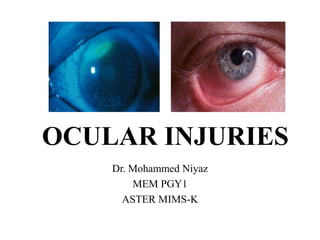
- 1. OCULAR INJURIES Dr. Mohammed Niyaz MEM PGY1 ASTER MIMS-K
- 2. What is ocular trauma? Damage or trauma inflicted to the eye by external means. The concept includes both surface injuries and intraocular injuries. During trauma soft tissues and bony structures around the eye maybe involved.
- 3. • Ocular injuries are classified as open globe, closed globe, and periocular . • Open globe — Open globe injuries have a full thickness break of the eye wall, which is composed of the sclera and the cornea. • These injuries are further described as follows: • ●Open globe ruptures – Full thickness eye injuries caused by blunt trauma • ●Open globe lacerations – Full thickness eye injuries caused by sharp objects
- 4. Closed globe injuries • Closed globe — Closed globe injuries do not have full thickness breaks of the eye wall. They are further divided into lamellar lacerations (partial thickness wound to the eye wall) or contusions (no eye wall wound). • Thus, the following injuries are also lamellar lacerations: • ●Conjunctival laceration – Full thickness break of the conjunctiva • ●Partial thickness scleral laceration – Incomplete scleral break not to the level of the choroid • ●Partial thickness corneal laceration – Incomplete corneal break without loss of aqueous humor
- 5. • Other closed globe injuries include: • ●Conjunctival abrasion – Injury to the epithelium of the conjunctiva • ●Corneal abrasion – Injury to the epithelium of the cornea • ●Hyphema – Blood in the anterior chamber of the eye • ●Traumatic iritis – Inflammation in the anterior chamber resulting from trauma • ●Traumatic mydriasis – Chronic pupil dilation usually from iris sphincter damage • ●Lens dislocation – Native or artificial lens implant displacement from its original location • ●Vitreous hemorrhage – Bleeding into the vitreous cavity • ●Commotio retinae – Retinal whitening due to trauma-associated retinal edema • ●Retinal detachment – Separation of the retina from the underlying choroid and sclera
- 6. Periocular injuries • The orbital septum divides the pre-septal tissues (eyelids, meibomian glands, lashes, nasolacrimal system , and the orbicularis oculi muscle) from the deeper orbital components (orbital bones, fat, extraocular muscles, nerves, and vessels). • Periocular injuries include: • ●Pre-septal: • •Eyelid abrasions – Superficial skin injury not requiring surgical repair • •Eyelid lacerations – Full thickness skin injury usually requiring surgical repair • •Canalicular lacerations – Full thickness eyelid skin injury, which includes the lacrimal drainage system; usually requires nasolacrimal cannulation for repair • •Periocular ecchymoses – Skin bruising, which may indicate more serious underlying injury
- 7. Orbital : • •Eyelid lacerations with fat prolapse – Full thickness eyelid skin injury with penetration through orbital septum ; requires complex repair by ophthalmology • •Eyelid lacerations with levator involvement – Full thickness eyelid skin injury, which may lead to ptosis if not repaired; requires ophthalmology consultation • •Orbital fractures – Fracture of any of the bones making up the socket and surrounding the eye • •Extraocular muscle entrapment – Prolapse of extraocular muscle(s) into the defect created by a fractured orbital bone; may induce muscle ischemia requiring more urgent repair
- 8. • •Orbital compartment syndrome (OCS) – Elevated intraorbital pressure from infection, bleeding or inflammation causing poor motility and possible ocular ischemia • •Orbital foreign bodies – Any post-septal foreign body present in the eye socket but outside the globe • •Traumatic optic neuropathy – Acute vision loss and afferent pupil defect from trauma-induced optic nerve damage. • •Optic nerve avulsion – Acute vision loss and afferent pupil defect from traumatic transection of the optic nerve • •Ophthalmic artery injuries – Ocular ischemia from compression or laceration of the ophthalmic artery
- 9. CONJUCTIVAL ABRASION • Conjunctiva has less innervation than the cornea • Less symptomatic than corneal abrasions. • Foreign body sensation, mild pain, tearing, photophobia • Conjunctival abrasion may be invisible without fluorescein staining.
- 10. CONJUCTIVAL LACERATION • Seidel test to exclude perforation of the globe (can be negative if a full-thickness laceration is small or has spontaneously closed) • Inspect the conjunctiva for a foreign body. • Conjunctival foreign bodies usually can be removed with a moistened, cotton tipped applicator . • Erythromycin ophthalmic ointment 0.5% four times a day for 2 to 3 days
- 11. CORNEALABRASION Cornea is richly innervated painful Corneal epithelium regenerates quickly, heals within 24 to 48 hours. Causes : Contact lens wear, fingernails makeup brushes, foreign objects blown into eyes while driving or on windy days, or which drop into the eye while working overhead (construction) or under a car (mechanics).
- 12. • Symptoms : Intense pain, foreign body sensation, photophobia, and tearing • Inspection : conjunctival injection, tearing, and lid swelling • Slit lamp : flare and cells from iritis if the abrasion is large and >24 hours old, but there is no corneal infiltrate • The abrasion usually appears as a superficial, irregular corneal defect appearing bright green under the cobalt blue light after instillation of fluorescein
- 13. • Key to pain control is adequate and persistent cycloplegia. • If an abrasion is >2 mm or very painful, instill a cycloplegic agent (cyclopentolate 1% or homatropine 5%) one drop every 6 to 8 hours • Avoid atropine because the effect lasts for approximately 2 weeks.
- 14. • Patients with large abrasions or abrasions in the central visual axis should follow up with ophthalmology in 24 hours, others at 48 to 72 hours. • Patching the eye does not promote healing, but some patients feel better with the eye patched
- 15. CORNEAL LACERATIONS • Misshapen iris, macro- or microhyphema, decrease in visual acuity, and shallow anterior chamber. • The Seidel test will be positive. • Causes : sharp sticks, fingernails, thorns, broken glass, or sharp toys • Pain out of proportion to physical findings, decrease in visual acuity, or other unexplained ocular symptoms. • CT of the orbit to identify changes in globe anatomy or contour or a foreign body within the globe, and consult ophthalmology.
- 16. ULTRAVIOLET KERATITIS Light in the UV range can cause death of corneal epithelial cells. Classically described as “snow blindness” Unprotected exposure to arc welders and tanning beds, and is often called “welder’s flash.” Blepharospasm, conjunctival injection, and prominent tearing. Slit lamp : diffuse punctate corneal edema, and instillation of fluorescein reveals diffuse punctate corneal abrasions. Treatment may include double patching of both eyes if the patient requests, use of cycloplegics, topical antibiotics (erythromycin), and oral analgesics. Healing occurs in 24 to 36 hours
- 17. CORNEAL FOREIGN BODIES • Penetration of a foreign body into the globe can cause loss of vision. • Metallic foreign bodies present on the cornea for more than a few hours will cause rust to diffuse into the cornea, termed a “rust ring.” • Inflammatory reaction, dilating blood vessels of the conjunctiva and causing edema of the lids, conjunctiva, and cornea • Tearing, blurred vision, and photophobia are common • The presence of hyphema (or microhyphema in the anterior chamber on slit lamp examination) suggests globe perforation
- 18. FOREIGN BODY REMOVAL • Irrigate with normal saline (NS) first • Local anesthetic such as 0.5% proparacaine • Full-thickness corneal foreign bodies should be removed by an ophthalmologist • For superficial foreign bodies, a 25-gauge needle (bevel up) or a sterile foreign body spud on an Alger brush (a low-speed, low-torque, battery operated hand-held drill) can be used • If a rust ring is present, the spud or an ophthalmic burr can remove superficial rust
- 19. LID LACERATIONS • Eyelid lacerations that involve the lid margin • those within 6 to 8 mm of the medial canthus • involving the lacrimal duct or sac, • those involving the inner surface of the lid, • those involving the lid margins • wounds associated with ptosis • those involving the tarsal plate or levator palpebrae muscle need repair by an oculoplastic specialist.
- 20. • Deep lacerations medial to the punctum potentially can transect the canalicular system • Instillation of fluorescein dye in the eye with subsequent appearance in the wound indicates loss of canalic ular integrity. • Canalicular laceration : Repair and Silastic tube stenting within 24 to 36 hours • Oral cephalexin (Keflex R), 500 mg twice or four times daily, and topical erythromycin ophthalmic ointment four times daily .
- 21. • Very small lacerations (<1 mm) at the lid edge only do not need suturing and can heal spontaneously. • Consider the possibility of corneal laceration and globe rupture in all full-thickness lid lacerations. • Partial-thickness lid lacerations usually be repaired in the ED, with referral for ophthalmologic evaluation in 2 to 3 days. • Use a soft, absorbable or nonabsorbable 6-0 or 7-0 suture. • Have the suture ends closest to the cornea tucked under more distant sutures to avoid corneal irritation
- 22. BLUNT EYE TRAUMA • Assessment of the visual acuity, anterior chamber, and integrity of the globe. • If the anterior chamber is flat, a ruptured globe is certain • Restricted upgaze or lateral gaze suggests a blow-out fracture with entrapment and a CT scan of facial bones should be obtained. • Feel the orbital rim above and below for step-off deformities. • Test for cutaneous sensation along the distribution of the inferior orbital nerve (below the eye and ipsilateral side of the nose). • Perform a slit lamp examination with fluorescein staining to check for abrasions, lacerations, foreign bodies, hyphema, iritis, and lens dislocation.
- 23. • If vision and ocular anatomy and function are preserved, outpatient follow-up by an ophthalmologist in the next 48 hours. • If a ruptured globe is suspected due to loss of visual acuity, flat anterior chamber, obvious full-thickness laceration, or intraocular foreign body, do not manipulate the eye or measure intraocular pressure. • Consult ophthalmology immediately.
- 26. HYPHEMA • Blood or blood clots in the anterior chamber are referred to as a hyphema • Traumatic hyphema usually : bleeding from a ruptured iris root vessel. • Spontaneous hyphemas : sickle cell disease • Treatment: Elevate the patient’s head to promote settling of suspended RBCs inferiorly to prevent occlusion of the trabecular meshwork. • After consultation with the ophthalmologist, dilate the pupil to avoid “pupillary play” (constriction and dilation movements of the iris in response to changing lighting conditions), which can stretch the involved iris vessel, causing additional bleeding
- 27. • Control of intraocular pressure consists of topical β-blockers, IV mannitol, topical α-adrenergic agonists (apraclonidine), and oral, topical, or IV carbonic anhydrase inhibitors (CAIs) such as Diamox. • Do not give CAIs to patients with sickle cell disease. • patients with hyphemas occupying one third or less of the anterior chamber can be followed closely as outpatients
- 29. BLOW-OUT FRACTURES • Most frequent sites : inferior wall (maxillary sinus) and medial wall (ethmoid sinus through the lamina papyracea). • Fractures of the inferior wall with entrapment of the inferior rectus muscle can cause restriction of upgaze and diplopia • Isolated blow-out fractures with or without entrapment can be referred to ophthalmology, plastic surgery, OMFS , ENT for repair within the next 3 to 10 days.
- 30. • A Waters view x-ray : cloudy maxillary sinus representing blood, fluid, and tissue on the side of the trauma. • Oral antibiotics (cephalexin, 250 to 500 milligrams PO four times daily for 10 days). • Refer to an ophthalmologist for an outpatient full dilated examination to rule out any unidentified retinal tears or detachments.
- 31. RUPTURED GLOBE • Mechanism of Injury Scleral rupture may occur from blunt or penetrating trauma • Penetrating trauma may occur from bullets, BB pellets, knives, sticks, darts, needles, hammering, and lawn mower projectiles. • Suspect globe penetration with any puncture or laceration of the eyelid or periorbital area. • Corneal abrasions occurring when hammering metal on metal, associated with use of high-speed machinery such as lawn mowers, line trimmers (weed whackers), grinders, or drills, and sustained during explosions should always be investigated for occult globe penetration.
- 32. • Whenever globe rupture is suspected, cover the eye with a metal eye shield and consult ophthalmology immediately • Clinical features : Decreased visual acuity, an irregular or teardrop-shaped pupil, an afferent pupillary defect, shallow anterior chamber, hyphema, positive Seidel test, and lens dislocation • Presence of a large subconjunctival hemorrhage involving the entire sclera or hemorrhagic chemosis (bullous, raised subconjunctival hemorrhage) is very suspicious for rupture of the globe . • Uveal prolapse through a scleral wound may appear as a brownish-black discoloration against the white sclera
- 33. • US as well as both direct and indirect ophthalmoscopy helpful in locating orbital and intraocular foreign bodies. • Immediate ophthalmologic consultation • Broad-spectrum IV antibiotics and TT administered • Provide sedation and analgesia • Administer antiemetic agents to prevent increased IOP and extrusion of intraocular contents from vomiting or retching. • The patient should be maintained NPO, anticipating surgery
- 34. RETROBULBAR HEMATOMA • Severe blunt trauma to the orbit can occasionally cause a retrobulbar hematoma. • Abrupt increase in IOP cause decreased blood flow to the optic nerve and loss of vision. • Pain, proptosis, and decreasing vision. • A CT scan of the orbit will demonstrate retrobulbar hemorrhage. • An intraocular pressure >40 mm Hg is a consideration for emergency canthotomy.
- 35. CHEMICAL OCULAR INJURY • Complications of chemical burns to the eye include scarring of the cornea with permanent loss of vision and loss of the eye due to corneal perforation. • Irrigation of the eyes with 1 to 2 L of NS must be done immediately and before any examination, including testing of vision.
- 37. ALKALI AND ACID INJURIES • The most serious alkali injuries are associated with ammonia found in many household cleaners, and lye, a common ingredient in drain cleaners. • Lye is also a component of concrete. • Alkali injuries cause a liquefaction necrosis, characterized by denaturing of proteins and saponification of fats, allowing deep penetration into tissue. • Acid causes coagulation necrosis, with denaturing of protein forming a coagulum that acts as a barrier to further tissue penetration
- 38. • Irrigation should be with sterile NS or other isotonic solution and may be instilled into the eye via a Morgan Lens • If the pH is >7.4, continue irrigation until the pH remains neutral 30 minutes after the last irrigation • Conjunctival injection and chemosis, scleral whitening, secondary to ischemia and blood vessel injury
- 39. • Patients with chemosis (edema of the bulbar conjunctiva overlying the white sclera) and no corneal or anterior chamber findings should be treated after irrigation with erythromycin ointment four times daily • Refer for an ophthalmologic examination in 24 to 48 hours. • These patients are considered to have “chemical conjunctivitis.” • A topical cycloplegic agent should be used three times daily for pain reduction if an epithelial defect is present.
- 40. CYANOACRYLATE (SUPER GLUE/CRAZY GLUE) • Accidental instillation into the eye and adnexa can cause adherence of the lids and clumps of adhesive to form on the cornea. • To remove crazy glue, instill generous amounts of erythromycin ointment onto the eye and on the surface of the eyelids to moisten, lubricate, and provide antibiotic coverage • The glue will loosen and become easier to remove in a few days. • Refer to an ophthalmologist within 24 hours for complete removal.
- 41. THE END