Biochemical tests for characterization of bacteria
•
32 j'aime•15,776 vues

Biochemical tests for characterization of bacteria

Recommandé
Contenu connexe
Tendances
Tendances (20)
Similaire à Biochemical tests for characterization of bacteria
Similaire à Biochemical tests for characterization of bacteria (20)
Plus de Dr. Pavan Kundur
Plus de Dr. Pavan Kundur (20)
Dernier
Dernier (20)
Biochemical tests for characterization of bacteria
- 1. Biochemical tests for Characterization of Bacteria By, Dr. Pavan K. J Assistant professor P C Jabin Science College Hubballi. Karnataka
- 2. IMViC Tests The IMViC series is a group of four individual tests that are commonly used to identify bacterial species, especially coliforms. A coliform is a gram negative, aerobic anaerobic rod which produces gas from lactose within 48 hours. The presence of some coliforms Each of the letters in “IMViC” stands for one of these tests. “I” is for indole; “M” is for methyl red; “V” is for Voges-Proskauer, and “C” is for citrate, lowercase “i” is added for the ease of pronunciation. IMViC is an acronym that stands for four different tests gas from lactose within 48 hours. The presence of some coliforms indicate fecal contamination.
- 3. To obtain the results of these four tests three test tubes are inoculated Indole test tryptone broth Methyl red test methyl red – (MR-VP broth) Voges-Proskauer test Voges Proskauer broth Citrate utilization test Simmons citrate agar IMViC tests are employed in the identification/differentiation of members of family enterobacteriaceae.members of family enterobacteriaceae. General procedure for performing IMViC Tests and their interpretations: Cultures of any members of enterobacteriaceae have to grow for 24 to 48 hours at 37°C and the respective tests can be performed:
- 4. Indole test It is performed on sulfide-indole-motility (SIM) medium or in tryptophan broth, or in motility urease indole (MIU) medium. Result is read after adding Kovac’s reagent. The positive result is indicated by the red layer at the top of the tube after the addition of Kovács reagent. A negative result is indicated by the lack of color change at the top of the tube after the addition of Kovács reagent.of the tube after the addition of Kovács reagent.
- 5. Voges-Proskauer (VP) test: Methyl red test and Voges-Proskauer test both are done in methyl red–Voges- Proskauer (MR-VP) broth, but the reagents that are added varies according to the test Methyl Red (MR)Test: Positive methyl red test are indicated by the development of red color after the addition of methyl red reagent. A negative methyl red test is indicated by no color change after the addition of methyl red reagent Voges-Proskauer (VP) test: Negative test is indicated by lack of color change after the addition of Barritt’s A and Barritt’s B reagents. A positive Voges-Proskauer test is indicated by the development of red-brown color after the addition of Barritt’s A and Barritt’s B reagents.
- 6. Citrate utilization test The test is performed on Simmons citrate agar: Negative citrate utilization test is indicated by the lack of growth and color change in the tube A positive citrate result as indicated by growth and a blue color change.
- 7. IMViC Test 1. IMViC tests of Escherichia coli 2.Indole: Positive 3.Methyl-Red: Positive 4.Voges-Proskauer test: Negative 5.Citrate test: Negative 2.IMViC tests of Enterobacter aerogenes 1.Indole: Negative 2.Methyl-Red: Negative 3.Voges-Proskauer test: Positive 4.Citrate test: Positive4.Citrate test: Positive 3.IMViC tests of Proteus vulgaris 1.Indole: Positive 2.Methyl-Red: Positive 3.Voges-Proskauer test: Negative 4.Citrate test: Negative 4.IMViC tests of Citrobacter freundii 1.Indole: Negative 2.Methyl-Red: Positive 3.Voges-Proskauer test: Negative 4.Citrate test: Positive
- 8. Oxidase test The oxidase test is used to identify bacteria that produce cytochrome c oxidase, an enzyme of the bacterial electron transport chain. When present, the cytochrome c oxidase oxidizes the reagent (tetramethyl-p-phenylenediamine) to (indophenols) purple color end product. When the enzyme is not present, the reagent remains reduced and is colorless.colorless.
- 10. Purpose of Oxidase test Oxidase test is most helpful in screening colonies suspected of being one of the Enterobacteriaceae (all negative) and in identifying colonies suspected of belonging to other genera such as Aeromonas, Pseudomonas, Neisseria, Campylobacter, and Pasteurella (positive). Test requirements for Oxidase test: Moist filter paper with the substrate (1% tetramethyl-p-phenylenediamine dihydrochloride), or commercially preparedtetramethyl-p-phenylenediamine dihydrochloride), or commercially prepared paper disk, wooden wire or platinum wire. Expected results of Oxidase test Positive: Development of dark purple color (indophenols) within 10 seconds Negative: Absence of color
- 11. Quality Control of Oxidase Test Bacterial species showing positive and negative reactions should be run as controls at frequent intervals. The following are suggested: A. Positive control: Pseudomonas aeruginosa B. Negative control: Escherichia coli Procedure of Oxidase test: Take a filter paper soaked with the substrate tetramethyl-p- phenylenediamine dihydrochloride Moisten the paper with a sterile distilled water Pick the colony to be tested with wooden or platinum loop and smear Pick the colony to be tested with wooden or platinum loop and smear in the filter paper Observe inoculated area of paper for a color change to deep blue or purple within 10-30 seconds Precaution to be taken while performing oxidase test: Do not use Nickel-base alloy wires containing chromium and iron (nichrome) to pick the colony and make smear as this may give false positive results Interpret the results within 10 seconds, timing is critical
- 12. Oxidase test results Bacterial genera characterized as oxidase positive include Neisseria and Pseudomonas etc. Genera of the Enterobacteriaceae family are characterized as oxidase negative. Name of Oxidase positive bacteria are: Mneomoics for Oxidase Positive Organisms- PVNCH (It’s just an acronym inspired by the famous mneomonic for Urease Positive organisms-PUNCH) P: Pseudomonas sppP: Pseudomonas spp V: Vibrio cholerae N: Neisseria spp C: Campylobacter spp H: Helicobacter spp/ Haemophilus spp. Aeromonas spp Alcaligens
- 13. Gelatin is a protein derived from the animal protein collagen– component of vertebrate connective tissue. It has been used as a solidifying agent in food for a long time. Gelatin hydrolysis test is a great way to highlight proteolysis by bacteria Gelatin hydrolysis test Principle of Gelatin hydrolysis test Gelatin hydrolysis test is used to detect the ability of an organismGelatin hydrolysis test is used to detect the ability of an organism to produce gelatinase (proteolytic enzyme) that liquefy gelatin. Hydrolysis of gelatin indicates the presence of gelatinases. This process takes place in two sequential reactions. In the first reaction, gelatinases degrade gelatin to polypeptides. Then, the polypeptides are further converted into amino acids. The bacterial cells can then take up these amino acids and use them in their metabolic processes.
- 14. Procedure /Method of Gelatin hydrolysis test There are several methods for determining gelatinase production, all of which make use of gelatin as the substrate. The standard and most commonly employed method is the nutrient gelatin stab method. Inoculate a heavy inoculum of test bacteria (18- to 24-hour-old) by stabbing 4-5 times (half inch) on the tube containing nutrient gelatin medium. Incubate the inoculated tube along with an uninoculated medium at 35°C, or at the test bacterium’s optimal growth temperature, for up to 2 weeks. Remove the tubes daily from the incubator and place in ice bath or refrigerator (4°C) for 15-30 minutes (until control is gelled) every day to check for gelatin liquefaction. (Gelatin normally liquefies at 28°C and above, so to confirm that liquefaction was due to gelatinase activity, the tubes are immersed in an ice bath or kept in refrigerator at 4°C). Tilt the tubes to observe if gelatin has been hydrolyzed.
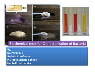
- 15. Expected results:- Positive: Partial or total liquefaction of the inoculated tube (uninoculated control medium must be completely solidified) even after exposure to cold temperature of ice bath or refrigerator (4°C) Negative: Complete solidification of the inoculated tube even after exposure to cold temperature of ice bath or refrigerator (4°C) Common bacteria and their reactions to the Gelatin Hydrolysis Test: Above tube: Positive Below Tube: NegativeCommon bacteria and their reactions to the gelatin hydrolysis test performed on nutrient gelatin. Below Tube: Negative Species Growth Liquefaction Bacillus subtilis + + Clostridium perfringens + + Escherichia coli + – Proteus vulgaris + + Serratia liquefaciens + + Staphylococcus aureus + +
- 16. Control organisms Positive Control: Proteus vulgaris Negative Control: Enterobacter aerogenes Uses of Gelatin Hydrolysis test Gelatin hydrolysis test is helpful in identifying and differentiating species of Bacillus, Clostridium, Proteus, Pseudomonas, and Serratia. It distinguishes the gelatinase-positive, pathogenic Staphylococcus aureus from the gelatinase-negative, non-pathogenic S. epidermidis . Gram-positive, spore-forming, rodshaped, aerobic or anaerobic bacteria such as Bacillus anthracis, Bacillus cereus, Bacillus subtilis, Clostridium perfringens and Clostridium tetani, are also positive for gelatin hydrolysis. The test can also be used to differentiate genera of gelatinase-producing bacteria such Serratia and Proteus from other members of the family Enterobacteriaceae.
- 17. Starch Hydrolysis Test Starch is a complex polysaccharide found abundantly in plants and usually deposited in the form of large granules in the cytoplasm of the cell. Starch consists of 2 components—amylose and amylopectin, which are present in various amounts. The amylose consists of D-glucose units linked in a linear fashion by α-1,4 linkages. It has 2 non-reducing ends and a reducing end.linkages. It has 2 non-reducing ends and a reducing end. Amylopectin is a branched polysaccharide. In these molecules, shorter chains of glucose units linked by α-1,4 are also joined to each other by α-1,6 linkages. The major component of starch can be hydrolyzed by a-amylase, which is present in some bacteria while well known in case of fungi. The ability to degrade starch is used as a criterion for the determination of amylase production by a microbe.
- 18. Objective To determine the ability of an organism to hydrolyze starch To differentiate organism based on their α- amylase enzyme activity Principle Many bacteria produce extracellular enzymes used to catalyze chemical reactions outside of the cell. In this manner, nutrient sources, such as starch, that are too large to be absorbed through the cell membrane can be broken down into smaller molecules and transported into the cell via diffusion. In the starch hydrolysis test, the test bacteria are grown on agar plates containing starch.starch. If the bacteria have the ability to hydrolyze starch, it does so in the medium, particularly in the areas surrounding their growth while the rest of the area of the plate still contain non-hydrolysed starch. Since no color change occurs in the medium when organisms hydrolyze starch, iodine solution is added as an indicator to the plate after incubation. While the non-hydrolysed starch forms dark blue color with iodine, its hydrolyzed end products do not acquire such dark blue color with iodine.
- 19. Consequently, transparent clear zones are formed around the colonies that hydrolyze starch while the rest of the plate show a dark blue coloration as iodine forms the colored complex with starch.
- 20. Media: Starch agar is a simple nutritive medium with starch added. Beef extract and pancreatic digest of gelatin provide nitrogen, vitamins, carbon and amino acids. Agar is the solidifying agent and starch is the carbohydrate. Composition: Peptic digest of animal tissue 5.000, Sodium chloride 5.000, Yeast extract 1.500, Beef extract 1.500 Starch, soluble 2.000 Agar 15.000 Final pH ( at 25°C) 7.4±0.2 Method Using a sterile technique, make a single streak inoculation of organism to be tested into the centre of labeled plate.tested into the centre of labeled plate. Incubate the bacterial inoculated plates for 48 hours at 37°C. Following incubation, flood the surface of the plates with iodine solution with a dropper for 30 seconds. Pour off the excess iodine. Examine for the clear zone around the line of bacterial growth.
- 21. Expected Results Positive test:A clear zone around the line of growth after addition of iodine solution indicates that the organism has hydrolyzed starch. Negative test:A blue, purple, or black coloration of the medium (depending on the concentration of iodine). Uses It aids in the differentiation of species of genera Corynebacterium, Clostridium, Bacillus, Bacteroides, Fusobacterium, and members ofEnterococcus spp. Limitations It is recommended that biochemical, immunological, molecular, or mass spectrometry testing be performed on colonies from pure culture for complete identification. Colonies cannot be subcultured from the medium after the addition of Gram’s iodine due to the oxidative nature of the reagent and the resulting cell death.