Cardiovascular System
•
5 j'aime•384 vues

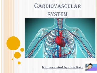
The circulatory system, also called the cardiovascular system or the vascular system, is an organ system that permits blood to circulate and transport nutrients (such as amino acids and electrolytes), oxygen, carbon dioxide, hormones, and blood cells to and from the cells in the body to provide nourishment and help in fighting diseases, stabilize temperature and pH, and maintain homeostasis.

Recommandé
Contenu connexe
Tendances
Tendances (20)
Similaire à Cardiovascular System
Similaire à Cardiovascular System (20)
Plus de Pawan Kumar Sahu
Plus de Pawan Kumar Sahu (13)
Dernier
Dernier (20)
Cardiovascular System
- 2. INTRODUCTION Cardiovascular system consist of heart and blood vessels. It is mainly a transport system. It transport respiratory gases, nutrients and excretory products to various part of the body. Blood is the medium through which these substance are transported. Cardiovascular system divided into two main parts: 1. Circulatory system: Circulator system consisting of heart which act as a pump and the blood vessel through which the blood circulates. 2. Lymphatic system: Lymphatic system consisting of lymph nodes and lymph vessel through which colourless lymph flows.
- 3. HUMAN HEART IN FRONT VIEW
- 4. HEART The heart is a muscular organ in most animals, which pumps blood through the blood vessels of the circulatory system. The pumped blood carries oxygen and nutrients to the body, while carrying metabolic waste such as carbon dioxide to the lungs. Heart is a thick, muscular, reddish brown, conical, hollow and musculotendinous organ. It lies in the thorax between lung and behind the sternum. It is about 10 cm long and weights about 300 gm. The heart is approximately the size of a closed fist and is located between the lungs, in the middle compartment of the chest.
- 5. FLOW OF BLOOD THROUGH THE HEART
- 6. STRUCTURE OF HEART The heart s composed of the three layers of the tissues:- 1. Pericardium 2. Myocardium 3. Endocardium
- 7. 1. PERICARDIUM Heart is surrounded by an outer covering layer known as pericardium. It is a thin, transport, two layered sac. The inner layer is called visceral pericardium and outer layer is called parietal pericardium. A narrow cavity between two layers is known pericardial cavity which is filled with a fluid called pericardial fluid. It performs two function:- a. It allows frictionless movements of heart. b. It protects the heart from mechanical shock.
- 8. PERICARDIUM
- 9. 2. MYOCARDIUM The middle layer is known as myocardium which made heart fibres. These muscle fibre found only in the heart. It is the muscular tissue of heart, it is not under voluntary control. The myocardium is thickest at the apex and thin out towards the base.
- 10. MYOCARDIUM
- 11. 3. ENDOCARDIUM The inner layer is known as endocardium. It is smooth thin membrane that permits smooth flow of blood inside the heart. This lines the chamber and valve of the heart.
- 12. ENDOCARDIUM
- 13. CHAMBERS OF HEART Heart is made of four chambers that is, two atria and two ventricles. Atria: The atria are smaller with their walls because they have to push the blood only upto ventricles. Both atria or auricles are separated by a inter- auricular septum which prevents mixing of oxygenated and deoxygenated blood in heart. Ventricles: The ventricles are larger and thicker. Left ventricle is much thicker than right ventricle (about three times) because ventricle has to push the blood to a body parts while right ventricle has to push the blood to closely lying lungs only. Both ventricle are separated by a thick inter-ventricular septum. So there is mixing of deoxygenated and oxygenated blood in heart.
- 15. A) RIGHT AURICLE It receives deoxygenated blood by three large veins- superior vena cava, inferior vena cava and coronary sinus. Right auricle opens in the right ventricle through the right atrioventricular valve or tricuspid valve. It regulates unidirectional flow of blood from right auricle to right ventricle but prevents the back flow of blood. (with the number of whitish elastic threads called chordae tendineae which extends from tricuspid valve to wall of right ventricle)
- 16. B) LEFT AURICLE It receives oxygenated blood from lungs by four pulmonary veins. (two each lungs) Left auricle open in left ventricle by the help of the left atrioventricular valve or bicuspid valve. It regulates the flow of blood from left auricle to left ventricle but prevents back flow of blood (with the help of chordae tendineae similar to those in right ventricles.)
- 17. A) RIGHT VENTRICLES From the right ventricles arise from the pulmonary artery or arch or aorta which carries deoxygenated blood to the lungs.
- 18. B) LEFT VENTRICLES Left ventricle originates aorta or arch which carries oxygenated blood to various parts of the body. At the base of each of pulmonary and aortic arches, there is a valve called pulmonary valve and aortic valve. These allow blood to enter the great artery from ventricle, but prevent the back flow of blood into the ventricle.
- 19. VALVES OF THE HEART The opening between right atrium and right ventricle is guarded by ‘tricuspid valves’. The opening between left atrium and left ventricle is guarded by ‘bicuspid valve’. The two atrioventricular valves are one way that keeps the flow of blood from the arteries to the ventricles and not the other way.
- 20. VALVES OF THE HEART
- 21. CIRCULATION OF BLOOD THROUGH HEART Heart is four-chambered and has complete double circulation which means blood possess twice through the heart to supply once to the body. There are two circulation:- A. Pulmonary circulation B. Systemic circulation
- 22. A. PULMONARY CIRCULATION In this blood complete its circulation from right ventricle to left auricle through the lungs. It is the part of circulation involving the purification (oxygenation) of blood in the lungs. Here, the right ventricle pumps deoxygenated blood into pulmonary artery. Then the pulmonary artery carries this blood to the lungs where it is oxygenated. Then the four pulmonary veins carry the purified blood from lungs to left auricle of the heart. Then left auricle pumps the blood to left ventricle.
- 23. B. SYSTEMIC CIRCULATION In this blood completes its circulation from left ventricle to right auricle through body organs. Here the left ventricle pumps oxygenated blood into systemic arch which supplies it to body organ other than the lungs through a number of arteries. Deoxygenated blood from these organs are returned to the right auricle through two large veins, superior and inferior vena cava. Right auricle pumps the deoxygenated blood in the right ventricle.
- 24. CIRCULATION OF BLOOD THROUGH HEART
- 25. ARTERIAL AND VENOUS SYSTEM The heart pumps blood into arteries. The arteries divide and subdivide and finally end in capillaries. The capillaries later unite to form veins. The veins returns blood to the heart. So arteries carry pure (oxygenated) blood from the heart. The veins carry impure (deoxygenated) blood to the heart. Capillaries are minute channels. They receive blood from smaller arteries (arterioles) and deliver into smaller veins (venules).
- 26. ARTERIAL AND VENOUS SYSTEM
- 27. ARTERIES & ARTERIOLES These are the blood vessel that transport blood away from the heart. They vary considerably in size and their wall consist of three layer of tissue. They are; arteries and veins have the same layer of structure. 1. Tunica adventitia: It is the outer layer of fibrous tissue. 2. Tunica media: It is the middle layer of smooth muscles and elastic. 3. Tunica intima: It is the inner layer of squamous epithelium called endothelium.
- 29. VEINS AND VENULES Veins are blood vessel that return blood at low pressure to the heart. The wall of the veins are thinner than those of arteries but have the same three layer of tissues. They are thinner because there is less muscle and elastic tissue in the tunica media. The veins carry blood at a lower pressure than arteries. The smallest veins are called venules.
- 31. CAPILLARIES The smallest arterioles break up into a number of a minute vessel called capillaries. Capillary well consist of a single layer of endothelial cell.
- 32. FUNCTION OF HEART The heart acts as a pump. It maintain a constant circulation of blood throughout the body. It is achieved as follows:- The superior vena cava and inferior vena cava bring venous parts of the body to the heart. The venous blood fills the right atrium. When it is full, the right atrium contracts sending blood to the right ventricle. Now, the right ventricles contracts. This send blood to the lungs through pulmonary trunk (which divides into right and left pulmonary arteries). The blood gets oxygenated in the lungs. The oxygenated blood is carried by pulmonary veins to the left atrium. Now the left atrium contracts and sends blood to the left ventricle. Now the left ventricle contracts and sends blood into the aorta. This blood is circulated throughout the body except lungs.
- 33. CARDIAC CYCLE It involves alternate contraction (called systole) and relaxation (called diastole) of heart at the rate of 70-72 times per minute at rest. All the chambers do not beat simultaneously. Right and left auricles contract simultaneously while both ventricle also work simultaneously. The sequence of events that occur during one heart beat is called cardiac cycle. Cardiac cycle occurs in two phase:- I. Systole: A period of contraction II. Diastole: A period of relaxation
- 34. CARDIAC CYCLE
- 35. HEART SOUNDS During a cardiac cycle two heart sound can be heard. Totally, four sounds are produced by the heart. The first sound as LUB and the second sound as DUB can be heard with a stethoscope. The third & fourth sounds cannot be heard.
- 36. 1. LUB OR SYSTOLIC SOUND It is heard during the beginning of ventricular systole and is produce due to rapid closing of atrioventricular valves to prevents back flow of blood from the ventricles of atria.
- 37. 2. DUB OR DIASTOLIC SOUND It is heard during the beginning of ventricular diastolic and is due to rapid closing of semi- lunar valves to prevents back flow of blood from the great arteries to the ventricle.
- 38. ELECTROCARDIOGRAM (ECG) It is the recording of electrical activity of the heart. ECG is the instruments which is used to record the electrical current generated in the heart. The heart current can be recorded by connecting any two parts of the body with this instruments. The connections are called as ‘Leads’. There are bipolar and unipolar leads. A. Bipolar leads: they are three type- i. Lead 1- Electrodes connecting to the right & left arm. ii. Lead 2- Right arm & left leg iii. Lead 3- Left arm & left leg B. Unipolar leads: they are two type- i. Unipolar limbs leads- VR, VL, VF ii. Unipolar chest leads- V1 , V2 , V3 , V4 , V5 & V6
- 39. ECG WAVES During recording electrical activity of the heart some waves are produced; These waves are known as P, Q, R, S and T waves. P-wave: is formed due to the stimulation and contraction of the atria. Q,R,S-wave: are formed by the contraction of the ventricles. T-wave: are formed by the relaxation of the ventricles. U-wave: are formed due to the continue electrical activity after the relaxation of the ventricles. It is very small. PR-interval: it represents the time taken by the impulses to travel from the SA node to AV node. It is measured from the P-wave. Note:- Normal time of PR-interval is 0.12 to 0.16 sec. If PR-interval is move than 0.20 seconds it means that there may be some heart disease.
- 40. NORMAL ECG GRAPH
- 42. HEART RATE Number of heart beats per minute is known as the heart rate. The normal heart rate is as follows; In an adult: 72/minute In old age: 75-80/minute In newborn child: 130-140/minute In children upto 5 years: 90-140/minute Generally the heart rate is increased during exercise, emotion & in some diseased conditions. Heart rate is decreased during sleep, shock and in some disease.
- 43. BLOOD PRESSURE It is the pressure exerted by the blood vessels known as B.P. The blood pressure which is normally expressed by arterial blood pressure. It has two phase:- A. Systolic blood pressure: It is the maximum blood pressure. This occurs during the systole of heart. (Range 100-120 mmHg) B. Diastolic blood pressure: It is the minimum blood pressure. It occurs during the diastole of heart. (Range 60-80 mmHg)
- 44. Cardiac output: It is defined as the quantity of blood pumped by the heart in one minute. Pulse pressure: It is the difference between systolic and diastolic blood pressure. It is nearly 40 mmHg.
- 45. DISORDERS Tachycardia: Increase in the heart rate. Bradycardia: Decrease in the heart rate. Cardiac arrhythmia: It is the disorder in cardiac rate and rhythm. It occurs due to defective impulse formation and conduction in the heart. Angina pectoris: It occurs due to narrowing of coronary arteries. Hypertension: It is a rise in BP above normal. Hypotension: It is occur due to low blood pressure. Atherosclerosis: It is the thickening of arterial walls due to the deposition of fat. Phlebitis: It is the infection of the vein wall due to inflammation or injury.