
Recommandé
Contenu connexe
Tendances
Tendances (20)
Similaire à A brief review of exosome & its detection
Similaire à A brief review of exosome & its detection (20)
Dernier
Dernier (20)
A brief review of exosome & its detection
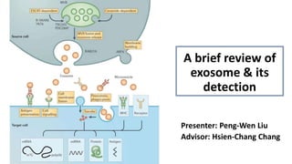
- 1. A brief review of exosome & its detection Presenter: Peng-Wen Liu Advisor: Hsien-Chang Chang
- 2. Introduction • Extracellular vesicles are lipid bilayer-closed structures derived from endocytosis and secreted by almost all types of cells, including ectosomes and exosomes. • The heterogeneity of exosomes is likely reflective of their size, content, functional impact on recipient cells, and cellular origin. • Research in this field is stimulated by the potential of exosomes as diagnostic and therapeutic tools for the treatment of various diseases, including neurodegeneration, cardiovascular dysfunction, and cancer. Membrane budding MVB fusion and exosome release Exosome vs. Ectosome: Some studies proposed that ectosomes bear CD9 or CD81 but not CD63 while exosomes bear CD63 and other tetranspanins.
- 3. Hallmarks of exosomes • Exosomes can contain membrane proteins, cytosolic and nuclear proteins metabolites, and nucleic acids, which renders exosome different regulatory functions. • CD9, CD63 and CD81 are the tetraspanins enriched in the membrane of exosomes, which are regarded as exosome biomarkers. ALIX: apoptosis-linked gene 2-interacting protein X HSP: heat shock protein
- 4. Zhu, L. et al. J Hematol Oncol 13, 152 (2020). • Exosomes can be detected in invasive liquid biopsies for disease diagnosis and prognosis: • Elevated urine-derived exosomal miR-21 has been associated with bladder and prostate cancer. • Exosomes for the delivery of drug payload(s) are being actively explored as therapeutic agents: • Clinical-grade MSC derived exosomes with KrasG12D siRNA payload (iExosomes) for pancreatic cancer treatment in multiple animal models without any obvious toxicity. Clinical applications of exosomes
- 5. Common exosomal separation techniques Fig. Schematic representation of common exosomal separation techniques. (A) Ultracentrifugation (Gold standard method) (B) Density gradient centrifugation (C) Dead-end filtration (DEF) (D)Tangential flow filtration (TFF) (E) Size-exclusion chromatography (F) Immunoaffinity Limitations: 1. Time-consuming 2. Requires large sample volume 3. Need specialized equipment 4. Low yield and purity Chen J, et al. Front Bioeng Biotechnol. 2022;9:811971 > 4 hr > 16 hr Prepare discontinuous gradient of sucrose or iodixanol
- 6. Common exosomal separation techniques- ExoQuik kit Manufacture: System Biosciences (SBI); 台灣代理商:騰達行 Peterson MF, Otoc N, Sethi JK, Gupta A, Antes TJ. Integrated systems for exosome investigation. Methods. 2015;87:31-45. • Captures and collects exosomes of a certain size range (60 – 150 nm) in ‘‘polymer nets’’ that can be recovered by a simple, low speed centrifugation on the bench top at 1500g. • Once the exosome pellet is obtained, the supernatant containing excess polymer is removed and the exosomes can then be resuspended in a suitable solution, such as PBS. • This resuspension process dilutes the residual polymer in the exosome pellet enough to dissolve the polymer net and liberate intact exosomes. The entire process can take as little at 30 min.
- 7. Methods for exosome quantification- Nanoparticle tracking analysis (NTA) Principle: Provides particle size and concentration in liquid suspension using the properties of Brownian motion and light scattering. Advantages: 1. Does not rely on detection of a specific marker 2. Direct quantification Disadvantages: 1. Expensive instrument 2. Photobleaching and potential background from dye aggregates Microscope (20x) Particles suspended in liquid Laser beam (Approx 50 μm) Metalized surface Glass Particles scattering from laser beam This instrument is available in NCKU center for Micro/Nano science technology.
- 8. Principle: Detects particles suspended in a fluid by their interaction with a laser beam as they flow through a detection cell. Advantages: 1. Direct quantification Disadvantages: 1. Insensitivity to smaller exosomes. 2. Requires binding to fuorophore- conjugated antibody-coated beads 3. Swarm effect that means multiple smaller vesicles are counted as single particle. This may provide false positive result Methods for exosome quantification- Flow cytometry Flow cytometry typically has a lower practical size limit of around 300 nm at which point the signal is indistinguishable from the baseline noise level.
- 9. Scheme 1. The electrochemical aptasensor for exosome detection based on click chemistry and HCR for signal amplification. An Y, Jin T, Zhu Y, Zhang F, He P. An ultrasensitive electrochemical aptasensor for the determination of tumor exosomes based on click chemistry. Biosens Bioelectron. 2019;142:111503. Signal amplification CuAAC click chemistry HRP catalysis 3 hr for hybridization chain reaction 0.5 hr for streptavidin- HRP conjugation OPD: o-phenylenediamine
- 10. Fig. 2. CV (A) and EIS (B) of different modified GCEs in 0.1M KCl containing 5.0mM [Fe(CN)6]3-/4- solution: bare GCE (a), DenAu/rGO/GCE (b), CD63 aptamer/DenAu/rGO/GCE (c), MCH/CD63 aptamer/DenAu/rGO/GCE (d), exosomes/MCH/CD63 aptamer/DenAu/rGO/GCE (e), and HCR-exosomes/MCH/CD63 aptamer/DenAu/rGO/GCE (f). An Y, Jin T, Zhu Y, Zhang F, He P. An ultrasensitive electrochemical aptasensor for the determination of tumor exosomes based on click chemistry. Biosens Bioelectron. 2019;142:111503. a. Bare GCE b. Reduction of graphene oxide and deposition of dendritic gold nanostructure c. CD63 aptamer immobilation d. MCH blocking e. Exosome binding f. HCR reaction
- 11. Fig. 3. (C) DPV responses with (a) and without (b) signal amplification by HCR; (D) DPV responses with (a) and without (b) the recognition of exosomes Fig. 4. (A) DPV responses of the electrochemical aptasensor for the exosomes at different concentrations; (B) linear relationship between the electrochemical signal and the logarithm of the exosome concentration. Error bars represent the relative standard deviation of measurements (%RSD ≤7.6%, n=3). An Y, Jin T, Zhu Y, Zhang F, He P. An ultrasensitive electrochemical aptasensor for the determination of tumor exosomes based on click chemistry. Biosens Bioelectron. 2019;142:111503. 1.12×10 2 1.12×10 8 The DPV was performed with HAc/NaAc buffer solution containing 2mM OPD and 4 mM H2O2.
- 12. Pang Y, Shi J, Yang X, Wang C, Sun Z, Xiao R. Personalized detection of circling exosomal PD-L1 based on Fe3O4@TiO2 isolation and SERS immunoassay. Biosens Bioelectron. 2020;148:111800. Scheme 1. A schematic view of the nanoparticles synthesis (a) and SERS tag-based exosomal PD-L1 detection (b). Exosome enrichment (5 min) Detection time: 40 min LoD: 1 PD-L1+ exosome/μl 4 μl TBOT: Tetrabutyl titanate (Titanium tetrabutoxide) The incubation time: 1. MBA immobilization: 2 hr 2. EDC/NHS activation: 15 min 3. Incubation with anti-PD-L1: overnight (785 nm) (300 nm) (20 nm)
- 13. Fig. 3. (c) Capture efficiency comparison of the Fe3O4@TiO2 nanospheres and UC in serum and PBS respectively. Fig. 4. SEM images and SERS signal of (a)Fe3O4@TiO2 (b) Fe3O4@TiO2/A549 exosome (c)Fe3O4@TiO2/A549 exosome/SERS tags. (Ultracentrifugation) Optimization for exosome isolation: 1. The quantity of Fe3O4@TiO2: 0.8 mg 2. Incubation time: 5 min
- 14. Fig. 5. (a) The SERS spectra with different A549 exosome concentrations (b) The correlation analysis of SERS intensity (at 1074 cm-1) and the logarithm of exosome concentrations. Blank represents SERS intensity without any exosome added. Fig. 6. (b) Scatter plots of the log [intensity] in the serum samples from the controls and the early-stage (stage I/II) and advanced (stage III/IV) patients measured by the SERS system. Error bars are the mean and one standard deviation of the mean (SD). Based on the personalized SERS (Surface-Enhanced Raman Scattering) signal analysis, NSCLC patients can be distinguished from the healthy controls easily.
- 15. Conclusion 1. Exosomes are extracellular vesicles generated by all cells and they carry nucleic acids, proteins, lipids, and metabolites. They are mediators of near and long- distance intercellular communication in health and disease and affect various aspects of cell biology. 2. The ExoQuik kit can greatly simply the isolation process and the operation time only requires 30 min. This method may be the suitable exosome separation method for our future study. 3. The nanoparticle tracking analysis is widely used as the comparison methods for exosome quantification. And the instrument is also available in NCKU. 4. The detection of exosomes depends on recognition of selected biomarkers, such as tetraspanin CD63 or PD-L1 on the exosome plasma membrane. Hence, the selection of target biomarkers should be taken seriously for future studies.
Notes de l'éditeur
- Ectosome vs exosome: Mathieu, M., Névo, N., Jouve, M. et al. Specificities of exosome versus small ectosome secretion revealed by live intracellular tracking of CD63 and CD9. Nat Commun 12, 4389 (2021).
- exosome大小會影響包含物的數量多寡,也許這是必須純化出相似大小exosome的重要原因 在乳癌細胞中,可透過exosome所含蛋白不同判斷是來自epithelial或mesenchymal cell,或許辨識癌症是哪種類型的好方法? 所收集的exosome可能同時包含剛釋出或準備進入細胞的exosome Tetraspanins are molecular scaffolds that distribute proteins into highly organized microdomains consisting of adhesion, signaling, and adaptor proteins. The C-terminal region usually presents crucial motifs involved in the sorting and targeting of tetraspanins to a determined intracellular location. Tetraspanins CD9, CD63, CD37, CD81, or CD82 are specially enriched in the membrane of exosomes.
- ExoQuick聚合物沉澱法分離原理是使用高分子量的PEG沉澱法,PEG為聚乙二醇,分子量區間為6000-30000,依照特定比例廠商獨家配方優化成ExoQuick系列產品,可與疏水性蛋白和脂質分子結合,並與其競爭游離的水分子,從而使溶解度較小的分子或外泌體(EVs)從溶液中析出,並透過低速離心後得到外泌體。不需要特殊的超高速離心儀器設備,即可從血液如血清、血漿樣品中得到外泌體。若需移除背景的雜蛋白(Immunoglobulin或Albumin),建議搭配ULTRA column,可有效去除70%的背景訊號。 https://ntuhmc.ntuh.gov.tw/epaper-100th.htm https://ntuhmc.ntuh.gov.tw/epaper-101th.htm
- 成大微奈米中心有NTA儀器 NTA provides information on particle size distribution of samples in liquid suspension using the properties of Brownian motion and light scattering. When particles in suspension are exposed to a laser beam traversing the sample chamber, they scatter the light, which makes them visible through a 20x magnification microscope. 目前已有多篇論文都是以此為對照方法,包含aptasensor那篇 可參考中文說明:https://ntuhmc.ntuh.gov.tw/epaper-47th.htm 其他參考資料: https://exosome-rna.com/nanosight-nanoparticle-tracking-analysis-nta-to-size-and-count-both-microvesicles-and-exosomes/ Nanoparticle Tracking Analysis (NTA) Measurements (azonano.com) Flow cytometry最多可測到300 nm的粒子,300 nm以下則測不到
- 成大微奈米中心有NTA儀器 NTA provides information on particle size distribution of samples in liquid suspension using the properties of Brownian motion and light scattering. When particles in suspension are exposed to a laser beam traversing the sample chamber, they scatter the light, which makes them visible through a 20x magnification microscope. 目前已有多篇論文都是以此為對照方法,包含aptasensor那篇 可參考中文說明:https://ntuhmc.ntuh.gov.tw/epaper-47th.htm Flow cytometry最多可測到300 nm的粒子,300 nm以下則測不到
- alkynyl-4-ONE, showing high reactivity toward proteins , was used to modify exosomes derived from MCF-7 cells, followed by the conjugation of an azide-labeled DNA probe as an anchor through a copper (I)-catalyzed click chemistry reaction. CuAAC: Copper -catalyzed azide – alkyne cycloaddition
- alkynyl-4-ONE, showing high reactivity toward proteins , was used to modify exosomes derived from MCF-7 cells, followed by the conjugation of an azide-labeled DNA probe as an anchor through a copper (I)-catalyzed click chemistry reaction. CuAAC: Copper -catalyzed azide – alkyne cycloaddition
- tetrabutyl titanate (TBOT A549 cell: A549 nonsmall cell lung cancer (NSCLC) cells and