SlideShare une entreprise Scribd logo
1  sur  41
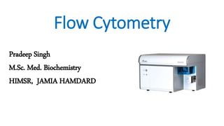
Flow Cytometry
Pradeep Singh
M.Sc. Med. Biochemistry
HIMSR, JAMIA HAMDARD
Contents
1. Introduction
2. Basic Principles of Flow Cytometry
3. Working of Flow Cytometer
4. Applications of Flow Cytometry
1. Introduction
• Flow cytometry is a technique of quantitative single cell analysis.
• This technique was first described by Wallace Coulter in the 1950s.
• The flow cytometer was developed in the 1970’s and applied to
automated cell counting.
• Flow cytometer count, examine and sort cells based on their optical
properties (Scattering and fluorescence).
• The present “state-of-the-art” flow cytometers are capable of
analyzing upto 13 parameters (forward scatter, side scatter, 11 colors
of immunofluorescence).
Components of flow cytometer:
1. Lasers
2. Dichroic mirrors
3. Filters
4. Detectors
2. Basic Principle of Flow Cytometry
• Prepared single cell or particle
suspension is necessary for flow
cytometric analysis.
• The suspension of cells or particles
is aspirated into a channel
surrounded by a narrow fluid
system.
• They pass one at a time through a
focused laser beam.
• The light is either scattered or
absorbed when it strikes a cell.
• Light scattering is dependent on
the internal structure of the cell
and its size and shape.
• Absorbed light of the appropriate
wavelength may be re-emitted as
fluorescence. (The cell may have a
naturally fluorescent substance or
one or more fluorochrome-labelled
antibodies are attached to surface
or internal cell structures).
• Light and/or fluorescence scatter signals are detected by a series of
photodiodes and amplified.
• Optical filters are essential to block unwanted light and permit light of
the desired wavelength to reach the photodetector.
• Fluorescein isothiocynate (FITS), Texas red and phycoerythrin (PE) are
the most common fluorescent dyes used in the biomedical sciences.
• Large number of cells are analysed in a short period of time
(>1,000/sec).
Sample
Loading
Flow
Chamber
Light
source
Scattering/
Absorption
Light
Detectors
Computer
3. Working of Flow Cytometer
• A flow cytometer is composed of three main systems:
1. Fluidics – Transport cells in a stream to the laser beam for interrogation.
2. Optics – Consist of lasers to illuminate the cells in the sample stream and
optical filters to direct the resulting light signals to the appropriate
detectors.
3. Electronics – Converts the detected light signals into electrical signals that
can be processed by computer.
3.1 Fluidics System
• Flow cytometers use the
principle of hydrodynamic
focusing for presenting cells one
at a time to a light source.
• The fluidics system consist of a
central channel through which
the sample is injected, enclosed
by an outer sheath that contains
faster flowing fluid.
• As the sheath fluid moves, it creates a massive drag effect on the
narrowing central chamber. This alters the velocity of the central fluid.
• The velocity of the central fluid becomes parabolic i.e., greatest
velocity at the center and zero velocity at the wall..
• This effect creates a single stream of particles or cells and is called
hydrodynamic focusing.
• Under laminar flow conditions, the fluid in the central chamber will
not mix with sheath fluid.
3.2 Optics
• Light scattering or fluorescence
emission provides information
about the cell’s properties.
• Light that is scattered by an object
is detected by different detectors.
• One detector is placed in line with
the beam to measure the forward
scatter (FSC) from the objects.
Detectors perpendicular to the
beam measure side scatter (SSC)
and fluorescence.
• Forward scatter is based on two
properties: size and refractive
index
• The FSC intensity roughly
equates to the particle’s size and
can also be used to distinguish
between cellular debris and
living cells.
• Dead cells have lower FSC and
higher SSC than living cells.
• Side scatter is based on the
granularity or internal
complexity.
• The more granular the cell, the
more side scatter light is
generated.
• Fluorescence measurements taken at different wavelength can
provide quantitative and qualitative data about fluorochrome-labelled
cell surface receptors or intracellular molecules such as DNA and
cytokines.
• When a fluorescent dye is conjugated to a monoclonal antibody, it
can be used to identify a particular cell type based on the individual
antigenic surface markers of the cell.
• The stating pattern of each subpopulation, combined with FSC and
SSC data, can be used to identify which cells are present in a sample
and to count their relative percentages.
Optical detectors
• Scattered and emitted light from cells are converted to electrical pulses by
optical detectors.
• Once a cell or particle passes through the laser light, the scattered and
fluorescence signals are diverted to the detectors.
• Detectors are either silicon photodiodes or photomultiplier tubes (PMTs).
• The photodiode is less sensitive to light signals than the PMTs and thus is
used to detect the stronger FSC.
• PMTs are used to detect the weaker signals generated by SSC and
fluorescence.
Optical
detectors
Photodiodes
Used to detect
FSC
Photomultiplier
tubes
Used to detect
SSC and
fluorescence
Optical
Detectors
Scattered
light detector
FSC and SSC
Fluorescence
light detector
FL-1, FL-2, FL-
3, FL-4
Filters
• All the signals are routed to their
detectors via a system of filters and
dichroic mirrors.
• Each PMT fluorescence detector is
placed behind a series of dichroic
mirrors and filters, so that it only
receives and detect light within a
particular range of wavelength.
• A particular color of light is split off
from the incoming mixture and
directed to the detectors by dichroic
mirrors.
Types of filters:
• There are three major type of
filters:
1. Longpass Filter: Transmits
wavelength of light equal to or
greater than the spectral band
of the filter.
2. Shortpass Filter: Transmits
wavelength of light equal to or
shorter than the spectral band
of the filter.
3. Bandpass Filter: Transmits
wavelength of light within a
specific range of wavelengths.
LP 500 SP 500
Longpass
480 500 520
Shortpass
480 500 520
Bandpass
480 520
460 500 540
BP 500
3.3 Electronics (Signal Processing)
• Scattered and emitted light data can
be converted to electrical pulses by
optical detectors.
• Flow cytometry data may be
represented as histograms or dot
plots.
Histogram: A histogram quantifies the
intensity of a single parameter, be it
fluorescence or scattering (SSC or FSC).
Dot plot: A dot plot is a two parameter
representation of a sample’s properties.
Gating
• Gating – It is a procedure to selectively visualize the cells of interest
while eliminating results from unwanted cells and debris.
• For example, if one has a heterogeneous population of cells which
contain lymphocytes, monocytes and granulocytes and is only
interested in evaluating the fluorescence of the lymphocytes
subpopulation.
• In this situation one could define an analysis gate around the
lymphocyte population. The resulting display would reflect the
fluorescence properties of only lymphocytes.
• For example, if we want to analyse a sample of peripheral blood that had been stained with
fluorochromes to identify the CD4 and CD14 surface markers but we are only interested in
knowing the percentage of monocytes that contain CD4 and CD14 surface markers. We will
place a gate around the monocyte population of the FSC versus SSC scatter plot.
Applications of Flow Cytometry
4.1 Cell Sorting (FCAS)
• A major application of flow
cytometry is the physical
separation of sub-population of
cells of interest from a
heterogeneous population.
• This process is known as cell
sorting of Fluorescence
Activated Cell Sorting (FACS)
New drop
Positive Charge
Negative Charge
No Charge
• Most commonly used cell sorting method is electrostatic deflection of droplets.
• In this method, the stream is focused in a vibrating nozzle and exits in a jet which
is broken into regularly spaced droplets.
• The cells of interest are charged electrically (positively or negatively). The
electrical charging actually occur at a precise moment called the ‘break-off point’.
• The cell of interest gets charged at the break-off point.
• When a charged droplet passes through a high voltage electrostatic field,
between the deflection plates, it is deflected and collected into the
corresponding collection tube.
• The deflection of the droplets is towards the oppositely charged plate, so that
this droplet is separated from uncharged and oppositely charged droplets.
4.2 Apoptosis
The following features of the apoptotic cascade
can be observed using flow cytometry:
• Altered phospholipid composition in the plasma membrane
• Activation of caspases
• Chromation condensation
• DNA fragmentation
• Expression of proteins involved in apoptosis
• Changes in mitochondrial membrane potential
• Decrease cytosolic pH
• Altered membrane permeability
Detection of apoptotic cells based on changes
in forward scattering
• During apoptosis there is an initial increase in SSC (probably due to
the chromatin condensation) with a reduction in FSC (due to cell
shrinkage).
• Drawback: In many cases, the forward light scattering histograms of
apoptotic and live cells overlap and make it difficult to discriminate
apoptotic cells based solely on this parameter.
Detection of apoptotic cells based on Annexin
V binding
• During early apoptosis cell lose
symmetry, phosphatidylserine
on the outer leaflet of the
plasma membrane.
• Annexin V is a calcium-
dependent phospholipid-binding
protein that binds preferentially
to negatively-charged
phosphatidylserine.
• The assay involves incubating cells briefly in a solution containing
fluorochrome conjugated Annexin V (FITC – Annexin V) in a buffer
that facilitates its binding.
• Apoptotic cells can be detected easily by flow cytometry on the basis
of fluorescence due to increased binding of FITC-conjugated Annexin
V.
• Drawback: Unfortunately, it is not specific only for apoptosis because
whenever cell membrane integrity is disrupted (even non-ionic
detergents), cells may stain with Annexin V.
Detection of apoptotic cells based on PI
binding
• The intact plasma membrane of live cells have a tendency to exclude
cationic dyes such as propidium iodide (PI) and 7-amino-
actinomycin D (7-AMD).
• PI is a good staining method to distinguish apoptotic, necrotic and
normal live cells.
• Apoptotic cells show an uptake of PI that is much lower than that of
necrotic cells. It is therefore possible to distinguish live (PI-negative),
apoptotic (PI-dim) and necrotic (PI-bright).
• Thus, the combined use of cationic dyes (e.g. PI) with annexin V
allows the discrimination between:
Live cells = Annexin V negative/PI negative
Early apoptotic cells = Annexin V positive/PI negative
Late apoptotic = Annexin V positive/PI positive
Necrotic cells = Annexin V negative/PI positive
Detection of apoptotic cells based on DNA
Fragmentation
• The late stages of apoptosis are characterized by changes in nuclear
morphology, including DNA fragmentation, chromatin condensation,
degradation of nuclear envelope, nuclear blebbing and DNA strand
breaks.
• Cells undergoing apoptosis display an increase in nuclear chromatin
condensation. As the chromatin condenses, cell-permeable nucleic
acid stains become hyperfluorescent, thus enabling the identification
of apoptotic cells.
Assessment of mitochondrial membrane
potential and caspases level
• Assessment of mitochondrial membrane potential and caspases level
within the cell through flow cytometry is also used to analyse
apoptotic cells.
• Cells undergoing apoptosis often lose the electric potential that
normally exists across the inner mitochondrial membrane.
• A distinctive feature of early stages of apoptosis is the activation of
caspases enzymes. These enzymes can be labelled with fluorophore
which can easily be detected by flow cytometry.
4.3 Cell Cycle Study
• One of the important application of flow cytometry is the
measurement of DNA content in cells.
• The duplication of the DNA occurs during the S-phase of cell cycle.
• There are two differnet methods to measure the DNA content:
1. The cells have to be stained with a fluorescent dye that binds DNA in a
stoichiometric manner.
2. Incorporation of thymidine analog bromodeoxyuridine (BrdU) during new
DNA synthesis.
Fluorescent dye that binds DNA in a
stoichiometric manner
• The cells are treated with a fluorescent dye that stains DNA
quantitatively. The fluorescence intensity of the stained cells at
certain wavelengths will therefore correlate with the amount of DNA
they contain.
• Dyes have different binding mechanism:
Intercalative binding dyes – Propidium iodide
A.T rich regions binding dyes – DAPI (4,6- Diamidion-2-phenylindole)
G.C rich regions binding dyes – Chromomycin A3
Incorporation of thymidine analog
bromodeoxyuridine (BrdU) during new DNA
synthesis
An accurate method for detection of
cell cycle progression also uses the
incorporation of thymidine analog
bromodeoxyuridine (BrdU) during
new DNA synthesis.
The incorporated BrdU is then stained
with specific fluorescently labelled
anti-BrdU antibodies, and the levels of
cell-associated BrdU measured using
flow cytometry.
By this method, the number of cells
that are proliferating rapidly & the
duration of S-phase can be calculated.
Clinical Applications of Flow Cytometry
1. Diagnosis of Hematologic
Malignancies
2. Detection of Minimal Residual
Disease
3. Lymphocyte Subset
Enumeration (HIV)
4. Efficacy of Cancer
Chemotherapy
5. Reticulocyte Enumeration
6. Cell Function Analysis
7. Application in Organ
Transplantation
Thank You !!!

Contenu connexe

Tendances

Flow Cytometry - basics, principles and applications
Flow Cytometry - basics, principles and applicationsFlow Cytometry - basics, principles and applications
Flow Cytometry - basics, principles and applicationsAnkit Raiyani
 
Flow cytometry in diagnostics
Flow cytometry in diagnosticsFlow cytometry in diagnostics
Flow cytometry in diagnosticsKaberi Nath
 
Flow cytometry definition, principle, parts, steps, types, uses
Flow cytometry definition, principle, parts, steps, types, usesFlow cytometry definition, principle, parts, steps, types, uses
Flow cytometry definition, principle, parts, steps, types, usesGayathri Devi S
 
Fluorescence-activated Cell Sorting (FACS).pptx
Fluorescence-activated Cell Sorting (FACS).pptxFluorescence-activated Cell Sorting (FACS).pptx
Fluorescence-activated Cell Sorting (FACS).pptxkalok4
 
Flow cytometry for cell componenet analysis
Flow cytometry for cell componenet analysisFlow cytometry for cell componenet analysis
Flow cytometry for cell componenet analysisRAJA GOPAL
 

Tendances (20)

Flow cytometry
Flow cytometryFlow cytometry
Flow cytometry
 
Introduction to Flow Cytometry
Introduction to Flow CytometryIntroduction to Flow Cytometry
Introduction to Flow Cytometry
 
Flow Cytometry - basics, principles and applications
Flow Cytometry - basics, principles and applicationsFlow Cytometry - basics, principles and applications
Flow Cytometry - basics, principles and applications
 
Flow Cytometry
Flow CytometryFlow Cytometry
Flow Cytometry
 
Flow cytometry in diagnostics
Flow cytometry in diagnosticsFlow cytometry in diagnostics
Flow cytometry in diagnostics
 
Immunofluorescence
ImmunofluorescenceImmunofluorescence
Immunofluorescence
 
Flow cytometry definition, principle, parts, steps, types, uses
Flow cytometry definition, principle, parts, steps, types, usesFlow cytometry definition, principle, parts, steps, types, uses
Flow cytometry definition, principle, parts, steps, types, uses
 
Fluorescence-activated Cell Sorting (FACS).pptx
Fluorescence-activated Cell Sorting (FACS).pptxFluorescence-activated Cell Sorting (FACS).pptx
Fluorescence-activated Cell Sorting (FACS).pptx
 
Flow cytometry
Flow cytometryFlow cytometry
Flow cytometry
 
Flow cytometry
Flow cytometry Flow cytometry
Flow cytometry
 
Flow Cytometry
Flow CytometryFlow Cytometry
Flow Cytometry
 
Flow cytometry
Flow cytometry Flow cytometry
Flow cytometry
 
Hla typing
Hla typingHla typing
Hla typing
 
Flow cytometry
Flow cytometryFlow cytometry
Flow cytometry
 
Fluorescence Microscopy
Fluorescence MicroscopyFluorescence Microscopy
Fluorescence Microscopy
 
Flow cytometry
Flow cytometryFlow cytometry
Flow cytometry
 
Flow cytometry for cell componenet analysis
Flow cytometry for cell componenet analysisFlow cytometry for cell componenet analysis
Flow cytometry for cell componenet analysis
 
MALDI - TOF
MALDI - TOFMALDI - TOF
MALDI - TOF
 
Flow cytometry
Flow cytometryFlow cytometry
Flow cytometry
 
Immunocytochemistry Protocol
Immunocytochemistry ProtocolImmunocytochemistry Protocol
Immunocytochemistry Protocol
 

Similaire à Flow cytometry

Flow cytometry in cell biology
Flow cytometry in cell biologyFlow cytometry in cell biology
Flow cytometry in cell biologyAmani Riyadh
 
FLOWCYTOMETRY........................pptx
FLOWCYTOMETRY........................pptxFLOWCYTOMETRY........................pptx
FLOWCYTOMETRY........................pptxHarishankarSharma27
 
flow cytometry.pptx
flow cytometry.pptxflow cytometry.pptx
flow cytometry.pptxjasveer15
 
flowcytometry-150403122734-conversion-gate01.pdf
flowcytometry-150403122734-conversion-gate01.pdfflowcytometry-150403122734-conversion-gate01.pdf
flowcytometry-150403122734-conversion-gate01.pdfdmfrmicro
 
Fluorescence- Activated Cell Sorter (FACS)
Fluorescence- Activated Cell Sorter (FACS)Fluorescence- Activated Cell Sorter (FACS)
Fluorescence- Activated Cell Sorter (FACS)Nidhi Parikh
 
flowcytometry-basic principle of flowcyto
flowcytometry-basic principle of flowcytoflowcytometry-basic principle of flowcyto
flowcytometry-basic principle of flowcytoDr. Ajit Surya Singh
 
Hematopoetic Stem Cell enumeration by Flowcytometry
Hematopoetic Stem Cell enumeration by FlowcytometryHematopoetic Stem Cell enumeration by Flowcytometry
Hematopoetic Stem Cell enumeration by FlowcytometrySusanta Das
 
FLOW CYTOMETRY -SUNANDA ARYA.pptx
FLOW CYTOMETRY -SUNANDA ARYA.pptxFLOW CYTOMETRY -SUNANDA ARYA.pptx
FLOW CYTOMETRY -SUNANDA ARYA.pptxSunandaArya
 
flow cytometry presentation
flow cytometry  presentationflow cytometry  presentation
flow cytometry presentationJamile Saberzade
 
Flow cytometry principle
Flow cytometry principleFlow cytometry principle
Flow cytometry principleJames Waita
 

Similaire à Flow cytometry (20)

Flow cytometry in cell biology
Flow cytometry in cell biologyFlow cytometry in cell biology
Flow cytometry in cell biology
 
FLOWCYTOMETRY........................pptx
FLOWCYTOMETRY........................pptxFLOWCYTOMETRY........................pptx
FLOWCYTOMETRY........................pptx
 
Flowcytometry
FlowcytometryFlowcytometry
Flowcytometry
 
flow cytometry.pptx
flow cytometry.pptxflow cytometry.pptx
flow cytometry.pptx
 
Flow cytometry
Flow cytometryFlow cytometry
Flow cytometry
 
Flow cytometry
Flow cytometryFlow cytometry
Flow cytometry
 
flow_cytometry.pdf
flow_cytometry.pdfflow_cytometry.pdf
flow_cytometry.pdf
 
Presentation 7
Presentation 7Presentation 7
Presentation 7
 
Flow cytometry
Flow cytometryFlow cytometry
Flow cytometry
 
Flow cytometry.pptx
Flow cytometry.pptxFlow cytometry.pptx
Flow cytometry.pptx
 
Flow cytometry
Flow cytometryFlow cytometry
Flow cytometry
 
flowcytometry-150403122734-conversion-gate01.pdf
flowcytometry-150403122734-conversion-gate01.pdfflowcytometry-150403122734-conversion-gate01.pdf
flowcytometry-150403122734-conversion-gate01.pdf
 
Fluorescence- Activated Cell Sorter (FACS)
Fluorescence- Activated Cell Sorter (FACS)Fluorescence- Activated Cell Sorter (FACS)
Fluorescence- Activated Cell Sorter (FACS)
 
flowcytometry-basic principle of flowcyto
flowcytometry-basic principle of flowcytoflowcytometry-basic principle of flowcyto
flowcytometry-basic principle of flowcyto
 
Hematopoetic Stem Cell enumeration by Flowcytometry
Hematopoetic Stem Cell enumeration by FlowcytometryHematopoetic Stem Cell enumeration by Flowcytometry
Hematopoetic Stem Cell enumeration by Flowcytometry
 
Flow cytometry
Flow cytometryFlow cytometry
Flow cytometry
 
FLOW CYTOMETRY -SUNANDA ARYA.pptx
FLOW CYTOMETRY -SUNANDA ARYA.pptxFLOW CYTOMETRY -SUNANDA ARYA.pptx
FLOW CYTOMETRY -SUNANDA ARYA.pptx
 
flow cytometry presentation
flow cytometry  presentationflow cytometry  presentation
flow cytometry presentation
 
Flow cytometry principle
Flow cytometry principleFlow cytometry principle
Flow cytometry principle
 
Flow Cytometry Principle
Flow Cytometry PrincipleFlow Cytometry Principle
Flow Cytometry Principle
 

Plus de Pradeep Singh Narwat

Comparative biology of rat and human
Comparative biology of rat and humanComparative biology of rat and human
Comparative biology of rat and humanPradeep Singh Narwat
 
Applications of Isotopes in Medicine
Applications of Isotopes in MedicineApplications of Isotopes in Medicine
Applications of Isotopes in MedicinePradeep Singh Narwat
 
Automation in Biochemistry (Autoanalyzers)
Automation in Biochemistry (Autoanalyzers)Automation in Biochemistry (Autoanalyzers)
Automation in Biochemistry (Autoanalyzers)Pradeep Singh Narwat
 
Major histocompatility complex (Antigen Presentation to T cells, Autoimmunity...
Major histocompatility complex (Antigen Presentation to T cells, Autoimmunity...Major histocompatility complex (Antigen Presentation to T cells, Autoimmunity...
Major histocompatility complex (Antigen Presentation to T cells, Autoimmunity...Pradeep Singh Narwat
 
Cell membrane (Plasma Membrane) & Cellular Junctions
Cell membrane (Plasma Membrane) & Cellular JunctionsCell membrane (Plasma Membrane) & Cellular Junctions
Cell membrane (Plasma Membrane) & Cellular JunctionsPradeep Singh Narwat
 
Junk DNA/ Non-coding DNA and its Importance (Regulatory RNAs, RNA interferen...
Junk DNA/ Non-coding DNA and its Importance  (Regulatory RNAs, RNA interferen...Junk DNA/ Non-coding DNA and its Importance  (Regulatory RNAs, RNA interferen...
Junk DNA/ Non-coding DNA and its Importance (Regulatory RNAs, RNA interferen...Pradeep Singh Narwat
 
Hemostatsis (Fibrinolysis - Plasminogen)
Hemostatsis (Fibrinolysis - Plasminogen)Hemostatsis (Fibrinolysis - Plasminogen)
Hemostatsis (Fibrinolysis - Plasminogen)Pradeep Singh Narwat
 
G protein coupled receptors (Type II Hormone Signaling)
G protein coupled receptors (Type II Hormone Signaling)G protein coupled receptors (Type II Hormone Signaling)
G protein coupled receptors (Type II Hormone Signaling)Pradeep Singh Narwat
 
Signal transduction processes connected to the changes in cytosolic calcium c...
Signal transduction processes connected to the changes in cytosolic calcium c...Signal transduction processes connected to the changes in cytosolic calcium c...
Signal transduction processes connected to the changes in cytosolic calcium c...Pradeep Singh Narwat
 
Purification of proteins (purification of enzymes)
Purification of proteins (purification of enzymes)Purification of proteins (purification of enzymes)
Purification of proteins (purification of enzymes)Pradeep Singh Narwat
 
Role of Nitric oxide & Nitric oxide synthases (NOS)
Role of Nitric oxide & Nitric oxide synthases (NOS)Role of Nitric oxide & Nitric oxide synthases (NOS)
Role of Nitric oxide & Nitric oxide synthases (NOS)Pradeep Singh Narwat
 
Metabolic syndrome & its complications
Metabolic syndrome & its complicationsMetabolic syndrome & its complications
Metabolic syndrome & its complicationsPradeep Singh Narwat
 
Determination of primary structure of proteins
Determination of primary structure of proteinsDetermination of primary structure of proteins
Determination of primary structure of proteinsPradeep Singh Narwat
 
Cytoskeleton & Extracellular matrix
Cytoskeleton & Extracellular matrixCytoskeleton & Extracellular matrix
Cytoskeleton & Extracellular matrixPradeep Singh Narwat
 

Plus de Pradeep Singh Narwat (20)

Intellectual property rights
Intellectual property rightsIntellectual property rights
Intellectual property rights
 
Comparative biology of rat and human
Comparative biology of rat and humanComparative biology of rat and human
Comparative biology of rat and human
 
Applications of Isotopes in Medicine
Applications of Isotopes in MedicineApplications of Isotopes in Medicine
Applications of Isotopes in Medicine
 
Human gene therapy
Human gene therapyHuman gene therapy
Human gene therapy
 
Chromatography
ChromatographyChromatography
Chromatography
 
Automation in Biochemistry (Autoanalyzers)
Automation in Biochemistry (Autoanalyzers)Automation in Biochemistry (Autoanalyzers)
Automation in Biochemistry (Autoanalyzers)
 
Major histocompatility complex (Antigen Presentation to T cells, Autoimmunity...
Major histocompatility complex (Antigen Presentation to T cells, Autoimmunity...Major histocompatility complex (Antigen Presentation to T cells, Autoimmunity...
Major histocompatility complex (Antigen Presentation to T cells, Autoimmunity...
 
Cell membrane (Plasma Membrane) & Cellular Junctions
Cell membrane (Plasma Membrane) & Cellular JunctionsCell membrane (Plasma Membrane) & Cellular Junctions
Cell membrane (Plasma Membrane) & Cellular Junctions
 
Junk DNA/ Non-coding DNA and its Importance (Regulatory RNAs, RNA interferen...
Junk DNA/ Non-coding DNA and its Importance  (Regulatory RNAs, RNA interferen...Junk DNA/ Non-coding DNA and its Importance  (Regulatory RNAs, RNA interferen...
Junk DNA/ Non-coding DNA and its Importance (Regulatory RNAs, RNA interferen...
 
Hemostatsis (Fibrinolysis - Plasminogen)
Hemostatsis (Fibrinolysis - Plasminogen)Hemostatsis (Fibrinolysis - Plasminogen)
Hemostatsis (Fibrinolysis - Plasminogen)
 
G protein coupled receptors (Type II Hormone Signaling)
G protein coupled receptors (Type II Hormone Signaling)G protein coupled receptors (Type II Hormone Signaling)
G protein coupled receptors (Type II Hormone Signaling)
 
Signal transduction processes connected to the changes in cytosolic calcium c...
Signal transduction processes connected to the changes in cytosolic calcium c...Signal transduction processes connected to the changes in cytosolic calcium c...
Signal transduction processes connected to the changes in cytosolic calcium c...
 
Purification of proteins (purification of enzymes)
Purification of proteins (purification of enzymes)Purification of proteins (purification of enzymes)
Purification of proteins (purification of enzymes)
 
Role of Nitric oxide & Nitric oxide synthases (NOS)
Role of Nitric oxide & Nitric oxide synthases (NOS)Role of Nitric oxide & Nitric oxide synthases (NOS)
Role of Nitric oxide & Nitric oxide synthases (NOS)
 
Sodium - Potassium Pump
Sodium - Potassium Pump Sodium - Potassium Pump
Sodium - Potassium Pump
 
Metabolic syndrome & its complications
Metabolic syndrome & its complicationsMetabolic syndrome & its complications
Metabolic syndrome & its complications
 
Transport across Plasma Membrane
Transport across Plasma Membrane Transport across Plasma Membrane
Transport across Plasma Membrane
 
Electrophoresis
ElectrophoresisElectrophoresis
Electrophoresis
 
Determination of primary structure of proteins
Determination of primary structure of proteinsDetermination of primary structure of proteins
Determination of primary structure of proteins
 
Cytoskeleton & Extracellular matrix
Cytoskeleton & Extracellular matrixCytoskeleton & Extracellular matrix
Cytoskeleton & Extracellular matrix
 

Dernier

💚Reliable Call Girls Chandigarh 💯Niamh 📲🔝8868886958🔝Call Girl In Chandigarh N...
💚Reliable Call Girls Chandigarh 💯Niamh 📲🔝8868886958🔝Call Girl In Chandigarh N...💚Reliable Call Girls Chandigarh 💯Niamh 📲🔝8868886958🔝Call Girl In Chandigarh N...
💚Reliable Call Girls Chandigarh 💯Niamh 📲🔝8868886958🔝Call Girl In Chandigarh N...Sheetaleventcompany
 
Ahmedabad Call Girls Book Now 9630942363 Top Class Ahmedabad Escort Service A...
Ahmedabad Call Girls Book Now 9630942363 Top Class Ahmedabad Escort Service A...Ahmedabad Call Girls Book Now 9630942363 Top Class Ahmedabad Escort Service A...
Ahmedabad Call Girls Book Now 9630942363 Top Class Ahmedabad Escort Service A...Genuine Call Girls
 
Gorgeous Call Girls Dehradun {8854095900} ❤️VVIP ROCKY Call Girls in Dehradun...
Gorgeous Call Girls Dehradun {8854095900} ❤️VVIP ROCKY Call Girls in Dehradun...Gorgeous Call Girls Dehradun {8854095900} ❤️VVIP ROCKY Call Girls in Dehradun...
Gorgeous Call Girls Dehradun {8854095900} ❤️VVIP ROCKY Call Girls in Dehradun...Sheetaleventcompany
 
Call Girls Shahdol Just Call 8250077686 Top Class Call Girl Service Available
Call Girls Shahdol Just Call 8250077686 Top Class Call Girl Service AvailableCall Girls Shahdol Just Call 8250077686 Top Class Call Girl Service Available
Call Girls Shahdol Just Call 8250077686 Top Class Call Girl Service AvailableDipal Arora
 
Chennai ❣️ Call Girl 6378878445 Call Girls in Chennai Escort service book now
Chennai ❣️ Call Girl 6378878445 Call Girls in Chennai Escort service book nowChennai ❣️ Call Girl 6378878445 Call Girls in Chennai Escort service book now
Chennai ❣️ Call Girl 6378878445 Call Girls in Chennai Escort service book nowtanudubay92
 
Call Girl in Chennai | Whatsapp No 📞 7427069034 📞 VIP Escorts Service Availab...
Call Girl in Chennai | Whatsapp No 📞 7427069034 📞 VIP Escorts Service Availab...Call Girl in Chennai | Whatsapp No 📞 7427069034 📞 VIP Escorts Service Availab...
Call Girl in Chennai | Whatsapp No 📞 7427069034 📞 VIP Escorts Service Availab...amritaverma53
 
ANATOMY AND PHYSIOLOGY OF REPRODUCTIVE SYSTEM.pptx
ANATOMY AND PHYSIOLOGY OF REPRODUCTIVE SYSTEM.pptxANATOMY AND PHYSIOLOGY OF REPRODUCTIVE SYSTEM.pptx
ANATOMY AND PHYSIOLOGY OF REPRODUCTIVE SYSTEM.pptxSwetaba Besh
 
tongue disease lecture Dr Assadawy legacy
tongue disease lecture Dr Assadawy legacytongue disease lecture Dr Assadawy legacy
tongue disease lecture Dr Assadawy legacyDrMohamed Assadawy
 
Premium Call Girls Dehradun {8854095900} ❤️VVIP ANJU Call Girls in Dehradun U...
Premium Call Girls Dehradun {8854095900} ❤️VVIP ANJU Call Girls in Dehradun U...Premium Call Girls Dehradun {8854095900} ❤️VVIP ANJU Call Girls in Dehradun U...
Premium Call Girls Dehradun {8854095900} ❤️VVIP ANJU Call Girls in Dehradun U...Sheetaleventcompany
 
Kolkata Call Girls Shobhabazar 💯Call Us 🔝 8005736733 🔝 💃 Top Class Call Gir...
Kolkata Call Girls Shobhabazar  💯Call Us 🔝 8005736733 🔝 💃  Top Class Call Gir...Kolkata Call Girls Shobhabazar  💯Call Us 🔝 8005736733 🔝 💃  Top Class Call Gir...
Kolkata Call Girls Shobhabazar 💯Call Us 🔝 8005736733 🔝 💃 Top Class Call Gir...Namrata Singh
 
Call Girls Mussoorie Just Call 8854095900 Top Class Call Girl Service Available
Call Girls Mussoorie Just Call 8854095900 Top Class Call Girl Service AvailableCall Girls Mussoorie Just Call 8854095900 Top Class Call Girl Service Available
Call Girls Mussoorie Just Call 8854095900 Top Class Call Girl Service AvailableJanvi Singh
 
Dehradun Call Girls Service {8854095900} ❤️VVIP ROCKY Call Girl in Dehradun U...
Dehradun Call Girls Service {8854095900} ❤️VVIP ROCKY Call Girl in Dehradun U...Dehradun Call Girls Service {8854095900} ❤️VVIP ROCKY Call Girl in Dehradun U...
Dehradun Call Girls Service {8854095900} ❤️VVIP ROCKY Call Girl in Dehradun U...Sheetaleventcompany
 
Circulatory Shock, types and stages, compensatory mechanisms
Circulatory Shock, types and stages, compensatory mechanismsCirculatory Shock, types and stages, compensatory mechanisms
Circulatory Shock, types and stages, compensatory mechanismsMedicoseAcademics
 
Call Girls Kathua Just Call 8250077686 Top Class Call Girl Service Available
Call Girls Kathua Just Call 8250077686 Top Class Call Girl Service AvailableCall Girls Kathua Just Call 8250077686 Top Class Call Girl Service Available
Call Girls Kathua Just Call 8250077686 Top Class Call Girl Service AvailableDipal Arora
 
❤️Chandigarh Escorts Service☎️9814379184☎️ Call Girl service in Chandigarh☎️ ...
❤️Chandigarh Escorts Service☎️9814379184☎️ Call Girl service in Chandigarh☎️ ...❤️Chandigarh Escorts Service☎️9814379184☎️ Call Girl service in Chandigarh☎️ ...
❤️Chandigarh Escorts Service☎️9814379184☎️ Call Girl service in Chandigarh☎️ ...Sheetaleventcompany
 
Pune Call Girl Service 📞9xx000xx09📞Just Call Divya📲 Call Girl In Pune No💰Adva...
Pune Call Girl Service 📞9xx000xx09📞Just Call Divya📲 Call Girl In Pune No💰Adva...Pune Call Girl Service 📞9xx000xx09📞Just Call Divya📲 Call Girl In Pune No💰Adva...
Pune Call Girl Service 📞9xx000xx09📞Just Call Divya📲 Call Girl In Pune No💰Adva...Sheetaleventcompany
 
Race Course Road } Book Call Girls in Bangalore | Whatsapp No 6378878445 VIP ...
Race Course Road } Book Call Girls in Bangalore | Whatsapp No 6378878445 VIP ...Race Course Road } Book Call Girls in Bangalore | Whatsapp No 6378878445 VIP ...
Race Course Road } Book Call Girls in Bangalore | Whatsapp No 6378878445 VIP ...dishamehta3332
 
(RIYA)🎄Airhostess Call Girl Jaipur Call Now 8445551418 Premium Collection Of ...
(RIYA)🎄Airhostess Call Girl Jaipur Call Now 8445551418 Premium Collection Of ...(RIYA)🎄Airhostess Call Girl Jaipur Call Now 8445551418 Premium Collection Of ...
(RIYA)🎄Airhostess Call Girl Jaipur Call Now 8445551418 Premium Collection Of ...TanyaAhuja34
 
ANATOMY AND PHYSIOLOGY OF RESPIRATORY SYSTEM.pptx
ANATOMY AND PHYSIOLOGY OF RESPIRATORY SYSTEM.pptxANATOMY AND PHYSIOLOGY OF RESPIRATORY SYSTEM.pptx
ANATOMY AND PHYSIOLOGY OF RESPIRATORY SYSTEM.pptxSwetaba Besh
 
Call girls Service Phullen / 9332606886 Genuine Call girls with real Photos a...
Call girls Service Phullen / 9332606886 Genuine Call girls with real Photos a...Call girls Service Phullen / 9332606886 Genuine Call girls with real Photos a...
Call girls Service Phullen / 9332606886 Genuine Call girls with real Photos a...call girls hydrabad
 

Dernier (20)

💚Reliable Call Girls Chandigarh 💯Niamh 📲🔝8868886958🔝Call Girl In Chandigarh N...
💚Reliable Call Girls Chandigarh 💯Niamh 📲🔝8868886958🔝Call Girl In Chandigarh N...💚Reliable Call Girls Chandigarh 💯Niamh 📲🔝8868886958🔝Call Girl In Chandigarh N...
💚Reliable Call Girls Chandigarh 💯Niamh 📲🔝8868886958🔝Call Girl In Chandigarh N...
 
Ahmedabad Call Girls Book Now 9630942363 Top Class Ahmedabad Escort Service A...
Ahmedabad Call Girls Book Now 9630942363 Top Class Ahmedabad Escort Service A...Ahmedabad Call Girls Book Now 9630942363 Top Class Ahmedabad Escort Service A...
Ahmedabad Call Girls Book Now 9630942363 Top Class Ahmedabad Escort Service A...
 
Gorgeous Call Girls Dehradun {8854095900} ❤️VVIP ROCKY Call Girls in Dehradun...
Gorgeous Call Girls Dehradun {8854095900} ❤️VVIP ROCKY Call Girls in Dehradun...Gorgeous Call Girls Dehradun {8854095900} ❤️VVIP ROCKY Call Girls in Dehradun...
Gorgeous Call Girls Dehradun {8854095900} ❤️VVIP ROCKY Call Girls in Dehradun...
 
Call Girls Shahdol Just Call 8250077686 Top Class Call Girl Service Available
Call Girls Shahdol Just Call 8250077686 Top Class Call Girl Service AvailableCall Girls Shahdol Just Call 8250077686 Top Class Call Girl Service Available
Call Girls Shahdol Just Call 8250077686 Top Class Call Girl Service Available
 
Chennai ❣️ Call Girl 6378878445 Call Girls in Chennai Escort service book now
Chennai ❣️ Call Girl 6378878445 Call Girls in Chennai Escort service book nowChennai ❣️ Call Girl 6378878445 Call Girls in Chennai Escort service book now
Chennai ❣️ Call Girl 6378878445 Call Girls in Chennai Escort service book now
 
Call Girl in Chennai | Whatsapp No 📞 7427069034 📞 VIP Escorts Service Availab...
Call Girl in Chennai | Whatsapp No 📞 7427069034 📞 VIP Escorts Service Availab...Call Girl in Chennai | Whatsapp No 📞 7427069034 📞 VIP Escorts Service Availab...
Call Girl in Chennai | Whatsapp No 📞 7427069034 📞 VIP Escorts Service Availab...
 
ANATOMY AND PHYSIOLOGY OF REPRODUCTIVE SYSTEM.pptx
ANATOMY AND PHYSIOLOGY OF REPRODUCTIVE SYSTEM.pptxANATOMY AND PHYSIOLOGY OF REPRODUCTIVE SYSTEM.pptx
ANATOMY AND PHYSIOLOGY OF REPRODUCTIVE SYSTEM.pptx
 
tongue disease lecture Dr Assadawy legacy
tongue disease lecture Dr Assadawy legacytongue disease lecture Dr Assadawy legacy
tongue disease lecture Dr Assadawy legacy
 
Premium Call Girls Dehradun {8854095900} ❤️VVIP ANJU Call Girls in Dehradun U...
Premium Call Girls Dehradun {8854095900} ❤️VVIP ANJU Call Girls in Dehradun U...Premium Call Girls Dehradun {8854095900} ❤️VVIP ANJU Call Girls in Dehradun U...
Premium Call Girls Dehradun {8854095900} ❤️VVIP ANJU Call Girls in Dehradun U...
 
Kolkata Call Girls Shobhabazar 💯Call Us 🔝 8005736733 🔝 💃 Top Class Call Gir...
Kolkata Call Girls Shobhabazar  💯Call Us 🔝 8005736733 🔝 💃  Top Class Call Gir...Kolkata Call Girls Shobhabazar  💯Call Us 🔝 8005736733 🔝 💃  Top Class Call Gir...
Kolkata Call Girls Shobhabazar 💯Call Us 🔝 8005736733 🔝 💃 Top Class Call Gir...
 
Call Girls Mussoorie Just Call 8854095900 Top Class Call Girl Service Available
Call Girls Mussoorie Just Call 8854095900 Top Class Call Girl Service AvailableCall Girls Mussoorie Just Call 8854095900 Top Class Call Girl Service Available
Call Girls Mussoorie Just Call 8854095900 Top Class Call Girl Service Available
 
Dehradun Call Girls Service {8854095900} ❤️VVIP ROCKY Call Girl in Dehradun U...
Dehradun Call Girls Service {8854095900} ❤️VVIP ROCKY Call Girl in Dehradun U...Dehradun Call Girls Service {8854095900} ❤️VVIP ROCKY Call Girl in Dehradun U...
Dehradun Call Girls Service {8854095900} ❤️VVIP ROCKY Call Girl in Dehradun U...
 
Circulatory Shock, types and stages, compensatory mechanisms
Circulatory Shock, types and stages, compensatory mechanismsCirculatory Shock, types and stages, compensatory mechanisms
Circulatory Shock, types and stages, compensatory mechanisms
 
Call Girls Kathua Just Call 8250077686 Top Class Call Girl Service Available
Call Girls Kathua Just Call 8250077686 Top Class Call Girl Service AvailableCall Girls Kathua Just Call 8250077686 Top Class Call Girl Service Available
Call Girls Kathua Just Call 8250077686 Top Class Call Girl Service Available
 
❤️Chandigarh Escorts Service☎️9814379184☎️ Call Girl service in Chandigarh☎️ ...
❤️Chandigarh Escorts Service☎️9814379184☎️ Call Girl service in Chandigarh☎️ ...❤️Chandigarh Escorts Service☎️9814379184☎️ Call Girl service in Chandigarh☎️ ...
❤️Chandigarh Escorts Service☎️9814379184☎️ Call Girl service in Chandigarh☎️ ...
 
Pune Call Girl Service 📞9xx000xx09📞Just Call Divya📲 Call Girl In Pune No💰Adva...
Pune Call Girl Service 📞9xx000xx09📞Just Call Divya📲 Call Girl In Pune No💰Adva...Pune Call Girl Service 📞9xx000xx09📞Just Call Divya📲 Call Girl In Pune No💰Adva...
Pune Call Girl Service 📞9xx000xx09📞Just Call Divya📲 Call Girl In Pune No💰Adva...
 
Race Course Road } Book Call Girls in Bangalore | Whatsapp No 6378878445 VIP ...
Race Course Road } Book Call Girls in Bangalore | Whatsapp No 6378878445 VIP ...Race Course Road } Book Call Girls in Bangalore | Whatsapp No 6378878445 VIP ...
Race Course Road } Book Call Girls in Bangalore | Whatsapp No 6378878445 VIP ...
 
(RIYA)🎄Airhostess Call Girl Jaipur Call Now 8445551418 Premium Collection Of ...
(RIYA)🎄Airhostess Call Girl Jaipur Call Now 8445551418 Premium Collection Of ...(RIYA)🎄Airhostess Call Girl Jaipur Call Now 8445551418 Premium Collection Of ...
(RIYA)🎄Airhostess Call Girl Jaipur Call Now 8445551418 Premium Collection Of ...
 
ANATOMY AND PHYSIOLOGY OF RESPIRATORY SYSTEM.pptx
ANATOMY AND PHYSIOLOGY OF RESPIRATORY SYSTEM.pptxANATOMY AND PHYSIOLOGY OF RESPIRATORY SYSTEM.pptx
ANATOMY AND PHYSIOLOGY OF RESPIRATORY SYSTEM.pptx
 
Call girls Service Phullen / 9332606886 Genuine Call girls with real Photos a...
Call girls Service Phullen / 9332606886 Genuine Call girls with real Photos a...Call girls Service Phullen / 9332606886 Genuine Call girls with real Photos a...
Call girls Service Phullen / 9332606886 Genuine Call girls with real Photos a...
 

Flow cytometry

  • 1. Flow Cytometry Pradeep Singh M.Sc. Med. Biochemistry HIMSR, JAMIA HAMDARD
  • 2. Contents 1. Introduction 2. Basic Principles of Flow Cytometry 3. Working of Flow Cytometer 4. Applications of Flow Cytometry
  • 3. 1. Introduction • Flow cytometry is a technique of quantitative single cell analysis. • This technique was first described by Wallace Coulter in the 1950s. • The flow cytometer was developed in the 1970’s and applied to automated cell counting. • Flow cytometer count, examine and sort cells based on their optical properties (Scattering and fluorescence). • The present “state-of-the-art” flow cytometers are capable of analyzing upto 13 parameters (forward scatter, side scatter, 11 colors of immunofluorescence).
  • 4. Components of flow cytometer: 1. Lasers 2. Dichroic mirrors 3. Filters 4. Detectors
  • 5. 2. Basic Principle of Flow Cytometry • Prepared single cell or particle suspension is necessary for flow cytometric analysis. • The suspension of cells or particles is aspirated into a channel surrounded by a narrow fluid system. • They pass one at a time through a focused laser beam. • The light is either scattered or absorbed when it strikes a cell.
  • 6. • Light scattering is dependent on the internal structure of the cell and its size and shape. • Absorbed light of the appropriate wavelength may be re-emitted as fluorescence. (The cell may have a naturally fluorescent substance or one or more fluorochrome-labelled antibodies are attached to surface or internal cell structures).
  • 7. • Light and/or fluorescence scatter signals are detected by a series of photodiodes and amplified. • Optical filters are essential to block unwanted light and permit light of the desired wavelength to reach the photodetector. • Fluorescein isothiocynate (FITS), Texas red and phycoerythrin (PE) are the most common fluorescent dyes used in the biomedical sciences. • Large number of cells are analysed in a short period of time (>1,000/sec).
  • 9. 3. Working of Flow Cytometer • A flow cytometer is composed of three main systems: 1. Fluidics – Transport cells in a stream to the laser beam for interrogation. 2. Optics – Consist of lasers to illuminate the cells in the sample stream and optical filters to direct the resulting light signals to the appropriate detectors. 3. Electronics – Converts the detected light signals into electrical signals that can be processed by computer.
  • 10. 3.1 Fluidics System • Flow cytometers use the principle of hydrodynamic focusing for presenting cells one at a time to a light source. • The fluidics system consist of a central channel through which the sample is injected, enclosed by an outer sheath that contains faster flowing fluid.
  • 11. • As the sheath fluid moves, it creates a massive drag effect on the narrowing central chamber. This alters the velocity of the central fluid. • The velocity of the central fluid becomes parabolic i.e., greatest velocity at the center and zero velocity at the wall.. • This effect creates a single stream of particles or cells and is called hydrodynamic focusing. • Under laminar flow conditions, the fluid in the central chamber will not mix with sheath fluid.
  • 12. 3.2 Optics • Light scattering or fluorescence emission provides information about the cell’s properties. • Light that is scattered by an object is detected by different detectors. • One detector is placed in line with the beam to measure the forward scatter (FSC) from the objects. Detectors perpendicular to the beam measure side scatter (SSC) and fluorescence.
  • 13. • Forward scatter is based on two properties: size and refractive index • The FSC intensity roughly equates to the particle’s size and can also be used to distinguish between cellular debris and living cells. • Dead cells have lower FSC and higher SSC than living cells.
  • 14. • Side scatter is based on the granularity or internal complexity. • The more granular the cell, the more side scatter light is generated.
  • 15. • Fluorescence measurements taken at different wavelength can provide quantitative and qualitative data about fluorochrome-labelled cell surface receptors or intracellular molecules such as DNA and cytokines. • When a fluorescent dye is conjugated to a monoclonal antibody, it can be used to identify a particular cell type based on the individual antigenic surface markers of the cell. • The stating pattern of each subpopulation, combined with FSC and SSC data, can be used to identify which cells are present in a sample and to count their relative percentages.
  • 16. Optical detectors • Scattered and emitted light from cells are converted to electrical pulses by optical detectors. • Once a cell or particle passes through the laser light, the scattered and fluorescence signals are diverted to the detectors. • Detectors are either silicon photodiodes or photomultiplier tubes (PMTs). • The photodiode is less sensitive to light signals than the PMTs and thus is used to detect the stronger FSC. • PMTs are used to detect the weaker signals generated by SSC and fluorescence.
  • 18. Optical Detectors Scattered light detector FSC and SSC Fluorescence light detector FL-1, FL-2, FL- 3, FL-4
  • 19. Filters • All the signals are routed to their detectors via a system of filters and dichroic mirrors. • Each PMT fluorescence detector is placed behind a series of dichroic mirrors and filters, so that it only receives and detect light within a particular range of wavelength. • A particular color of light is split off from the incoming mixture and directed to the detectors by dichroic mirrors.
  • 20. Types of filters: • There are three major type of filters: 1. Longpass Filter: Transmits wavelength of light equal to or greater than the spectral band of the filter. 2. Shortpass Filter: Transmits wavelength of light equal to or shorter than the spectral band of the filter. 3. Bandpass Filter: Transmits wavelength of light within a specific range of wavelengths. LP 500 SP 500 Longpass 480 500 520 Shortpass 480 500 520 Bandpass 480 520 460 500 540 BP 500
  • 21. 3.3 Electronics (Signal Processing) • Scattered and emitted light data can be converted to electrical pulses by optical detectors. • Flow cytometry data may be represented as histograms or dot plots.
  • 22. Histogram: A histogram quantifies the intensity of a single parameter, be it fluorescence or scattering (SSC or FSC). Dot plot: A dot plot is a two parameter representation of a sample’s properties.
  • 23. Gating • Gating – It is a procedure to selectively visualize the cells of interest while eliminating results from unwanted cells and debris. • For example, if one has a heterogeneous population of cells which contain lymphocytes, monocytes and granulocytes and is only interested in evaluating the fluorescence of the lymphocytes subpopulation. • In this situation one could define an analysis gate around the lymphocyte population. The resulting display would reflect the fluorescence properties of only lymphocytes.
  • 24. • For example, if we want to analyse a sample of peripheral blood that had been stained with fluorochromes to identify the CD4 and CD14 surface markers but we are only interested in knowing the percentage of monocytes that contain CD4 and CD14 surface markers. We will place a gate around the monocyte population of the FSC versus SSC scatter plot.
  • 25. Applications of Flow Cytometry
  • 26. 4.1 Cell Sorting (FCAS) • A major application of flow cytometry is the physical separation of sub-population of cells of interest from a heterogeneous population. • This process is known as cell sorting of Fluorescence Activated Cell Sorting (FACS) New drop Positive Charge Negative Charge No Charge
  • 27. • Most commonly used cell sorting method is electrostatic deflection of droplets. • In this method, the stream is focused in a vibrating nozzle and exits in a jet which is broken into regularly spaced droplets. • The cells of interest are charged electrically (positively or negatively). The electrical charging actually occur at a precise moment called the ‘break-off point’. • The cell of interest gets charged at the break-off point. • When a charged droplet passes through a high voltage electrostatic field, between the deflection plates, it is deflected and collected into the corresponding collection tube. • The deflection of the droplets is towards the oppositely charged plate, so that this droplet is separated from uncharged and oppositely charged droplets.
  • 29. The following features of the apoptotic cascade can be observed using flow cytometry: • Altered phospholipid composition in the plasma membrane • Activation of caspases • Chromation condensation • DNA fragmentation • Expression of proteins involved in apoptosis • Changes in mitochondrial membrane potential • Decrease cytosolic pH • Altered membrane permeability
  • 30. Detection of apoptotic cells based on changes in forward scattering • During apoptosis there is an initial increase in SSC (probably due to the chromatin condensation) with a reduction in FSC (due to cell shrinkage). • Drawback: In many cases, the forward light scattering histograms of apoptotic and live cells overlap and make it difficult to discriminate apoptotic cells based solely on this parameter.
  • 31. Detection of apoptotic cells based on Annexin V binding • During early apoptosis cell lose symmetry, phosphatidylserine on the outer leaflet of the plasma membrane. • Annexin V is a calcium- dependent phospholipid-binding protein that binds preferentially to negatively-charged phosphatidylserine.
  • 32. • The assay involves incubating cells briefly in a solution containing fluorochrome conjugated Annexin V (FITC – Annexin V) in a buffer that facilitates its binding. • Apoptotic cells can be detected easily by flow cytometry on the basis of fluorescence due to increased binding of FITC-conjugated Annexin V. • Drawback: Unfortunately, it is not specific only for apoptosis because whenever cell membrane integrity is disrupted (even non-ionic detergents), cells may stain with Annexin V.
  • 33. Detection of apoptotic cells based on PI binding • The intact plasma membrane of live cells have a tendency to exclude cationic dyes such as propidium iodide (PI) and 7-amino- actinomycin D (7-AMD). • PI is a good staining method to distinguish apoptotic, necrotic and normal live cells. • Apoptotic cells show an uptake of PI that is much lower than that of necrotic cells. It is therefore possible to distinguish live (PI-negative), apoptotic (PI-dim) and necrotic (PI-bright).
  • 34. • Thus, the combined use of cationic dyes (e.g. PI) with annexin V allows the discrimination between: Live cells = Annexin V negative/PI negative Early apoptotic cells = Annexin V positive/PI negative Late apoptotic = Annexin V positive/PI positive Necrotic cells = Annexin V negative/PI positive
  • 35. Detection of apoptotic cells based on DNA Fragmentation • The late stages of apoptosis are characterized by changes in nuclear morphology, including DNA fragmentation, chromatin condensation, degradation of nuclear envelope, nuclear blebbing and DNA strand breaks. • Cells undergoing apoptosis display an increase in nuclear chromatin condensation. As the chromatin condenses, cell-permeable nucleic acid stains become hyperfluorescent, thus enabling the identification of apoptotic cells.
  • 36. Assessment of mitochondrial membrane potential and caspases level • Assessment of mitochondrial membrane potential and caspases level within the cell through flow cytometry is also used to analyse apoptotic cells. • Cells undergoing apoptosis often lose the electric potential that normally exists across the inner mitochondrial membrane. • A distinctive feature of early stages of apoptosis is the activation of caspases enzymes. These enzymes can be labelled with fluorophore which can easily be detected by flow cytometry.
  • 37. 4.3 Cell Cycle Study • One of the important application of flow cytometry is the measurement of DNA content in cells. • The duplication of the DNA occurs during the S-phase of cell cycle. • There are two differnet methods to measure the DNA content: 1. The cells have to be stained with a fluorescent dye that binds DNA in a stoichiometric manner. 2. Incorporation of thymidine analog bromodeoxyuridine (BrdU) during new DNA synthesis.
  • 38. Fluorescent dye that binds DNA in a stoichiometric manner • The cells are treated with a fluorescent dye that stains DNA quantitatively. The fluorescence intensity of the stained cells at certain wavelengths will therefore correlate with the amount of DNA they contain. • Dyes have different binding mechanism: Intercalative binding dyes – Propidium iodide A.T rich regions binding dyes – DAPI (4,6- Diamidion-2-phenylindole) G.C rich regions binding dyes – Chromomycin A3
  • 39. Incorporation of thymidine analog bromodeoxyuridine (BrdU) during new DNA synthesis An accurate method for detection of cell cycle progression also uses the incorporation of thymidine analog bromodeoxyuridine (BrdU) during new DNA synthesis. The incorporated BrdU is then stained with specific fluorescently labelled anti-BrdU antibodies, and the levels of cell-associated BrdU measured using flow cytometry. By this method, the number of cells that are proliferating rapidly & the duration of S-phase can be calculated.
  • 40. Clinical Applications of Flow Cytometry 1. Diagnosis of Hematologic Malignancies 2. Detection of Minimal Residual Disease 3. Lymphocyte Subset Enumeration (HIV) 4. Efficacy of Cancer Chemotherapy 5. Reticulocyte Enumeration 6. Cell Function Analysis 7. Application in Organ Transplantation

Notes de l'éditeur

  1. Flow = Motion Cyto = Cell Metry = Measurement
  2. After hydrodynamic focusing, each cell passes through a beam of light.
  3. The number of detectors will vary according to the machine.
  4. A particular color of light is split off from the incoming mixture and directed to the detectors by dichroic mirrors.
  5. ‘Y’ = Counts ‘X’ = Fluorescence Intensity Subpopulations are defined as peaks in the histogram. FITC – Fluorescein isothiocyanate dye
  6. Light scatter plot Fluorescence data from the gated region of monocytes population clarifies which cells contain surface surface markers (CD14 and CD4) FITC – Fluoroscein isothiocyanate dye PE – Phycoerythrin dye
  7. Most commonly used cell sorting method is electrostatic deflection of droplets.
  8. Apoptosis is a form of programmed cell death that occurs in multicellular organisms. Biochemical events lead to characteristic cell changes (morphology) and death.
  9. PI = Propidium iodide
  10. In addition to surface immunophenotyping and cytoplasmic characterization, flow cytometry is also used in cell cycle analysis. Stoichiometric means the stain is directly proportional to the amount of DNA within the cell.