Parts and Functions of a Petrological Microscope
•Télécharger en tant que DOC, PDF•
0 j'aime•710 vues

Description of Petrological Microscope

Recommandé
Recommandé
Contenu connexe
Tendances
Tendances (20)
Similaire à Parts and Functions of a Petrological Microscope
Similaire à Parts and Functions of a Petrological Microscope (20)
Plus de Pramod Hanamgond
Dernier
Dernier (20)
Parts and Functions of a Petrological Microscope
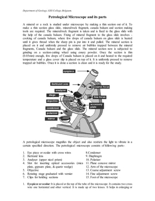
- 1. Department of Geology, GSS College,Belgaum. Petrological Microscope and its parts A mineral or a rock is studied under microscope by making a thin section out of it. To make a thin section glass slide, mineral/rock fragment, canada balsam and section making tools are required. The mineral/rock fragment is taken and is fixed to the glass slide with the help of the canada balsam. Fixing of mineral fragment to the glass slide involves - cooking of canada balsam, where few drops of canada balsam on glass slide is heated until it gives thread when the sharp pin is put into it and pulled. The mineral section is placed on it and uniformly pressed to remove air bubbles trapped between the mineral fragments, Canada balsam and the glass slide. The mineral section now is subjected to grinding on a section-cutting wheel using emery powder. Once the section is thin (0.03mm) enough, few drops of Canada balsam is placed on it and heated to the required temperature and a glass cover slip is placed on top of it. It is uniformly pressed to remove trapped air bubbles. Once it is done a section is clean and it is ready for the study. A petrological microscope magnifies the object and also restricts the light to vibrate in a certain specified direction. The petrological microscope consists of following parts- 1. Eye piece or ocular with cross wires 8.Condenser 2. Bertrand lens 9. Diaphragm 3. Analyser (upper nicol prism) 10. Polarizer 4. Slot for inserting optical accessories (mica plate, gypsum plate, & quartz wedge) 11. Plane concave mirror 12. Arm of the microscope 5. Objective 13. Coarse adjustment screw 6. Rotating stage graduated with vernier 14. Fine adjustment screw 7. Clips for holding sections 15. Foot of the microscope 1. Eyepiece or ocular:It is placed at the top of the tube of the microscope. It contains two cross wire one horizontal and other vertical. It is made up of two lenses. It helps in enlarging or
- 2. Department of Geology, GSS College,Belgaum. magnifying image of the mineral section. The ocular contains a lens, which is usually a 10x lens. The magnification of your thin section is the product of the magnification of the ocular and the objective. For example, if you are looking at a section using the 4x objective, and your ocular is 10x then the total magnification is 40x. 2. Bertrand lens: It is placed in the tube of the microscope. It can be brought in and out of the path of rays. This is used when examining interference figures and while studying the proprties of minerals like sign of the mineral and finding out biaxial and uniaxial nature of the mineral under conoscopic condition. 3. Analyser (Nicol Prism): This is similar to polarizer made up of transparent calcite crystal called as Iceland spar in the form of a prism. Two rhombohedral parts of a calcite cut diagonally, are joined by means of Canada balsam. This can be put into or out of the path of rays of light as and when required. Below the analyser there is a slot or opening in which optical accessories like mica plate, gypsum plate and quartz wedge are inserted. 4. Objective: This is placed at the other end of the tube of the microscope and can be removed easily whenever required. Obectives are used for magnification. There are three types of objectives- low power, high power and medium. Low power objectives magnify or enlarge 12 to 14 times of the original diameter. Medium and high power objectives magnify 40 to 80 times the diameter. 5. Tube of the microscope: Ocular, Bertrand lens, analyser and objective are fixed in the tube of the microscope. This tube can be raised or lowered as and when focusing is required by means of screw. 6. Rotating stage: This is provided just below the objective, which is graduated with vernier scale. This can be rotated either clockwise or anticlock wise. For holding the thin section on the stage it is provided with 2 clips. The stage can be fixed by operating a screw. 7. Clips for holding the section: These are two steel clips, which are fitted on the stage of the microscope. Their one end is fixed to the stage and the other is moveable. These clips hold the section in place. 8. Condenser: It is a lens, which condenses the rays and illuminates a particular point of the mineral or rock section on the stage and is fixed below the stage of the microscope. 9. Iris Diaphragm: It is made up of metal piece. It works as a shutter and controls the amount of light that falls on the object. 10. Polarizer: It is similar to analyser in construction. It is fixed at the lower end of sub stage and is rotatable. It is also called as nicol prism.When both polarizer and analyser is in the path of rays they are said to be under cross nicols (under analysed light). With the help of both the nicol prisms (analyser & polarizer) the light can be made to vibrate in any specified plane. 11. Mirror: This is placed below the sub stage and can be tilted. It is a Plano-concave mirror. Light can be reflected and made to fall on thin section. Plane mirror is used for studying sections under low power objective. Below this there is a base which is the heaviest part of the microscope. 12. Arm of the microscope: One end of the arm of the microscope is attached to the base with the help of the fastening screw. The other end is attached to the tube of the microscope. The arm can be moved in back to front for inclined position. 13. Coarse adjustment screws: Rotating clockwise or anti clockwise can bring the object into rough focus. 14. Fine adjustment screws: By operating these screws the objective studied under investigation is brought under sharp focus. 15. Subs-tage: The sub-stage arrangement consists of condenser, diaphragm, and polarizer. This sub-stage can be raised or lowered by operating screws.