SlideShare une entreprise Scribd logo
1  sur  30
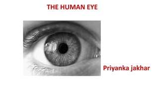
THE HUMAN EYE
Priyanka jakhar
THE HUMAN EYE
• The human eye is one of the most valuable and sensitive sense organs.
• It enables us to see the wonderful world and the colours around us.
• On closing the eyes, we can identify objects to some extent by their smell,
taste, sound they make or by touch.
• It is, however, impossible to identify colours while closing the eyes.
• Thus, of all the sense organs, the human eye is the most significant one as it
enables us to see the beautiful, colourful world around us.
• The human eye is like a camera.
• Its lens system forms an image on a light-sensitive screen called the retina.
• Light enters the eye through a thin membrane called the cornea. It forms the
transparent bulge on the front surface of the eyeball.ss
• It is like a camera which has a lens and screen system.
• The human eye are located in the specialized sockets carved out in
the human skull.
• Each human eye sizes for approximately 2.5 cm in diameter.
S.No. Human Eye Part Functions
1. Pupil
Opens and closes in order to regulate and control the amount of
light.
2. Iris Controls light level similar to the aperture of a camera.
3. Sclera Protects the outer coat.
4. Cornea
A thin membrane which provides 67% of the eye’s focusing
power.
5. Crystalline lens Helps to focus light into the retina.
6. Conjunctive Covers the outer surface (visible part) of the eye.
7. Aqueous humour Provides power to the cornea.
8. Vitreous humour Provides the eye with its form and shape.
9. Retina
Captures the light rays focussed by the lens and sends impulses
to the brain via the optic nerve.
10. Optic nerve Transmits electrical signals to the brain.
11. Ciliary muscles
Contracts and extends in order to change the lens shape for
Anterior Chambers
The anterior chamber is the front part of the eye between the cornea and the iris.
Posterior Chambers
The posterior chamber is between the iris and lens.
Posterior chamber is an important structure involved in production and circulation of a watery fluid
known as the aqueous humor, or aqueous.
WALL OF THE EYEBALL :
OUTER LAYER (FIBROUS COAT) : SCLERA AND CORNEA
MIDDLE LAYER (VASCULAR COAT) : CHOROID ,CILIARYBODY AND IRIS
INNER LAYER (NERVOUS COAT) : RETINA
The main parts of a human eye are:s
Sclera
• The white outer region of our eye which protects the internal parts of
our eyes is known as ‘sclera‘.
• Its made up of fibrous tissues.
• It’s continuous with the cornea.
• It resists intraocular pressure.
• It provides protection to the delicate structure within the eye.
• It maintains shape of the eyeball
• The smooth external surface allow easy eye movement
Cornea
The cornea is described as the “window of the eye”
• The anterior one sixth part of the sclera is transparent and is known as cornea.
• Light rays pass through the cornea to reach the retina.
• The transparent portion of our eye that allows the light to enter our
eye is known as the ‘cornea’ and is made up of transparent tissue.
• The cornea covers the pupil, anterior chamber, and the iris.
• Along with the anterior chamber and lens, cornea refracts light and
accounts for two-thirds of the eye’s total power.
• Usually, the refractive power of the cornea is approximately 43
dioptres.
Iris
A circular, thin structure made up of contracting and relaxing muscles
in the eye that controls the size of the pupil and the light reaching the
retina are known as the ‘iris’.
Iris defines a person’s eye color.
If we define the human eye as a camera then the iris becomes aperture
of the eye.
• Iris is the pigmented membrane surrounds the pupil
• It arises from the margin of ciliary body and forms a dark centered opening
called pupil
• The space between cornea (in front) and the lens (behind) is the anterior
segment
• It is again divided into two parts by the iris;
• Anterior chamber -The space between the iris and cornea is the anterior
chamber
• Posterior chamber -The space between iris and lens is posterior chamber
• They are filled with a clear fluid, the aqueous humor
Choroid
Choroid is a thin pigmented membrane, dark brown in color which is situated in
between sclera (externally) and retina (internally).
CILIARY BODY
Ciliary body is the continuation of choroid consisting of smooth muscle fibers,
i.e., the ciliary muscle.
• Ciliary body contains suspensory ligament for attaching the lens in position
• The ciliary muscles help in accommodation by adjusting the thickness of lens
Pupil
• The part of the eye located in the center of the iris allowing light to
reach the retina.
• The pupil appears black in color since the eye tissues absorb or
diffusely reflect the light entering the pupil. Iris controls the pupil.
Lens
• The lens is a biconvex, transparent structure present in the eye
behind the pupil.
• The lens along with the cornea refracts the light, so as to focus it on
the retina. • By changing its shape, the lens is capable of changing the
focal distance of the eye.
Retina
• Retina is the inner most layer of the eyeball
• It is a thin delicate layer continuous posteriorly with optic nerve
• The outer surface of the retina, formed by pigment cells, is attached to choroid.
• Its inner surface is in contact with the hyaloid membrane of the vitreous.
• The small area of retina where the optic nerve leaves the eye is the optic disc
or the blind spot.
• It has no light sensitive cells (Rods or Cones).
• The retina is a light-sensitive tissue in the inner coat of the eye that
sends electrical signals after converting them from light to the brain
for processing.
• When light strikes the retina, two types of cells are activated.
• Rods detect light and dark and help form images under dim conditions.
• Cones are responsible for color vision.
• The three types of cones are called red, green, and blue, but each actually
detects a range of wavelengths and not these specific colors.
• When you focus clearly on an object, light strikes a region called the fovea.
• The fovea is packed with cones and allows sharp vision.
• Rods outside the fovea are largely responsible for peripheral vision.
• Rods and cones convert light into an electric signal that is carried from the
• The brain translates nerve impulses. to form an image.
• Three-dimensional information comes from comparing the differences
between the images formed by each eye.
Rods and cones are the two light-sensitive types of cells present in
the retina.
Rods help us for night-time vision and cones help us see colors.
The rods and cones are the receptors of light and sight
These cells contains photosensitive pigments (Rods Rhodopsin, Cones – Iodopsin)
involved in the conversion of light rays into nerve impulses
Optic Nerve
The optic nerve sends electrical impulses from the retina, at the back
of the eyes to the brain.
• LIGHT TRANSMITTING MEDIA (OR) REFRACTIVE MEDIA
•Aqueous humor
•Vitreous humor
•Lens
• Aqueous humor is a clear fluid fills the space between cornea and lens
• It is secreted by capillaries of ciliary process
• From here the fluid reaches to the anterior chamber which finally reaches to
the canal of Schlemm.
Interference with drainage of aqueous humor results in an increase of intraocular
pressure (glaucoma) (Normal IOP 10 to 20 mmHg)
This leads to atrophy of the retina, leading to blindness
FUNCTIONS
• It helps to maintain intraocular pressure and thus maintains the shape of
eyeball
• It is rich in ascorbic acid, glucose and amino acids and nourishes the cornea and
lens
VITREOUS HUMOR
Vitreous humor or vitreous body is a colorless, transparent, jelly-like substance
which fills the posterior segment of the eye (i.e., behind the lens).
It is enclosed in a delicate hyaloid membrane
FUNCTIONS
• It helps to preserve the spherical shape of the eyeball and to support the
retina.
Lens .
• The lens of the eyeball is crystalline in nature .
• It is situated behind the pupil .
• It is biconvex, transparent, and elastic in structure.
• Lens refracts light rays and helps to focus the image of the object on retina.
• Lens is supported by suspensory ligaments (Zonular fibers) which are attached
with ciliary bodies.
• The lens obtains nutrients from the fluid, aqueous humour because it does not have any blood
supply.
• Waste products are removed through these fluids as well.
Macula
The macula, a small part which is located in the center of the retina that gives central
vision.
Conjunctiva
The conjunctiva is the clear, thin membrane that covers part of the front surface of
the eye and the inner surface of the eyelids . It has two segments:
• Bulbar conjunctiva
 This portion of the conjunctiva covers the anterior part of the sclera.
 The bulbar conjunctiva stops at the junction between the sclera and cornea; it
does not cover the cornea.
• Palpebral conjunctiva.
 This portion covers the inner surface of both the upper and lower eyelids.
(Another term for the palpebral conjunctiva is tarsal conjunctiva.)
The bulbar and palpebral conjunctiva are continuous (see illustration).
This feature makes it impossible for a contact lens (or anything else) to get lost
behind your eye.
Conjunctiva Function
The primary functions of the conjunctiva are:
•Keep the front surface of the eye moist and lubricated.
•Keep the inner surface of the eyelids moist and lubricated so they open and close
easily without friction or causing eye irritation.
•Protect the eye from dust, debris and infection-causing microorganisms.
•The conjunctiva has many small blood vessels that provide nutrients to the eye
and lids.
•It also contains special cells that secrete a component of the tear film to help
prevent dry eye syndrome.
Blind Spot
• This is a small area of the retina where the optic nerve connects.
• In this area there are no light-sensitive cells i.e. no rods and cones.
• Due to which the retina can not see at that spot.
• This is called a Blind Spot.
Tear Glands
• The tear gland located above each eyeball and inside your upper
eyelid.
• This gland is responsible for making a fluid that is mostly salt and
water (tears) to keep the surface of your eyeball clean and moist.
• sIt also protects your eye from damage.
Yellow spot
• The yellow spot, also known as macula, is the centre of the eye and sharpest sight place.
• In fact, it’s the centre of our eye placed on the background of the eye and it’s around 5 milli
metres big.
• Yellow spot is a part of inner layer of the eye called the retina.
• The nerves inside the macula are rich with lutein and zeaxanthin pigment, which makes it look
yellow.
• It’s also the reason it is called the yellow spot.
• In order to provide clear vision, the yellow spot must be very efficiently organized.
• There are millions of tiny nerves converting light to electric impulse in a very small area of only a
few milli metres.
• This process requires a lot of energy and oxygen.
Colour Blindness:
• A person having defective cone cells is not able to distinguish between the different colours.
• This defect is known as Colour Blindness.
Defects of The Eye and Their Corrections
As perfect the human eye may seem; it’s not.
If the human eye isn’t perfect, which means it has its share of defects of the
human eye.
Here are few common defects of the human eye:
a. Myopia or Near-Sightedness
Myopia is a defect of vision where in far-off objects appear blurred and objects
near are seen clearly.
Since the eyeball is too long or the eye lens’s refractive power is too high; the
image forms in front of the retina rather than forming on it.
Myopia is a defect of vision in which a person can see nearby objects clearly but
cannot see distant objects clearly because the image is formed in front of the
retina.
This may be due to:-
i) Increase in curvature of the eye lens
ii) Increase in the length of the eye ball
Correction of myopia can happen by wearing glasses/contacts made
of concave lenses to help focus the image on the retina.
b. Hypermetropia or Longsightedness
Hypermetropia is a defect of vision wherein there is difficulty in viewing objects
that are near but one can view far objects easily.
Since the eyeball is too short or eye lens’s refractive power is too weak hence the
image instead is of being forming upon the retina, its forms behind the retina.
Hypermetropia is a defect of vision in which a person can see distant objects
clearly but cannot see nearby objects clearly because the image is formed behind
the retina.
This may be due to:-
i) Decrease in curvature of eye lens
ii) Decrease in the length of the eye ball
Correction of hypermetropia can happen by wearing glasses/contacts containing
convex lenses.
c. Cataract
Cataract is the clouding of the lens, that prevents the formation of a clear, sharp
image.
A cataract forms when old cells after they die, stick in a capsule wherein with time
a clouding over lens happens. Because of this clouding blurred images are
formed.
Other factors that may increase the risk of developing cataracts :
•A family history of cataracts
•Diabetes
•High blood pressure
•Other eye conditions such as uveitis
•Previous eye surgery, injury or inflammation
•Long-term use of corticosteroid medication (eg : prednisone, prednisolone)
•Excessive exposure to sunlight
•Smoking
•Drinking too much alcohol
•Poor diet.
Correction of cataract can happen through a surgery. An artificial lens in place of
d. Presbyopia or Old-age Longsightedness
• Presbyopia is a natural defect that occurs with the age.
• In presbyopia, the ciliary muscles become weak and are no longer able to adjust
the eye lens.
• The eye muscles become so weak that no longer can a person see nearby
objects clearly.
• The near point of a person with presbyopia is more than 25cm.
• Presbyopia is a defect of vision in old people in which they are not able to see
nearby objects clearly due to the increase in the distance of near point.
• This is due to the weakening of the ciliary muscles and decrease in the flexibility
of the eye lens. It can be
• corrected by using suitable convex lens.
• Sometimes they are not able to see both nearby and distant objects clearly.
• It can be corrected by using bifocal lenses consisting of both concave and
convex lenses.
• The upper part is concave for correction of distant vision and the lower part is
convex for correction of near vision.
• Correction of presbyopia can happen by wearing bifocal glasses or Progressive
portion contains a convex lens.
• A person with presbyopia can also have just myopia or just
hypermetropia.
e. Astigmatism
Astigmatism is a defect where in the light rays entering the eye do not focus light
evenly to a single focal point on the retina but instead scatter away.
The light rays in a way where some focus on the retina and some focus in front of
or behind it. This happens because of non-uniform curvature of the cornea;
resulting in a distorted or blurry vision at any distance.
Correction of astigmatism can happen by using a special spherical cylindrical
lens.
Power of Accommodation
Power of accommodation is the process by which ciliary muscles
function, to adjust the focal length of the eyes so that clear image
forms on the retina.
This varies far or nearby objects. For a normal eyesight, the power of
accommodation is 4 dioptre.
Near point :-
The minimum distance at wh ich the eye can see objects clearly is called the near
point or least distance of distinct vision.
For a normal eye it is 25cm.
Far point :-
The farthest distance up to which the eye can see objects clearly is

Contenu connexe

Tendances (20)

The human eye amatomy
The human eye amatomyThe human eye amatomy
The human eye amatomy
 
ppt on Eye anatomy
ppt on Eye anatomy ppt on Eye anatomy
ppt on Eye anatomy
 
Human eye
Human eyeHuman eye
Human eye
 
Eye Anatomy & Physiology
Eye Anatomy & Physiology Eye Anatomy & Physiology
Eye Anatomy & Physiology
 
PHYSIOLOGY OF VISION
PHYSIOLOGY OF VISIONPHYSIOLOGY OF VISION
PHYSIOLOGY OF VISION
 
Sense organs
Sense organsSense organs
Sense organs
 
Physiology of the eye
Physiology of the eye Physiology of the eye
Physiology of the eye
 
1. physiology of eye
1. physiology of eye1. physiology of eye
1. physiology of eye
 
Eye Anatomy & Physiology
Eye Anatomy & Physiology Eye Anatomy & Physiology
Eye Anatomy & Physiology
 
Anatomy of the eye presentation
Anatomy of the eye presentationAnatomy of the eye presentation
Anatomy of the eye presentation
 
anatomy and physiology of eye
anatomy and physiology of eyeanatomy and physiology of eye
anatomy and physiology of eye
 
Anatomy of the eye
Anatomy of the eyeAnatomy of the eye
Anatomy of the eye
 
Chapter 8 - Special Senses
Chapter 8 - Special SensesChapter 8 - Special Senses
Chapter 8 - Special Senses
 
Anatomy of the eye for studentss
Anatomy of the eye for studentssAnatomy of the eye for studentss
Anatomy of the eye for studentss
 
Anatomy and physiology of the Eye-Nursing
Anatomy and physiology of the Eye-NursingAnatomy and physiology of the Eye-Nursing
Anatomy and physiology of the Eye-Nursing
 
Anatomy of eye ppt
Anatomy of eye pptAnatomy of eye ppt
Anatomy of eye ppt
 
Aqueous humor
Aqueous humorAqueous humor
Aqueous humor
 
Anatomy of sight and the eye
Anatomy of sight and the eyeAnatomy of sight and the eye
Anatomy of sight and the eye
 
Structure and function of eye
Structure and function of eyeStructure and function of eye
Structure and function of eye
 
The human eye
The human eyeThe human eye
The human eye
 

Similaire à The Human Eye

EYE ANATOMY AND PHYSIOLOGY ppt.pptx
EYE  ANATOMY AND PHYSIOLOGY ppt.pptxEYE  ANATOMY AND PHYSIOLOGY ppt.pptx
EYE ANATOMY AND PHYSIOLOGY ppt.pptxNitin Sharma
 
Presentation on eye
Presentation on eyePresentation on eye
Presentation on eyePreem Sharma
 
vision.pptx
vision.pptxvision.pptx
vision.pptxLuzSan3
 
The eye (sense of sight organ)
The eye (sense of sight organ)The eye (sense of sight organ)
The eye (sense of sight organ)Jordan Mwelwa
 
Anatomy and physilogy of eye,nose and throat
Anatomy and physilogy of eye,nose and throatAnatomy and physilogy of eye,nose and throat
Anatomy and physilogy of eye,nose and throatDeeps Gupta
 
anatomyofeye-180217183456.pdf
anatomyofeye-180217183456.pdfanatomyofeye-180217183456.pdf
anatomyofeye-180217183456.pdfEidTahir
 
Anatomy of eye neelesh
Anatomy of eye neeleshAnatomy of eye neelesh
Anatomy of eye neeleshNeelesh Kumar
 
structureofeyeppt-190302154704 (1).pdf
structureofeyeppt-190302154704 (1).pdfstructureofeyeppt-190302154704 (1).pdf
structureofeyeppt-190302154704 (1).pdfShivamsharma15812
 
ANATOMY OF EYE n.pdf
ANATOMY OF EYE n.pdfANATOMY OF EYE n.pdf
ANATOMY OF EYE n.pdfPooja Rani
 
SENSORY ORGANS...(EYE).pptx
SENSORY ORGANS...(EYE).pptxSENSORY ORGANS...(EYE).pptx
SENSORY ORGANS...(EYE).pptxAgravatDarshan1
 
The human eye 10 13-2011
The human eye 10 13-2011The human eye 10 13-2011
The human eye 10 13-2011Varunitya
 
Anatomy and Physiology of Special senses
Anatomy and Physiology of Special senses Anatomy and Physiology of Special senses
Anatomy and Physiology of Special senses DR .PALLAVI PATHANIA
 
ANATOMY & PHYSIOLOGY OF EYE.pptx
ANATOMY  & PHYSIOLOGY OF EYE.pptxANATOMY  & PHYSIOLOGY OF EYE.pptx
ANATOMY & PHYSIOLOGY OF EYE.pptxSaili Gaude
 
ANATOMY OF EYE.pptx
ANATOMY OF EYE.pptxANATOMY OF EYE.pptx
ANATOMY OF EYE.pptxSaili Gaude
 

Similaire à The Human Eye (20)

EYE ANATOMY AND PHYSIOLOGY ppt.pptx
EYE  ANATOMY AND PHYSIOLOGY ppt.pptxEYE  ANATOMY AND PHYSIOLOGY ppt.pptx
EYE ANATOMY AND PHYSIOLOGY ppt.pptx
 
Presentation on eye
Presentation on eyePresentation on eye
Presentation on eye
 
The Eye.pdf
The Eye.pdfThe Eye.pdf
The Eye.pdf
 
vision.pptx
vision.pptxvision.pptx
vision.pptx
 
The eye (sense of sight organ)
The eye (sense of sight organ)The eye (sense of sight organ)
The eye (sense of sight organ)
 
Mammalian eye
Mammalian eyeMammalian eye
Mammalian eye
 
Anatomy and physilogy of eye,nose and throat
Anatomy and physilogy of eye,nose and throatAnatomy and physilogy of eye,nose and throat
Anatomy and physilogy of eye,nose and throat
 
anatomyofeye-180217183456.pdf
anatomyofeye-180217183456.pdfanatomyofeye-180217183456.pdf
anatomyofeye-180217183456.pdf
 
Anatomy of eye
Anatomy of eyeAnatomy of eye
Anatomy of eye
 
Anatomy of eye neelesh
Anatomy of eye neeleshAnatomy of eye neelesh
Anatomy of eye neelesh
 
structureofeyeppt-190302154704 (1).pdf
structureofeyeppt-190302154704 (1).pdfstructureofeyeppt-190302154704 (1).pdf
structureofeyeppt-190302154704 (1).pdf
 
eye ppt.pptx
eye ppt.pptxeye ppt.pptx
eye ppt.pptx
 
ANATOMY OF EYE n.pdf
ANATOMY OF EYE n.pdfANATOMY OF EYE n.pdf
ANATOMY OF EYE n.pdf
 
structure of eye
structure of eyestructure of eye
structure of eye
 
SENSORY ORGANS...(EYE).pptx
SENSORY ORGANS...(EYE).pptxSENSORY ORGANS...(EYE).pptx
SENSORY ORGANS...(EYE).pptx
 
The human eye 10 13-2011
The human eye 10 13-2011The human eye 10 13-2011
The human eye 10 13-2011
 
Anatomy and Physiology of Special senses
Anatomy and Physiology of Special senses Anatomy and Physiology of Special senses
Anatomy and Physiology of Special senses
 
ANATOMY & PHYSIOLOGY OF EYE.pptx
ANATOMY  & PHYSIOLOGY OF EYE.pptxANATOMY  & PHYSIOLOGY OF EYE.pptx
ANATOMY & PHYSIOLOGY OF EYE.pptx
 
ANATOMY OF EYE.pptx
ANATOMY OF EYE.pptxANATOMY OF EYE.pptx
ANATOMY OF EYE.pptx
 
Comparative study of vertebrates
Comparative  study of vertebratesComparative  study of vertebrates
Comparative study of vertebrates
 

Plus de PriyankaJakhar9

Plus de PriyankaJakhar9 (7)

Electrostatic part 2
Electrostatic part 2Electrostatic part 2
Electrostatic part 2
 
Electrostatic part 1 class 12th
Electrostatic part 1      class 12th Electrostatic part 1      class 12th
Electrostatic part 1 class 12th
 
emw
emwemw
emw
 
digital learning
digital learningdigital learning
digital learning
 
Compound microscope
Compound microscopeCompound microscope
Compound microscope
 
Motion in a straight line(part 1)
Motion in a straight line(part 1)Motion in a straight line(part 1)
Motion in a straight line(part 1)
 
Motion in straight line ( part 1)
Motion in straight line ( part 1)Motion in straight line ( part 1)
Motion in straight line ( part 1)
 

Dernier

FSB Advising Checklist - Orientation 2024
FSB Advising Checklist - Orientation 2024FSB Advising Checklist - Orientation 2024
FSB Advising Checklist - Orientation 2024Elizabeth Walsh
 
TỔNG ÔN TẬP THI VÀO LỚP 10 MÔN TIẾNG ANH NĂM HỌC 2023 - 2024 CÓ ĐÁP ÁN (NGỮ Â...
TỔNG ÔN TẬP THI VÀO LỚP 10 MÔN TIẾNG ANH NĂM HỌC 2023 - 2024 CÓ ĐÁP ÁN (NGỮ Â...TỔNG ÔN TẬP THI VÀO LỚP 10 MÔN TIẾNG ANH NĂM HỌC 2023 - 2024 CÓ ĐÁP ÁN (NGỮ Â...
TỔNG ÔN TẬP THI VÀO LỚP 10 MÔN TIẾNG ANH NĂM HỌC 2023 - 2024 CÓ ĐÁP ÁN (NGỮ Â...Nguyen Thanh Tu Collection
 
Graduate Outcomes Presentation Slides - English
Graduate Outcomes Presentation Slides - EnglishGraduate Outcomes Presentation Slides - English
Graduate Outcomes Presentation Slides - Englishneillewis46
 
SOC 101 Demonstration of Learning Presentation
SOC 101 Demonstration of Learning PresentationSOC 101 Demonstration of Learning Presentation
SOC 101 Demonstration of Learning Presentationcamerronhm
 
ICT Role in 21st Century Education & its Challenges.pptx
ICT Role in 21st Century Education & its Challenges.pptxICT Role in 21st Century Education & its Challenges.pptx
ICT Role in 21st Century Education & its Challenges.pptxAreebaZafar22
 
Google Gemini An AI Revolution in Education.pptx
Google Gemini An AI Revolution in Education.pptxGoogle Gemini An AI Revolution in Education.pptx
Google Gemini An AI Revolution in Education.pptxDr. Sarita Anand
 
80 ĐỀ THI THỬ TUYỂN SINH TIẾNG ANH VÀO 10 SỞ GD – ĐT THÀNH PHỐ HỒ CHÍ MINH NĂ...
80 ĐỀ THI THỬ TUYỂN SINH TIẾNG ANH VÀO 10 SỞ GD – ĐT THÀNH PHỐ HỒ CHÍ MINH NĂ...80 ĐỀ THI THỬ TUYỂN SINH TIẾNG ANH VÀO 10 SỞ GD – ĐT THÀNH PHỐ HỒ CHÍ MINH NĂ...
80 ĐỀ THI THỬ TUYỂN SINH TIẾNG ANH VÀO 10 SỞ GD – ĐT THÀNH PHỐ HỒ CHÍ MINH NĂ...Nguyen Thanh Tu Collection
 
On_Translating_a_Tamil_Poem_by_A_K_Ramanujan.pptx
On_Translating_a_Tamil_Poem_by_A_K_Ramanujan.pptxOn_Translating_a_Tamil_Poem_by_A_K_Ramanujan.pptx
On_Translating_a_Tamil_Poem_by_A_K_Ramanujan.pptxPooja Bhuva
 
How to Create and Manage Wizard in Odoo 17
How to Create and Manage Wizard in Odoo 17How to Create and Manage Wizard in Odoo 17
How to Create and Manage Wizard in Odoo 17Celine George
 
HMCS Vancouver Pre-Deployment Brief - May 2024 (Web Version).pptx
HMCS Vancouver Pre-Deployment Brief - May 2024 (Web Version).pptxHMCS Vancouver Pre-Deployment Brief - May 2024 (Web Version).pptx
HMCS Vancouver Pre-Deployment Brief - May 2024 (Web Version).pptxmarlenawright1
 
COMMUNICATING NEGATIVE NEWS - APPROACHES .pptx
COMMUNICATING NEGATIVE NEWS - APPROACHES .pptxCOMMUNICATING NEGATIVE NEWS - APPROACHES .pptx
COMMUNICATING NEGATIVE NEWS - APPROACHES .pptxannathomasp01
 
Accessible Digital Futures project (20/03/2024)
Accessible Digital Futures project (20/03/2024)Accessible Digital Futures project (20/03/2024)
Accessible Digital Futures project (20/03/2024)Jisc
 
ICT role in 21st century education and it's challenges.
ICT role in 21st century education and it's challenges.ICT role in 21st century education and it's challenges.
ICT role in 21st century education and it's challenges.MaryamAhmad92
 
Sociology 101 Demonstration of Learning Exhibit
Sociology 101 Demonstration of Learning ExhibitSociology 101 Demonstration of Learning Exhibit
Sociology 101 Demonstration of Learning Exhibitjbellavia9
 
Fostering Friendships - Enhancing Social Bonds in the Classroom
Fostering Friendships - Enhancing Social Bonds  in the ClassroomFostering Friendships - Enhancing Social Bonds  in the Classroom
Fostering Friendships - Enhancing Social Bonds in the ClassroomPooky Knightsmith
 
Understanding Accommodations and Modifications
Understanding  Accommodations and ModificationsUnderstanding  Accommodations and Modifications
Understanding Accommodations and ModificationsMJDuyan
 
NO1 Top Black Magic Specialist In Lahore Black magic In Pakistan Kala Ilam Ex...
NO1 Top Black Magic Specialist In Lahore Black magic In Pakistan Kala Ilam Ex...NO1 Top Black Magic Specialist In Lahore Black magic In Pakistan Kala Ilam Ex...
NO1 Top Black Magic Specialist In Lahore Black magic In Pakistan Kala Ilam Ex...Amil baba
 
Application orientated numerical on hev.ppt
Application orientated numerical on hev.pptApplication orientated numerical on hev.ppt
Application orientated numerical on hev.pptRamjanShidvankar
 
How to Manage Global Discount in Odoo 17 POS
How to Manage Global Discount in Odoo 17 POSHow to Manage Global Discount in Odoo 17 POS
How to Manage Global Discount in Odoo 17 POSCeline George
 

Dernier (20)

FSB Advising Checklist - Orientation 2024
FSB Advising Checklist - Orientation 2024FSB Advising Checklist - Orientation 2024
FSB Advising Checklist - Orientation 2024
 
TỔNG ÔN TẬP THI VÀO LỚP 10 MÔN TIẾNG ANH NĂM HỌC 2023 - 2024 CÓ ĐÁP ÁN (NGỮ Â...
TỔNG ÔN TẬP THI VÀO LỚP 10 MÔN TIẾNG ANH NĂM HỌC 2023 - 2024 CÓ ĐÁP ÁN (NGỮ Â...TỔNG ÔN TẬP THI VÀO LỚP 10 MÔN TIẾNG ANH NĂM HỌC 2023 - 2024 CÓ ĐÁP ÁN (NGỮ Â...
TỔNG ÔN TẬP THI VÀO LỚP 10 MÔN TIẾNG ANH NĂM HỌC 2023 - 2024 CÓ ĐÁP ÁN (NGỮ Â...
 
Graduate Outcomes Presentation Slides - English
Graduate Outcomes Presentation Slides - EnglishGraduate Outcomes Presentation Slides - English
Graduate Outcomes Presentation Slides - English
 
SOC 101 Demonstration of Learning Presentation
SOC 101 Demonstration of Learning PresentationSOC 101 Demonstration of Learning Presentation
SOC 101 Demonstration of Learning Presentation
 
ICT Role in 21st Century Education & its Challenges.pptx
ICT Role in 21st Century Education & its Challenges.pptxICT Role in 21st Century Education & its Challenges.pptx
ICT Role in 21st Century Education & its Challenges.pptx
 
Google Gemini An AI Revolution in Education.pptx
Google Gemini An AI Revolution in Education.pptxGoogle Gemini An AI Revolution in Education.pptx
Google Gemini An AI Revolution in Education.pptx
 
80 ĐỀ THI THỬ TUYỂN SINH TIẾNG ANH VÀO 10 SỞ GD – ĐT THÀNH PHỐ HỒ CHÍ MINH NĂ...
80 ĐỀ THI THỬ TUYỂN SINH TIẾNG ANH VÀO 10 SỞ GD – ĐT THÀNH PHỐ HỒ CHÍ MINH NĂ...80 ĐỀ THI THỬ TUYỂN SINH TIẾNG ANH VÀO 10 SỞ GD – ĐT THÀNH PHỐ HỒ CHÍ MINH NĂ...
80 ĐỀ THI THỬ TUYỂN SINH TIẾNG ANH VÀO 10 SỞ GD – ĐT THÀNH PHỐ HỒ CHÍ MINH NĂ...
 
On_Translating_a_Tamil_Poem_by_A_K_Ramanujan.pptx
On_Translating_a_Tamil_Poem_by_A_K_Ramanujan.pptxOn_Translating_a_Tamil_Poem_by_A_K_Ramanujan.pptx
On_Translating_a_Tamil_Poem_by_A_K_Ramanujan.pptx
 
How to Create and Manage Wizard in Odoo 17
How to Create and Manage Wizard in Odoo 17How to Create and Manage Wizard in Odoo 17
How to Create and Manage Wizard in Odoo 17
 
HMCS Vancouver Pre-Deployment Brief - May 2024 (Web Version).pptx
HMCS Vancouver Pre-Deployment Brief - May 2024 (Web Version).pptxHMCS Vancouver Pre-Deployment Brief - May 2024 (Web Version).pptx
HMCS Vancouver Pre-Deployment Brief - May 2024 (Web Version).pptx
 
COMMUNICATING NEGATIVE NEWS - APPROACHES .pptx
COMMUNICATING NEGATIVE NEWS - APPROACHES .pptxCOMMUNICATING NEGATIVE NEWS - APPROACHES .pptx
COMMUNICATING NEGATIVE NEWS - APPROACHES .pptx
 
Accessible Digital Futures project (20/03/2024)
Accessible Digital Futures project (20/03/2024)Accessible Digital Futures project (20/03/2024)
Accessible Digital Futures project (20/03/2024)
 
ICT role in 21st century education and it's challenges.
ICT role in 21st century education and it's challenges.ICT role in 21st century education and it's challenges.
ICT role in 21st century education and it's challenges.
 
Sociology 101 Demonstration of Learning Exhibit
Sociology 101 Demonstration of Learning ExhibitSociology 101 Demonstration of Learning Exhibit
Sociology 101 Demonstration of Learning Exhibit
 
Fostering Friendships - Enhancing Social Bonds in the Classroom
Fostering Friendships - Enhancing Social Bonds  in the ClassroomFostering Friendships - Enhancing Social Bonds  in the Classroom
Fostering Friendships - Enhancing Social Bonds in the Classroom
 
Understanding Accommodations and Modifications
Understanding  Accommodations and ModificationsUnderstanding  Accommodations and Modifications
Understanding Accommodations and Modifications
 
NO1 Top Black Magic Specialist In Lahore Black magic In Pakistan Kala Ilam Ex...
NO1 Top Black Magic Specialist In Lahore Black magic In Pakistan Kala Ilam Ex...NO1 Top Black Magic Specialist In Lahore Black magic In Pakistan Kala Ilam Ex...
NO1 Top Black Magic Specialist In Lahore Black magic In Pakistan Kala Ilam Ex...
 
Application orientated numerical on hev.ppt
Application orientated numerical on hev.pptApplication orientated numerical on hev.ppt
Application orientated numerical on hev.ppt
 
How to Manage Global Discount in Odoo 17 POS
How to Manage Global Discount in Odoo 17 POSHow to Manage Global Discount in Odoo 17 POS
How to Manage Global Discount in Odoo 17 POS
 
Mehran University Newsletter Vol-X, Issue-I, 2024
Mehran University Newsletter Vol-X, Issue-I, 2024Mehran University Newsletter Vol-X, Issue-I, 2024
Mehran University Newsletter Vol-X, Issue-I, 2024
 

The Human Eye

  • 2.
  • 3. THE HUMAN EYE • The human eye is one of the most valuable and sensitive sense organs. • It enables us to see the wonderful world and the colours around us. • On closing the eyes, we can identify objects to some extent by their smell, taste, sound they make or by touch. • It is, however, impossible to identify colours while closing the eyes. • Thus, of all the sense organs, the human eye is the most significant one as it enables us to see the beautiful, colourful world around us. • The human eye is like a camera. • Its lens system forms an image on a light-sensitive screen called the retina. • Light enters the eye through a thin membrane called the cornea. It forms the transparent bulge on the front surface of the eyeball.ss • It is like a camera which has a lens and screen system. • The human eye are located in the specialized sockets carved out in the human skull. • Each human eye sizes for approximately 2.5 cm in diameter.
  • 4. S.No. Human Eye Part Functions 1. Pupil Opens and closes in order to regulate and control the amount of light. 2. Iris Controls light level similar to the aperture of a camera. 3. Sclera Protects the outer coat. 4. Cornea A thin membrane which provides 67% of the eye’s focusing power. 5. Crystalline lens Helps to focus light into the retina. 6. Conjunctive Covers the outer surface (visible part) of the eye. 7. Aqueous humour Provides power to the cornea. 8. Vitreous humour Provides the eye with its form and shape. 9. Retina Captures the light rays focussed by the lens and sends impulses to the brain via the optic nerve. 10. Optic nerve Transmits electrical signals to the brain. 11. Ciliary muscles Contracts and extends in order to change the lens shape for
  • 5. Anterior Chambers The anterior chamber is the front part of the eye between the cornea and the iris. Posterior Chambers The posterior chamber is between the iris and lens. Posterior chamber is an important structure involved in production and circulation of a watery fluid known as the aqueous humor, or aqueous. WALL OF THE EYEBALL : OUTER LAYER (FIBROUS COAT) : SCLERA AND CORNEA MIDDLE LAYER (VASCULAR COAT) : CHOROID ,CILIARYBODY AND IRIS INNER LAYER (NERVOUS COAT) : RETINA The main parts of a human eye are:s Sclera • The white outer region of our eye which protects the internal parts of our eyes is known as ‘sclera‘. • Its made up of fibrous tissues. • It’s continuous with the cornea. • It resists intraocular pressure.
  • 6. • It provides protection to the delicate structure within the eye. • It maintains shape of the eyeball • The smooth external surface allow easy eye movement Cornea The cornea is described as the “window of the eye” • The anterior one sixth part of the sclera is transparent and is known as cornea. • Light rays pass through the cornea to reach the retina. • The transparent portion of our eye that allows the light to enter our eye is known as the ‘cornea’ and is made up of transparent tissue. • The cornea covers the pupil, anterior chamber, and the iris. • Along with the anterior chamber and lens, cornea refracts light and accounts for two-thirds of the eye’s total power. • Usually, the refractive power of the cornea is approximately 43 dioptres.
  • 7.
  • 8. Iris A circular, thin structure made up of contracting and relaxing muscles in the eye that controls the size of the pupil and the light reaching the retina are known as the ‘iris’. Iris defines a person’s eye color. If we define the human eye as a camera then the iris becomes aperture of the eye. • Iris is the pigmented membrane surrounds the pupil • It arises from the margin of ciliary body and forms a dark centered opening called pupil • The space between cornea (in front) and the lens (behind) is the anterior segment • It is again divided into two parts by the iris; • Anterior chamber -The space between the iris and cornea is the anterior chamber
  • 9. • Posterior chamber -The space between iris and lens is posterior chamber • They are filled with a clear fluid, the aqueous humor Choroid Choroid is a thin pigmented membrane, dark brown in color which is situated in between sclera (externally) and retina (internally). CILIARY BODY Ciliary body is the continuation of choroid consisting of smooth muscle fibers, i.e., the ciliary muscle.
  • 10. • Ciliary body contains suspensory ligament for attaching the lens in position • The ciliary muscles help in accommodation by adjusting the thickness of lens Pupil • The part of the eye located in the center of the iris allowing light to reach the retina. • The pupil appears black in color since the eye tissues absorb or diffusely reflect the light entering the pupil. Iris controls the pupil. Lens • The lens is a biconvex, transparent structure present in the eye behind the pupil. • The lens along with the cornea refracts the light, so as to focus it on the retina. • By changing its shape, the lens is capable of changing the focal distance of the eye. Retina • Retina is the inner most layer of the eyeball • It is a thin delicate layer continuous posteriorly with optic nerve
  • 11. • The outer surface of the retina, formed by pigment cells, is attached to choroid. • Its inner surface is in contact with the hyaloid membrane of the vitreous. • The small area of retina where the optic nerve leaves the eye is the optic disc or the blind spot. • It has no light sensitive cells (Rods or Cones). • The retina is a light-sensitive tissue in the inner coat of the eye that sends electrical signals after converting them from light to the brain for processing. • When light strikes the retina, two types of cells are activated. • Rods detect light and dark and help form images under dim conditions. • Cones are responsible for color vision. • The three types of cones are called red, green, and blue, but each actually detects a range of wavelengths and not these specific colors. • When you focus clearly on an object, light strikes a region called the fovea. • The fovea is packed with cones and allows sharp vision. • Rods outside the fovea are largely responsible for peripheral vision. • Rods and cones convert light into an electric signal that is carried from the
  • 12. • The brain translates nerve impulses. to form an image. • Three-dimensional information comes from comparing the differences between the images formed by each eye. Rods and cones are the two light-sensitive types of cells present in the retina. Rods help us for night-time vision and cones help us see colors. The rods and cones are the receptors of light and sight These cells contains photosensitive pigments (Rods Rhodopsin, Cones – Iodopsin) involved in the conversion of light rays into nerve impulses Optic Nerve The optic nerve sends electrical impulses from the retina, at the back of the eyes to the brain. • LIGHT TRANSMITTING MEDIA (OR) REFRACTIVE MEDIA •Aqueous humor •Vitreous humor •Lens
  • 13. • Aqueous humor is a clear fluid fills the space between cornea and lens • It is secreted by capillaries of ciliary process • From here the fluid reaches to the anterior chamber which finally reaches to the canal of Schlemm. Interference with drainage of aqueous humor results in an increase of intraocular pressure (glaucoma) (Normal IOP 10 to 20 mmHg) This leads to atrophy of the retina, leading to blindness FUNCTIONS • It helps to maintain intraocular pressure and thus maintains the shape of eyeball • It is rich in ascorbic acid, glucose and amino acids and nourishes the cornea and lens VITREOUS HUMOR Vitreous humor or vitreous body is a colorless, transparent, jelly-like substance which fills the posterior segment of the eye (i.e., behind the lens). It is enclosed in a delicate hyaloid membrane
  • 14. FUNCTIONS • It helps to preserve the spherical shape of the eyeball and to support the retina. Lens . • The lens of the eyeball is crystalline in nature . • It is situated behind the pupil . • It is biconvex, transparent, and elastic in structure. • Lens refracts light rays and helps to focus the image of the object on retina. • Lens is supported by suspensory ligaments (Zonular fibers) which are attached with ciliary bodies. • The lens obtains nutrients from the fluid, aqueous humour because it does not have any blood supply. • Waste products are removed through these fluids as well. Macula The macula, a small part which is located in the center of the retina that gives central vision.
  • 15. Conjunctiva The conjunctiva is the clear, thin membrane that covers part of the front surface of the eye and the inner surface of the eyelids . It has two segments: • Bulbar conjunctiva  This portion of the conjunctiva covers the anterior part of the sclera.  The bulbar conjunctiva stops at the junction between the sclera and cornea; it does not cover the cornea. • Palpebral conjunctiva.  This portion covers the inner surface of both the upper and lower eyelids. (Another term for the palpebral conjunctiva is tarsal conjunctiva.) The bulbar and palpebral conjunctiva are continuous (see illustration). This feature makes it impossible for a contact lens (or anything else) to get lost behind your eye. Conjunctiva Function The primary functions of the conjunctiva are: •Keep the front surface of the eye moist and lubricated.
  • 16. •Keep the inner surface of the eyelids moist and lubricated so they open and close easily without friction or causing eye irritation. •Protect the eye from dust, debris and infection-causing microorganisms. •The conjunctiva has many small blood vessels that provide nutrients to the eye and lids. •It also contains special cells that secrete a component of the tear film to help prevent dry eye syndrome.
  • 17. Blind Spot • This is a small area of the retina where the optic nerve connects. • In this area there are no light-sensitive cells i.e. no rods and cones. • Due to which the retina can not see at that spot. • This is called a Blind Spot. Tear Glands • The tear gland located above each eyeball and inside your upper eyelid. • This gland is responsible for making a fluid that is mostly salt and water (tears) to keep the surface of your eyeball clean and moist. • sIt also protects your eye from damage. Yellow spot • The yellow spot, also known as macula, is the centre of the eye and sharpest sight place. • In fact, it’s the centre of our eye placed on the background of the eye and it’s around 5 milli metres big. • Yellow spot is a part of inner layer of the eye called the retina.
  • 18. • The nerves inside the macula are rich with lutein and zeaxanthin pigment, which makes it look yellow. • It’s also the reason it is called the yellow spot. • In order to provide clear vision, the yellow spot must be very efficiently organized. • There are millions of tiny nerves converting light to electric impulse in a very small area of only a few milli metres. • This process requires a lot of energy and oxygen.
  • 19. Colour Blindness: • A person having defective cone cells is not able to distinguish between the different colours. • This defect is known as Colour Blindness. Defects of The Eye and Their Corrections As perfect the human eye may seem; it’s not. If the human eye isn’t perfect, which means it has its share of defects of the human eye. Here are few common defects of the human eye: a. Myopia or Near-Sightedness Myopia is a defect of vision where in far-off objects appear blurred and objects near are seen clearly. Since the eyeball is too long or the eye lens’s refractive power is too high; the image forms in front of the retina rather than forming on it. Myopia is a defect of vision in which a person can see nearby objects clearly but cannot see distant objects clearly because the image is formed in front of the retina. This may be due to:- i) Increase in curvature of the eye lens
  • 20.
  • 21. ii) Increase in the length of the eye ball Correction of myopia can happen by wearing glasses/contacts made of concave lenses to help focus the image on the retina.
  • 22. b. Hypermetropia or Longsightedness Hypermetropia is a defect of vision wherein there is difficulty in viewing objects that are near but one can view far objects easily. Since the eyeball is too short or eye lens’s refractive power is too weak hence the image instead is of being forming upon the retina, its forms behind the retina. Hypermetropia is a defect of vision in which a person can see distant objects clearly but cannot see nearby objects clearly because the image is formed behind the retina. This may be due to:- i) Decrease in curvature of eye lens ii) Decrease in the length of the eye ball Correction of hypermetropia can happen by wearing glasses/contacts containing convex lenses.
  • 23.
  • 24.
  • 25. c. Cataract Cataract is the clouding of the lens, that prevents the formation of a clear, sharp image. A cataract forms when old cells after they die, stick in a capsule wherein with time a clouding over lens happens. Because of this clouding blurred images are formed. Other factors that may increase the risk of developing cataracts : •A family history of cataracts •Diabetes •High blood pressure •Other eye conditions such as uveitis •Previous eye surgery, injury or inflammation •Long-term use of corticosteroid medication (eg : prednisone, prednisolone) •Excessive exposure to sunlight •Smoking •Drinking too much alcohol •Poor diet. Correction of cataract can happen through a surgery. An artificial lens in place of
  • 26. d. Presbyopia or Old-age Longsightedness • Presbyopia is a natural defect that occurs with the age. • In presbyopia, the ciliary muscles become weak and are no longer able to adjust the eye lens. • The eye muscles become so weak that no longer can a person see nearby objects clearly. • The near point of a person with presbyopia is more than 25cm. • Presbyopia is a defect of vision in old people in which they are not able to see nearby objects clearly due to the increase in the distance of near point. • This is due to the weakening of the ciliary muscles and decrease in the flexibility of the eye lens. It can be • corrected by using suitable convex lens. • Sometimes they are not able to see both nearby and distant objects clearly. • It can be corrected by using bifocal lenses consisting of both concave and convex lenses. • The upper part is concave for correction of distant vision and the lower part is convex for correction of near vision. • Correction of presbyopia can happen by wearing bifocal glasses or Progressive
  • 27. portion contains a convex lens. • A person with presbyopia can also have just myopia or just hypermetropia.
  • 28. e. Astigmatism Astigmatism is a defect where in the light rays entering the eye do not focus light evenly to a single focal point on the retina but instead scatter away. The light rays in a way where some focus on the retina and some focus in front of or behind it. This happens because of non-uniform curvature of the cornea; resulting in a distorted or blurry vision at any distance. Correction of astigmatism can happen by using a special spherical cylindrical lens.
  • 29.
  • 30. Power of Accommodation Power of accommodation is the process by which ciliary muscles function, to adjust the focal length of the eyes so that clear image forms on the retina. This varies far or nearby objects. For a normal eyesight, the power of accommodation is 4 dioptre. Near point :- The minimum distance at wh ich the eye can see objects clearly is called the near point or least distance of distinct vision. For a normal eye it is 25cm. Far point :- The farthest distance up to which the eye can see objects clearly is