
Recommandé
Recommandé
Contenu connexe
Tendances
Tendances (20)
Similaire à Comparative Anatomy of Respiratory System of Vertebrates
Similaire à Comparative Anatomy of Respiratory System of Vertebrates (20)
Plus de RameshPandi4
Plus de RameshPandi4 (19)
Dernier
Dernier (20)
Comparative Anatomy of Respiratory System of Vertebrates
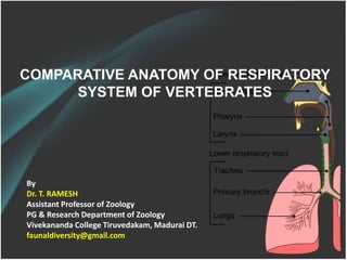
- 1. COMPARATIVE ANATOMY OF RESPIRATORY SYSTEM OF VERTEBRATES By Dr. T. RAMESH Assistant Professor of Zoology PG & Research Department of Zoology Vivekananda College Tiruvedakam, Madurai DT. faunaldiversity@gmail.com
- 2. RESPIRATORY SYSTEM It is the process of obtaining oxygen from the external environment & eliminating CO2 from the body of an organisms. Kind of respiration 1. External respiration - oxygen and carbon dioxide exchanged between the external environment & the body cells 2. Internal respiration - cells use oxygen for ATP production (& produce carbon dioxide in the process)
- 3. Respiratory organs 1. CUTANEOUS RESPIRATION- respiration through the skin can take place in air, water, or both most important among amphibians (especially the family Plethodontidae). 2. GILLS- Vertebrate gills are designed for water breathing Mechanism of ventilation depends on whether the gills are located internally or externally 1. Internal Gills- Lie within the head 2. External Gills-Develop from surface ectoderm and extend beyond the head
- 4. 3. LUNGS Designed for air breathing It is elastic bags that lie within the body Volume expands when air is inhaled and decrease When air is exhaled 4. GAS BALDDERS Are air filled with the air Swim bladders are used to control the buoyancy of a fish Gas bladders differ in lungs in two ways Gas bladder are usually situated dorsal to the digestive tracts Gas bladders are not paired
- 5. KEY DIFFERENCE IN FISHES 1. Cartilaginous fishes: •5 ‘naked’ gill slits •Anterior & posterior walls of the 1st 4 gill chambers have a gill surface (demibranch). Posterior wall of last (5th) chamber has no demibranch. •Interbranchial septum lies between 2 demibranchs of a gill arch •2 demibranchs + septum & associated cartilage, blood vessels, muscles, & nerves = holobranch 2. Bony fishes (teleosts): (See 'Ventilation in Teleost Fishes') •usually have 5 gill slits •operculum projects backward over gill chambers •interbranchial septa are very short or absent 3. Agnathans: •6 - 15 pairs of gill pouches.. Pouches connected to pharynx by afferent branchial (or gill) ducts & to exterior by efferent branchial (or gill) ducts.
- 6. MECHANISMS OF EXCHANGE OF GASES IN FISHES
- 7. Swim bladder & origin of lungs Swim bladder & origin of lungs Most vertebrates develop an out pocketing of pharynx or oesophagus that becomes one or a pair of sacs (swim bladders or lungs) filled with gases derived directly or indirectly from the atmosphere. Similarities between swim bladders & lungs indicate they are the same organs.
- 8. LUNGS 1. It is respiratory organ of higher vertebrates. 2. Lungs are developed from pharynx, arise in embryo an endo dermal diverticulam from the ventral wall of pharynx 3. The diverticulam divided into two portions- Right & Left lungs 4. Windpipe trachea connects the lungs and pharynx 5. The birds has sound producing organ is called Syrinx. 6. Lungs is branched- Primary Secondary bronchi Tertiary and Bronchides
- 9. RESPIRATION OF AMPHIBIANS It has respiratory surfaces on its body that it uses to exchange gas with the surroundings: the skin, in the lungs and on the lining of the mouth. A frog may also breathe much like a human, by taking air in through their nostrils and down into their lungs. The mechanism of taking air into the lungs is however slightly different than in humans. Frogs do not have ribs nor a diaphragm, which in humans helps serve in expand the chest and thereby decreasing the pressure in the lungs allowing outside air to flow in. Respiratory organs of Amphibians
- 10. RESPIRATION OF AMPHIBIANS 1. In order to draw air into its mouth the frog lowers the floor of its mouth, which causes the throat to expand. 2. Then the nostrils open allowing air to enter the enlarged mouth. 3. The nostrils then close and the air in the mouth is forced into the lungs by contraction of the floor of the mouth. 4. To eliminate the carbon dioxide in the lungs the floor of the mouth moves down, drawing the air out of the lungs and into the mouth. 5. Finally the nostrils are opened and the floor of the mouth moved up pushing the air out of the nostrils. 6. 2-simple, long spindle shaped, semi transparent, elastic, delicate, thin walled and sac like structure. 7. Internal lining may be smooth or have simple scaculation or packets 8. Air exchanged via positive pressure ventilation.
- 12. Four stages of frog lung ventilation 1. Buccal cavity expands to draw fresh air through the open nares 2. Glottis opens rapidly, releasing spent air from the elastic lungs. 3. Nares close, floor the bauccal cavity rises forcing the fresh air held in this cavity into the lung through the open glottis 4. Glottis closes retaining the air that has just filled the lungs and nares open again.
- 13. SKIN RESPIRATION Respiration through the skin can take place in air & water or both. It is very common in the family of Plethodontidae Cutaneous respiration is the absorption of oxygen and disposal of carbon dioxide thorough the skin. While completely submerged all of the frog's respiration takes place through the skin. The skin is composed of thin membranous tissue that is quite permeable to water and contains a large network of blood vessels. The thin membranous skin is allows the respiratory gases to readily diffuse directly down their gradients between the blood vessels and the surroundings. When the frog is out of the water, mucus glands in the skin keep the frog moist, which helps absorb dissolved oxygen from the air.
- 14. Reptilian lungs Simple sacs in Sphenodon & snakes Lizards, crocodilians, & turtles - lining is septets, with lots of chambers & sub chambers Air exchanged via positive-pressure ventilation. Specialized adaptation exist depending on the lizard's natural habitat way of life etc Crocodilians - bony secondary palate for breathing underwater Snakes- Tracheal extension for protection against asphyxiation while swallowing prey buccal pumping allows some lizards to increase stamina and oxygen capacity
- 15. Reptilian lungs Inspiration is caused by the movement of intercostals muscles, raising the ribs that increases the volume of the thorax and reduces the lung pressure causing the inflow of air into the lung. Oxygen of the air enters the blood of the blood capillaries and CO2 of the blood enters the alveoli. Expiration is done by lowering the ribs that decreases the volume of the thoracic cavity, flows back to the exterior.
- 16. Avian lungs The avian respiratory system delivers oxygen from the air to the tissues and also removes carbon dioxide. In addition, the respiratory system plays an important role in thermoregulation (maintaining normal body temperature). The avian respiratory system is different from that of other vertebrates, with birds having relatively small lungs plus nine air sacs that play an important role in respiration (but are not directly involved in the exchange of gases).
- 17. Avian lungs Birds must be capable of high rates of gas exchange because their oxygen consumption at rest is higher than that of all other vertebrates and it increases many times during flight. The air sacs permit a unidirectional flow of air through the lungs. Unidirectional flow means that air moving through bird lungs is largely 'fresh' air & has a higher oxygen content. So, in bird lungs, more oxygen is available to diffuse into the blood (avian respiratory system).
- 19. Avian lungs The air sacs permit a unidirectional flow of air through the lungs. Unidirectional flow means that air moving through bird lungs is largely 'fresh' air & has a higher oxygen content. In contrast, air flow is 'bidirectional' in mammals, moving back and forth into and out of the lungs. As a result, air coming into a mammal's lungs is mixed with 'old' air (air that has been in the lungs for a while) & this 'mixed air' has less oxygen. So, in bird lungs, more oxygen is available to diffuse into the blood (avian respiratory system).
- 20. Mammalian lungs Air is inhaled through the lungs (breathing). Mammalian lungs are subdivided internally. The repetitive subdivisions of the lung airways provide gas to the tiny alveoli (gas sacs) that form the functional gas-exchange surface area of the lungs. Haemoglobin molecules inside red blood cells capture oxygen. Blood with oxygen is pumped through the body to all tissues. In capillaries, cells release carbon dioxide into the blood and pick up fresh oxygen. Oxygen diffuses into the cell and is used in the mitochondria to break down glucose molecules and make ATP.
- 21. Human lungs
- 26. Thank you