
Recommandé
Contenu connexe
Tendances
Tendances (20)
Similaire à Keratoconus...
Similaire à Keratoconus... (20)
Dernier
Dernier (20)
Keratoconus...
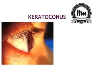
- 1. KERATOCONUS REEMA V. DANDAVATE S.Y. B.OPTOMETRY
- 2. Introduction… Kerato- cornea ; conus- cone shaped. Keratoconus is a non-inflammatory bilateral ectatic condition of cornea in its axial part. It usually starts at puberty and progresses slowly. Prevalence of keratoconus in India is upto 2300 per 1,00,000.
- 3. Etiopathogenesis… Still not clear Various theories proposed, labeled it as: developmental condition degenerative condition hereditary dystrophy endocrine anomaly Essential pathological changes are progressive thinning and ectasia which occurs as a result of defective synthesis of mucopolysaccharide and collagen tissue.
- 4. Clinical features… Symptoms : Frequent changing of glasses. Glare. Photophobia. Distorted vision.
- 5. Signs… Torch light examination: Rizzutti sign Munson’s sign Placido disc Keratometer reaings.
- 6. Retinoscopy Topography Ophthalmoscope examination Slit-lamp examination. Vogt striae. Copper ring. Hydrops.
- 7. Morphological classification… conesize size size in mm Nipple cone Small size & steep curvature <5 mm Oval cone Larger & ellipsoid in shape 5-6 mm Globus cone Very large & globe like >6mm Depending upon size and shape of the cone keratoconus is classifies into threee types…
- 9. Classification based on severity… Curvature Power in diopters Mild <45 D Moderate 45D – 52D Advanced 52D – 62D Severe >62D
- 10. Complications… It may be complicated by development of acute hydrops due to rupture of descemet’s membrane. The condition is characterized by sudden development of corneal oedema associated with marked defective vision, pain, photophobia, lacrimation.
- 11. Associations… Keratoconus may be associated with : OCULAR CONDITIONS SYSTEMIC CONDITIONS
- 12. Ocular conditions… Ectopia lentis Congenital cataract Aniridia Retinitis pigmentosa Vernal keratoconjunctivitis (VKC)
- 13. Systemic conditions… Marfan’s syndrome Atopy Down’s syndrome Ehlers-Danols syndrome Osteogenesis imperfecta Mitral valve prolapse
- 14. treatment… Spectacle correction. Contact lenses. Intacts. Corneal collagen cross-linking with riboflavin (CXL or C3R) and UV-A rays may slow the progression of disease. Keratoplasty.
- 15. Reference… A.K. Khurana book of ophthalmology. Kanski
Notes de l'éditeur
- Developmental cond : Degenerative cond : cone shape increases coz of irregular corneal fibers. : Once initiated, the disease normally develops by progressive dissolution of Bowman&apos;s layer,[4] which lies between the corneal epithelium and stroma. As the two come into contact, cellular and structural changes in the cornea adversely affect its integrity and lead to the bulging and scarring characteristic of the disorder. Within any individual keratoconic cornea, regions of degenerative thinning coexisting with regions undergoing wound healing may be found. Scarring appears to be an aspect of the corneal degradation; however, a recent, large, multicenter study suggests abrasion by contact lenses may increase the likelihood of this finding by a factor over two.[20][21] Heri : Endocrine anomaly :
- Munson sign :a protrusion of the lower eyelid in downgaze. Placido disc exa:A handheld keratoscope, sometimes known as &quot;Placido&apos;s disk&quot;, can provide a simple noninvasive visualization of the surface of the cornea by projecting a series of concentric rings of light onto the cornea. K readings :The eye examination may proceed to measurement of the localized curvature of the cornea with a manual keratometer,[7] with detection of irregular astigmatism suggesting a possibility of keratoconus. Severe cases can exceed the instrument&apos;s measuring ability.
- Retino :A further indication can be provided by retinoscopy, in which a light beam is focused on the person&apos;s retina and the reflection, or reflex, observed as the examiner tilts the light source back and forth. Keratoconus is amongst the ophthalmic conditions that exhibit a scissor reflex action of two bands moving toward and away from each other like the blades of a pair of scissors Topo :A more definitive diagnosis can be obtained using corneal topography, in which an automated instrument projects the illuminated pattern onto the cornea and determines its topology from analysis of the digital image. The topographical map indicates any distortions or scarring in the cornea, with keratoconus revealed by a characteristic steepening of curvature which is usually below the centreline of the eye Slit lamp : Vogt strie:Similarly, around 50% of subjects exhibit Vogt&apos;s striae, fine stress lines within the cornea caused by stretching and thinning.[10] The striae temporarily disappear while slight pressure is applied to the eyeball. Copper ring: Under close examination, a ring of yellow-brown to olive-green pigmentation known as a Fleischer ring can be observed in around half of keratoconic eyes.[10] The Fleischer ring, caused by deposition of the iron oxide hemosiderin within the corneal epithelium, is subtle and may not be readily detectable in all cases, but becomes more evident when viewed under a cobalt blue filter.[4 Hydropes :
- Ectopia lentis is a displacement or malposition of the eye&apos;s crystalline lens from its normal location.
- atopy : Keratoconus has been associated with atopic diseases,[31] which include asthma, allergies, and eczema, and it is not uncommon for several or all of these diseases to affect one person