1. Necrolytic acral erythema =اﻠﺤﻤاﻤﻰ اﻠﻨﺨرﻴﺔ اﻠاﻨﺤﻠاﻠﻴﺔ ﻋﻠﻰ اﻠﻨﻬاﻴاﺖ
Necrolytic acral erythema
Necrolytic acral erythema (NAE) was first described in 1996 by physicians in Egypt, M. El
Darouti and M. Abu El Ela.1 Reports have continued to link hepatitis C with NAE.2 Necrolytic
acral erythema manifests as well-circumscribed, dusky erythematous plaques with adherent
scale. While the plaques are psoriasiform, they do not manifest an Auspitz sign as would be
seen with psoriasis. Patients with active necrolytic acral erythema report burning or pruritus. It is
limited to an acral distribution and, in most cases, is associated with hepatitis C infection.
3,4
Several cases of necrolytic acral erythema have occurred in patients without hepatitis C.
5
This suggests that necrolytic acral erythema might be a result of zinc dysregulation, rather than
1 / 13
2. Necrolytic acral erythema =اﻠﺤﻤاﻤﻰ اﻠﻨﺨرﻴﺔ اﻠاﻨﺤﻠاﻠﻴﺔ ﻋﻠﻰ اﻠﻨﻬاﻴاﺖ
a result of hepatitis C infection itself.
Authors debate whether necrolytic acral erythema is a distinct entity or a subtype of necrolytic
migratory erythema. However, the distinct appearance and usual coincidence with hepatitis C
infections suggests that it is a unique entity. It has been speculated that viral load and viral
genotype might play a role in NAE.6
Pathophysiology
The pathophysiology of this condition is uncertain. Proposed theories for the cause of necrolytic
acral erythema describe alterations in some metabolic factor, many of which are seen in other
necrolytic erythemas, including necrolytic migratory erythema, pellagra, essential fatty acid and
biotin deficiency, and acrodermatitis enteropathica. The hypothesized causes for the metabolic
alteration include hypoalbuminemia, hypoaminoacidemia, low zinc level, increased glucagon,
liver dysfunction, or diabetes. Only hepatitis C is universally present in all persons with
necrolytic acral erythema.
An odd fact is that no cases of necrolytic acral erythema have been reported in Japan, which
has a high seroprevalence rate of hepatitis C.
History
Patients with necrolytic acral erythema present with a 1-month to 2-year history of a rash on the
dorsum of the feet, which may or may not also involve the hands. In many cases, it has been
unresponsive to topical steroids.
Patients do not necessarily give a history of hepatitis C. Necrolytic acral erythema may be the
presenting sign. Patients may report a burning sensation, especially when walking or standing.
Sometimes, necrolytic acral erythema lesions are pruritic. The most common area of origination
of the rash is the dorsal aspect of the great toe. It also occurs on the shins. In 43 of 44 reported
cases, necrolytic acral erythema did not affect the palms or soles, the nail bed, nail plate, or
mucous membranes.
Williams8 reported a case of necrolytic acral erythema in an adolescent boy with a history of
infection with hepatitis C virus; he had hepatic fibrosis, hypertension, and thrombocytopenia.
2 / 13
3. Necrolytic acral erythema =اﻠﺤﻤاﻤﻰ اﻠﻨﺨرﻴﺔ اﻠاﻨﺤﻠاﻠﻴﺔ ﻋﻠﻰ اﻠﻨﻬاﻴاﺖ
The patient developed a pruritic eruption on his face, trunk, genitals, and extremities, and the
eruption had a predilection for bony prominences. This patient did not respond to topical
steroids, antihistamines, topical barrier repair creams, narrow-band UV-B therapy, or tar baths.
A skin biopsy specimen demonstrated a nonspecific psoriasiform dermatitis consistent with
necrolytic acral erythema. Hepatitis C virus RNA polymerase chain reaction studies showed
974,000 IU/mL (<50) and hepatitis C virus RNA genotype 1b.
Janjua9 noted a 45-year-old man who developed well-defined erythematous hyperpigmented
keratotic pruritic plaques on the dorsa of his feet and the lateral aspect of his ankles; he was
determined to have necrolytic acral erythema.
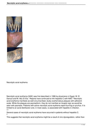
Physical
Dusky, red erythematous plaques are present bilaterally on the dorsum of the feet and toes.
These may extend around to the skin overlying the Achilles tendon and up the lower leg, as
depicted in the image below
The dorsum of the hands may or may not be involved. The lesions are clearly demarcated from
uninvolved skin by a dark red border. The surface may be scaly, eroded, or velvety. Thick
hyperkeratosis is sometimes present. Active lesions often include flaccid blisters. Edema may or
may not be present.
Abdallah et al10 describe stages of the lesions:
1. Initial stage: Erythematous papules or plaque with scale are present and have a dusky or
eroded center.
2. Fully developed stage: A confluence of papules and plaques with sharply defined margins
and adherent scale develops. Increased hyperpigmentation and decreased redness may be
present. Lesions may be lichenified. Pustules also may occur at this stage.
3. Late stage: Thinning of lesions occurs, with continued hyperpigmentation. Demarcation
continues, followed by spontaneous relapse and remission.
3 / 13
4. Necrolytic acral erythema =اﻠﺤﻤاﻤﻰ اﻠﻨﺨرﻴﺔ اﻠاﻨﺤﻠاﻠﻴﺔ ﻋﻠﻰ اﻠﻨﻬاﻴاﺖ
As a general rule, necrolytic acral erythema does not affect the palms or soles, the nail bed, nail
plate, or mucous membranes. Hivnor et al11 reported a single case in which the plaques
extended proximally to the thighs. This patient also had hyperkeratosis of the palms and soles
and involvement of the face. While multiple biopsy specimens confirmed necrolytic acral
erythema histologically, it is not clear whether these biopsy specimens were taken from the
typical locations of necrolytic acral erythema or if the palms and soles were also included in the
biopsy specimen.
Other disorders that can be seen in persons with hepatitis C should be considered as possible
epiphenomena. These include the following:
- Lichen planus
- Porphyria cutanea tarda
- Palpable purpura/leukocytoclastic vasculitis
- Spider telangiectasias
- Vitiligo
- Polyarteritis nodosa
- Erythema multiforme
- Urticaria
- Erythema nodosum
Causes
The cause is uncertain, but a metabolic alteration due to hepatocellular degeneration from
hepatitis C infection is proposed. Many hypotheses describe deficiencies similar to those of the
other necrolytic erythemas; histological features are similar among necrolytic acral erythema
and other nutrient deficiencies. Note the following:
- el Darouti and Abu El Ela1 suggested that the increased levels of glucagon seen in
persons with liver disease allow for "potentiation" of arachidonic acid after trauma.
- High glucagon levels alone may allow for greater arachidonic acid potentiation, which may
induce inflammatory changes and necrosis in the epidermis.
- Low amino acid levels may lead to epidermal protein depletion and necrolysis.
- Low albumin levels have also been postulated as causative. Albumin sequesters fatty
acids and helps regulate prostaglandin levels. High levels of prostaglandins as a result of low
levels of albumin may induce inflammation.
- Low zinc levels have also been proposed as a cause. Because albumin is the main carrier
of zinc in plasma, these 2 may be interrelated.
- Diabetic microangiopathy also may play a role; 4 of 5 necrolytic acral erythema patients
described by Nofal et al 12 also had diabetes.
4 / 13
5. Necrolytic acral erythema =اﻠﺤﻤاﻤﻰ اﻠﻨﺨرﻴﺔ اﻠاﻨﺤﻠاﻠﻴﺔ ﻋﻠﻰ اﻠﻨﻬاﻴاﺖ
Laboratory Studies
Hepatitis A, B, and C serologies: These are the most important tests because necrolytic acral
erythema (NAE) has a high association with hepatitis C.
Assess liver function, including aspartate aminotransferase, alanine aminotransferase, bilirubin,
protein, albumin, and alkaline phosphatase values. Some authors believe the lesions are most
active during the active stage of hepatitis. The severity of the lesions does not correlate with the
severity of the hepatitis.
Serum zinc, serum glucagon, and serum amino acid levels: These values have been reduced in
some reported cases. Zinc deficiency associated with necrolytic acral erythema was noted by
Najarian et al.13
Serum glucose and hemoglobin A1C levels: Consider performing these tests because necrolytic
acral erythema may be more common in persons with diabetes and hepatitis C.
Najarian et al has associated hypozincemia and hyperzincuria with necrolytic acral erythema.14
Imaging Studies
CT scanning of the liver and pancreas can be considered as an ancillary assessment. In some
reported cases, the pancreas was imaged, most likely because some believe necrolytic acral
erythema is a subset of necrolytic migratory erythema, a condition indicative of pancreatic
glucagonoma. No pancreatic masses were found any of the patients imaged.
Procedures
5 / 13
6. Necrolytic acral erythema =اﻠﺤﻤاﻤﻰ اﻠﻨﺨرﻴﺔ اﻠاﻨﺤﻠاﻠﻴﺔ ﻋﻠﻰ اﻠﻨﻬاﻴاﺖ
Consider skin biopsy to substantiate the diagnosis of necrolytic acral erythema. Consider liver
biopsy to assess the status of hepatitis and before the institution of hepatitis C treatment.
Histologic Findings
Necrolytic acral erythema is perhaps best defined clinically rather than histologically. Bentley et
al noted a case of necrolytic acral erythema without classic histology findings, which responded
to oral zinc therapy.15
In 2004, Abdallah et al10 noted the following histopathological features:
- Early stage - Nummular dermatitis–like moderate and regular acanthosis, as follows:
- Variable spongiosis
- Inflammatory infiltrate
- Pigment incontinence
- Fully evolved stage - Psoriasiform epidermal hyperplasia, as follows:
- Marked papillomatosis
- Parakeratosis
- Focal hypergranulosis
- Pigment incontinence
- Occasional subcorneal pustules
- Epidermal pallor
- Vascular ectasia
- Infiltrate of inflammatory cells in the papillary dermis
- Necrotic keratinocytes - Sometimes become confluent in the upper epidermis, sometimes
tracking along the acrosyringium
- Late stage samples - Minimal-to-moderate acanthosis, as follows:
- Minimal-to-moderate inflammatory cell infiltrate
- Pigment incontinence
Others have noted epidermal necrosis with or without blister formation; vacuolar degeneration
of the basal layer; and psoriatic changes with acanthosis, papillomatosis, hyperkeratosis, and
parakeratosis. All of these are commonly found in persons in deficient nutritional states. Dermal
findings have included a nonspecific superficial perivascular and interstitial lymphocytic
dermatitis.
El-Ghandour et al7 noted that electron microscope examination of patients with necrolytic acral
6 / 13
7. Necrolytic acral erythema =اﻠﺤﻤاﻤﻰ اﻠﻨﺨرﻴﺔ اﻠاﻨﺤﻠاﻠﻴﺔ ﻋﻠﻰ اﻠﻨﻬاﻴاﺖ
erythema demonstrated clumped tonofilaments in the keratinocytes, yet hepatitis C virus RNA
could not be detected in the plaques of necrolytic acral erythema.
Medical Care
The optimal treatment for necrolytic acral erythema (NAE) is the optimal treatment of hepatitis
C—combination therapy with interferon and ribavirin. In one patient, ribavirin in addition to the
interferon alfa therapy improved the necrolytic acral erythema despite the presence of a
continued high viral load.
Oral zinc replacement has been successful in some cases.4,16 Interferon alfa monotherapy has
been reported to be effective treatment.
4
Amino acid replacement therapy, both orally and parenterally, has yielded some improvement.
Disease response to corticosteroid therapy (ie, topical, intralesional, and systemic) has been
poor. No benefit has been seen with topical tar or tetracycline.
El-Ghandour et al7 noted in their series of 23 Egyptian patients that interferon alfa combined
with ribavirin caused regression of the plaques of necrolytic acral erythema in 3 patients and
complete clearance in 1 patient. Oral zinc administration induced some improvement in the
appearance of the skin in some patients.
Surgical Care
A surgical approach has not been shown to be therapeutically beneficial.
Consultations
Consultation with an infectious disease specialist, gastroenterologist, or hepatologist may be
helpful for management of the hepatitis C. Consultation with a dermatologist may be helpful for
diagnosis and management of cutaneous findings.
Diet
Supplementation with zinc and amino acids has shown benefit.
7 / 13
8. Necrolytic acral erythema =اﻠﺤﻤاﻤﻰ اﻠﻨﺨرﻴﺔ اﻠاﻨﺤﻠاﻠﻴﺔ ﻋﻠﻰ اﻠﻨﻬاﻴاﺖ
Activity
No restrictions on activity are needed for persons with this condition.
Medication
The goal of treatment is to decrease the burning and pruritus and improve the appearance of
the lesions of necrolytic acral erythema. Zinc sulfate,17 amino acid supplementation, and
interferon alfa have been successful in treating necrolytic acral erythema. Topical tacrolimus
has been reported as an effective treatment for necrolytic acral erythema.
18
Minerals
Act as enzyme cofactors and are required in metabolic processing.
Zinc supplements
Use sulfate or gluconate zinc salt. Zinc sulfate 4.4 mg = 1 mg of elemental zinc. Zinc gluconate
7.1 mg = 1 mg of elemental zinc.
- Dosing
- Interactions
- Contraindications
- Precautions
Adult
Effective zinc sulfate dose used in studies: 440 mg PO qd (single dose or divided) for 100 mg/d
of elemental zinc
Pediatric
General supplementation: 1-2 mg/kg/d elemental zinc; dose not documented in 12-year-old
treated with zinc
8 / 13
9. Necrolytic acral erythema =اﻠﺤﻤاﻤﻰ اﻠﻨﺨرﻴﺔ اﻠاﻨﺤﻠاﻠﻴﺔ ﻋﻠﻰ اﻠﻨﻬاﻴاﺖ
- Dosing
- Interactions
- Contraindications
- Precautions
May reduce penicillamine and tetracycline effects
- Dosing
- Interactions
- Contraindications
- Precautions
Documented hypersensitivity
- Dosing
- Interactions
- Contraindications
- Precautions
Pregnancy
C - Fetal risk revealed in studies in animals but not established or not studied in humans; may
use if benefits outweigh risk to fetus
Precautions
Caution in patients with renal impairment
Interferons
Naturally occurring compounds that have both antiviral and immunomodulatory effects.
Peginterferon alfa-2a (Pegasys)
Used in combination with ribavirin to treat patients with chronic hepatitis C who have
9 / 13
10. Necrolytic acral erythema =اﻠﺤﻤاﻤﻰ اﻠﻨﺨرﻴﺔ اﻠاﻨﺤﻠاﻠﻴﺔ ﻋﻠﻰ اﻠﻨﻬاﻴاﺖ
compensated liver disease and have not previously received interferon alfa. Consists of
interferon alfa-2a attached to a 40-kd branched PEG molecule. Predominantly metabolized by
the liver.
- Dosing
- Interactions
- Contraindications
- Precautions
Adult
180 mcg SC qwk
Pediatric
Not established
- Dosing
- Interactions
- Contraindications
- Precautions
Theophylline may increase toxicity by reducing clearance; cimetidine may increase antitumor
effects; zidovudine and vinblastine may increase toxicity
- Dosing
- Interactions
- Contraindications
- Precautions
Documented hypersensitivity; decompensated liver disease; significant preexisting psychiatric
disease; ongoing or recent alcohol use; platelet count <70,000/µL
- Dosing
- Interactions
- Contraindications
10 / 13
11. Necrolytic acral erythema =اﻠﺤﻤاﻤﻰ اﻠﻨﺨرﻴﺔ اﻠاﻨﺤﻠاﻠﻴﺔ ﻋﻠﻰ اﻠﻨﻬاﻴاﺖ
- Precautions
Pregnancy
C - Fetal risk revealed in studies in animals but not established or not studied in humans; may
use if benefits outweigh risk to fetus
Precautions
Insomnia; mental dysfunction (eg, mood dysfunction, depression, psychosis, aggressive
behavior, hallucinations, violent behavior, suicidal ideation, suicide attempt, suicide, homicidal
ideation [rare]), even without previous history of psychiatric illness; flulike symptoms; rash and
pruritus; anorexia; neutropenia; thrombocytopenia; thyroid dysfunction; retinal abnormalities
Peginterferon alfa-2b (PEG Intron)
Escherichia coli recombinant product. Used to treat chronic hepatitis C in patients not previously
treated with interferon alfa who have compensated liver disease. Exerts cellular activities by
binding to specific membrane receptors on cell surface, which, in turn, may suppress cell
proliferation and may enhance phagocytic activity of macrophages. May also increase
cytotoxicity of lymphocytes for target cells and inhibit virus replication in virus-infected cells.
- Dosing
- Interactions
- Contraindications
- Precautions
Adult
Inject SC qwk for 1 y using weight-based dosing
37-45 kg: 40 mcg (0.4 mL of 100 mcg/mL)
46-56 kg: 50 mcg (0.5 mL of 100 mcg/mL)
57-72 kg: 64 mcg (0.4 mL of 160 mcg/mL)
73-88 kg: 80 mcg (0.5 mL of 160 mcg/mL)
89-106 kg: 96 mcg (0.4 mL of 240 mcg/mL)
107-136 kg: 120 mcg (0.5 mL of 240 mcg/mL)
137-160 kg: 150 mcg (0.5 mL of 300 mcg/mL)
Pediatric
11 / 13
12. Necrolytic acral erythema =اﻠﺤﻤاﻤﻰ اﻠﻨﺨرﻴﺔ اﻠاﻨﺤﻠاﻠﻴﺔ ﻋﻠﻰ اﻠﻨﻬاﻴاﺖ
Not established
- Dosing
- Interactions
- Contraindications
- Precautions
Concurrent administration with IL-2 may increase nephrotoxicity; theophylline, zidovudine, and
vinblastine may increase toxicity; cimetidine may increase antitumor effects
- Dosing
- Interactions
- Contraindications
- Precautions
Documented hypersensitivity; autoimmune hepatitis; pancreatitis; colitis
- Dosing
- Interactions
- Contraindications
- Precautions
Pregnancy
C - Fetal risk revealed in studies in animals but not established or not studied in humans; may
use if benefits outweigh risk to fetus
Precautions
Considered to have abortifacient potential; reduce starting dose by 50% or discontinue if serious
adverse reactions develop during course of treatment (may reinitiate treatment if adverse
reaction abates or decreases in severity); fatal and nonfatal pancreatitis or ulcerative and
hemorrhagic colitis reported; life-threatening or fatal neuropsychiatric events may occur; severe
suppression of bone marrow function may occur, sometimes resulting in severe cytopenias;
may cause, headache, flulike symptoms, or myelosuppressive, pulmonary, thyroid,
cardiovascular, or infectious disorders
12 / 13
13. Necrolytic acral erythema =اﻠﺤﻤاﻤﻰ اﻠﻨﺨرﻴﺔ اﻠاﻨﺤﻠاﻠﻴﺔ ﻋﻠﻰ اﻠﻨﻬاﻴاﺖ
Nucleoside Analogs
Drugs that interfere with the production of DNA and RNA.
Ribavirin (Virazole)
Antiviral nucleoside analog. Chemical name is D
-ribofuranosyl-1H-1,2,4-triazole-3-carboxamide. Given alone, has little effect on the course of
hepatitis C. When given with interferon, significantly augments rate of sustained virologic
response.
- Dosing
- Interactions
- Contraindications
- Precautions
Adult
10.6 mg/kg PO qd or divided bid
Pediatric
Not established
.
13 / 13