Developmental reorganization of the human cerebral cortex
•
1 j'aime•805 vues

Developmental reorganization of the human cerebral cortex

Recommandé
Contenu connexe
Tendances
Tendances (9)
En vedette
En vedette (7)
Similaire à Developmental reorganization of the human cerebral cortex
Similaire à Developmental reorganization of the human cerebral cortex (20)
Plus de South Dakota Pain Capable Unborn Child Protection Act
Plus de South Dakota Pain Capable Unborn Child Protection Act (20)
Dernier
Dernier (20)
Developmental reorganization of the human cerebral cortex
- 1. Paediatr Croat 2007; 51 (Supl 1): 93-98 Pregled Review DEVELOPMENTAL REORGANIZATION OF THE HUMAN CEREBRAL CORTEX IVICA KOSTOVIĆ1, ZDRAVKO PETANJEK1, 2 This paper reveals data on developmental reorganization of the human cortex. Three criteria were used to determine reorga- nization: (1) presence of transient cellular zones: ventricular (VZ), subventricular (SVZ), intermediate (IZ) zone, subplate (SP), cortical plate (CP) and marginal zone (MZ); (2) intensity of specific neurodevelopmental cellular events: proliferation, migration, differentiation, growth of axon and synaptogenesis; (3) pattern of functional organization: (a) endogenous, transient circuitry and (b) permanent, sensory driven circuitry. First half of gestation is characterized by proliferation and migration. During the second half of gestation major axonal pathways grow through the intermediate zone and "wait" in the subplate. Initial synaptogenesis begins during third month of gestation and is related to the endogenous, spontaneous circuitry. In early preterm thalamocortical fibers relocate from the subplate (after 24th postconceptional weeks) and first evoked potentials may be recorded. The fundamental pattern is coexistence of endogenous and permanent, sensory driven circuitry. In neonatal brain synaptogenesis is main neuroge- netic event and there is gradual disappearance of transient endogenous circuitry and transient cellular zones. The review support ideas of developmental "windows", selective vulnerability of specific transient cellular zones (subplate) and increased vulnerability of the human fetal and neonatal brain. Descriptors: PREMATURUS, KORTIKALNE VEZE, TALAMOKORTIKALNA VLAKNA, SINAPTOGENEZA, HISTOGENEZA Acknowledgements: This work was ● presence of transient circuitry and ● Functional patterns in developing supported by grants 108-1081870-1876 transient functions (1). brain might be (a) endogenous (spon- (I.K.) and 108-1081870-1932 (Z.P.) from taneous), transient circuitry, mostly the Croatian Ministry of Science, Educa- In this review we will discuss reor- characterized with oscillatory prop- tion & Sport. ganization of developing brain based on erties and (b) sensory driven, perma- these three basic patterns of organiza- nent circuitry (2, 3). Introduction tion. Our focus will be on fetal, early pre- term, late preterm and neonatal period. Transient patterns and their reorganization during Fetal cortex differs from the neona- In order to describe each of this periods prenatal and perinatal developmental "windows" tal in three different aspects of organiza- of development it is necessarily to define tion: transient zones, neurogenetic events and Fetal period ● presence of transient cell zones in types of transient functional patterns. which cellular events takes place; Fetal period is dominated by two ● Transient zones (Figure 1) in which neurodevelopment events, proliferation ● intensity of specific neurodevelop- neurodevelopmental events take and migration (4, 5). It is also a period mental cellular events; places are ventricular zone (VZ), when synaptogenesis begins and axonal subventricular zone (SVZ), interme- pathways (projections) establish. Prolif- 1 Department of Neuroscience diate zone (IZ), subplate (SP), cor- Croatian Institute for Brain Research eration: Neurons are generated in ven- tical plate (CP) and marginal zone School of Medicine, University of Zagreb tricular and subventricular zone from (MZ). 2 Department of Anatomy the neuronal stem cells by asymmetric School of Medicine, University of Zagreb and symmetric divisions. The period of ● The neurodevelopmental cellular Address: events which will be considered in proliferation of neurons is between 4th Zdravko Petanjek, MD, PhD, Professor of this review are proliferation, migra- and 28th postconceptional week (Rakic Anatomy & Neuroscience 2006, Cerebral Cortex). According to re- Department of Neuroscience tion and differentiation of neurons, Croatian Institute for Brain Research axonal growth and synaptogenesis. cent evidence radial glia cells also serve School of Medicine, University of Zagreb Myelinization and cell death will be as neural stem cell and might produce 10000 Zagreb, Šalata 12, Croatia not discussed in this review. pyramidal neurons (Rakic 2006). The E-mail: zpetanjek@net.hr GABA-ergic (inhibitory interneurons) in 93
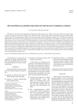
- 2. I. Kostović et al. Developmental reorganization... Paediatr Croat 2007; 51 (Supl 1): 93-98 Migration: Newly generated neu- Pattern of functional organization: rons migrate along radial glia cells In the fetal cortex there is no sensory through intermediate zone and form em- driven activity. However, synapse seems bryonic column (4). The disturbances of to be very active (3). This type of oscilla- proliferation and migration might result tory activity was described as spontane- in so called migratory disorders, which ous activity (2, 3, 5, 17). Early thalamic are frequently associated with epilepsy input to subplate might participate in this and mental retardation. Transient lami- circuitry, as a transient input. Subplate nar pattern: After 13 postconceptional neurons have also efferent projection to week a new lamina develops below cor- thalamus and subcortical centers and tical plate and became very soon thickest this presumably glutamatergic output lamina of the telencephalon. This lamina might contribute to generation of fetal is subplate zone which can be seen on general movements (18). In conclusion, both Nissl preparations and magnetic early fetal circuitry is transient and it is resonance images in vivo and in vitro (6, related to transient pattern of structural 7). Prominent subplate zone is the main organization. This concept is accepted characteristic of fetal pattern of laminar by most modern neurophysiologist and it organization. It is easy to distinguish the is in contrast with some classical opin- following layers (from ventricle to pia): ions about reflex type generation of fetal VZ, SVZ, IZ, SP, CP and MZ (Figure 1). motility. The subplate zone is site of early syn- aptogenesis, endogenous neural activity Early preterm period and neuronal differentiation. Figure 1 During early preterm period several Transient laminar organization of the Growth of axonal pathways: The reorganizational events take place. This telencephalic wall: Cresyl violet (Nissl) staining of the earliest pathways which arrive in fe- period is characterized by development telencephalic wall in the human fetus during tal cortex are monoaminergic afferents of primary gyri and sulci (19-21). Tran- midgestation; transient zones in which from brain stem tegmentum and cholin- sient laminar organization: For the first neurodevelopmental events take place are ergic afferents from the basal nucleus time there is initial lamination in corti- ventricular zone (VZ), subventricular zone (SVZ), intermediate zone (IZ), subplate (SP), of Maynert (8-10). Next afferents in se- cal plate, which coexists with extremely cortical plate (CP) and marginal zone (MZ) quentional growth are massive pathways prominent subplate zone. This is a spe- Slika 1. originating in thalamus. Thalamic fibers cial feature of preterm cortex and mix- Prolazna laminarna organizacija stjenke originate not only from sensory tha- ture of fetal and permanent patterns (2, telencefalona: lamic nuclei, but also from associative 14). Neurogenetic events: Intensity of Krezil violet (Nisslovo) bojanje stjenke proliferation and migration during pre- thalamic nuclei (2, 5, 11-13). This most telencefalona u fetusa čovjeka tijekom srednje trećine trudnoće: prolazni stanični slojevi koji massive input grows throughout subplate term period decreased significantly. Ven- su mjesto specifičnih neurorazvojnih događaja zone during prolonged period of axonal tricular zone become thinner and neural su: ventrikularni (VZ), subventrikularni pathfinding (14). At the end of fetal pe- stem cells gradually stop producing neu- (SVZ), intermedijarni (IZ) sloj (zona), sloj rons and continue to produce glia cell riod that is between 21-23 postconcep- ispod ploče (subplate-SP), kortikalna ploča (CP) i marginalni sloj (zona) (MZ) tional weeks, thalamocortical afferents lines (proolygodendrocites, astrocytes). accumulate in the superficial part of the Migration decreases in intensity and late subplate zone (5, 13). These fibers are de- born neurons only might be found to mi- humans originate in the pallial (cortical) scribed as "waiting" fibers. grate through intermediate zone. ventricular zone, while in rodent brain main source of GABA-ergic neurons is Synaptogenesis: Early synapses de- Axonal growth: The crucial event in in another germinal structure called gan- velop above and below cortical plate. axonal growth is relocation of thalamic glionic eminence. Several distinct fea- Below cortical plate is plexiform pre- afferents from subplate zone and their tures distinguish human brain from other subplate layer and above cortical plate is ingrowth into cortical plate. That event species, especially rodents (4). This is on marginal zone. This early fetal pattern of occurs almost simultaneously in primary the first place increased number of mitot- synaptic distribution might be described and associative cortex (11-13). Parallel to ic cycles (35 in human compared to 11 in as period of two synaptic strata (15, 16). the thalamo-cortical ingrowth there is the rodents). Second, there is local gen- The early postsynaptic elements are pre- rapid areal differentiation (14, 22). Syn- eration of the GABA-ergic interneurons plate and marginal zone neurons, as well aptogenesis: In the early preterm infant (as stated above). Third, ganglionic emi- as branches of cortical plate neurons synapses are formed, for the first time, nence which is the basal enlargement of which are distributed in marginal zone within deep part of the cortical plate (15, the basal telencephalic ventricular zone and preplate (15). 16). This intracortical synaptogenesis is generates also neurons for thalamus (4). related to development of thalamo-corti- cal circuitry (12-14). 94
- 3. I. Kostović et al. Developmental reorganization... Paediatr Croat 2007; 51 (Supl 1): 93-98 Functional pattern of organization: still waiting in subplate, and majority as it was show by Burkhalter et al. (42). Development of thalamocortical connec- of "permanent" axons are already in the Functional pattern: The main character- tivity explains early evoked potential in cortical plate. istic of neonatal period is establishment preterm infant (23-27). The early devel- of sensory driven activity. This is par- opment of evoked potential in preterm Synaptogenesis and neuronal differ- ticularly important for development of infant together with transient circuitry in entiation: Synaptogenesis is proceeding columnar organization in sensory corti- subplate shows that there are interactions very fast in superficial part of cortical ces (3). Also, there is synchronization in between endogenous circuitry of the sub- plate paralleled with accelerated devel- EEG and sleep pattern, transient general plate zone, and thalamic sensory driven opment of dendrites pyramidal neurons movements still persist in the neonatal circuitry (2, 3). Same thalamic terminals (35, 36). Functional pattern: Due to the period indicating immaturity of cortical might activate cell in the cortical plate fast synaptogenesis in superficial cortex, and subcortical circuitry (18). and form synapses in subplate. This co- cortical electrical dipole changes and existence with transient endogens and surface negative response dominates in electrical recordings. There is gradual Discussion permanent driven circuitry might exist in the human preterm for a prolonged disappearance of giant potentials and In this review we have presented time (2). This combined circuitry seems synchronization of EEG (23, 37). evidence that fetal and preterm cortex to be essential feature of the preterm in- shows transient pattern of organiza- fant and underlie transient electrophysi- Neonatal period tion and permanent developmental re- ological and behavioral phenomena (23, organization. These phenomena were 28-30). Establishment of thalamo-corti- During neonatal period fetal and also observed using different structural cal connection with somatosensory cor- preterm patterns are reorganized dra- approaches and physiological record- tex attracted recently a great attention matically. This reorganization from tran- ings (1, 4). Neurodevelopmental cellu- among anesthesiologists, due to the fact sitional patterns to final organization is lar event occur with different intensity that this pathway may represent anatom- crucial for understanding physiological throughout development. The periods of ical substrate for pain input to cortex in and behavioral phenomena in neonatal increased cellular activity might also be early preterm infants (7, 14, 16, 31, 32). period. Laminar organization: Neocor- described as developmental "windows". The final prove that pain stimuli can tex develops into typical six layered pat- Throughout each developmental window reach human cortex in early preterm in- tern. However, layer IV (granular layer) transient zones display characteristic fants comes from study of M. Fitcgerald is still present in motor cortex (in adult structural and chemical properties. In showing changes in cortical blood flow motor cortex is agranular) (14). Second, our review we have emphasized out that detected by infrared monitoring after subplate zone is not different cytoarchi- transient zones are essential spatial pa- pain peripheral stimuli (33). tectonic layer due to the full development rameters for cellular events (4, 6). Using of white matter in cortical gyri. Howev- imaging techniques it is possible to visu- er, subplate neurons remain as the inter- alize all transient cellular zones (7). Ex- Late preterm face between layer VI and white matter. ception is marginal zone which is visible Transient laminar pattern: In the In the associative cortex neurons of the in the hippocampal cortex only, while in late preterm transient laminar pattern subplate might exist as a distinct popula- neocortical areas is beyond resolution of gradually disappear and six layer Brod- tion as long as 6 months (38). Some of 1.5 T imaging. mann grundtypus appeared (1, 14). De- the cells die by naturally occurring cell velopmental events: There is no produc- death (4, 15). The demonstration of transient cel- tion of neurons, except in hippocampal lular zones is important for in-vivo as- Neurogenetic events: Two neuro- sessment of structural and functional cellular formation and olfactory region. genetic events dominate early postna- All neurons are in final position and mi- development and reorganization of the tal development. First this is explosive human fetal and preterm brain (5). In ad- gratory processes stopped. Radial glial development of synapses and second cells which were main guides for radial dition, magnetic resonance imaging of is the extensive production of postsyn- transient patterns of organization is im- migratory neurons undergone transfor- aptic spines (39, 40). Axons: The major mation in atrocities (4). Axonal growth: portant for contemporary diagnostic pro- reorganization during neonatal period cedures (20, 21, 43-45). We believe that Callosal fibers show overgrowth phe- is related to exuberant callosal axons. nomena (exuberance) and the number of abnormalities of transient cellular zones The newborn monkey shows three times might be objectively analyzed in differ- axons in corpus callosum is higher then grater number of axons than adult mon- in adult brain. The overgrowth of corpus ent genetic and epigenetic pathologies. key (34). The same phenomena have The knowledge about reorganization of callosum was proven by counting axons been described in the human brain (41). on the midsagital sections (34). Within developing cortex is of great significance However, the exact time of axon reduc- of our understanding of structural plas- the hemisphere there are three major tion in man is not known, but is expected patterns of callosal distribution. Some ticity and vulnerability of the human to occur during early postnatal time (41). brain. For example, thalamocortical af- axons are distributed within the white During neonatal period there is continua- matter around ventricles, other axons are ferents are in "waiting" position in the tion of short corticocortical fiber growth 95
- 4. I. Kostović et al. Developmental reorganization... Paediatr Croat 2007; 51 (Supl 1): 93-98 poxia-ischemia (44, 49). It was proposed growing axons and subplate zone, pos- that subplate neural elements might be sibilities of neuroprotection and their vi- partially involved in diffuse periven- sualization with modern magnetic reso- tricular "lesion", so called DEHSI (5, 44, nance imaging techniques, is the most 48, 50). It was found using diffusion ten- challenging task in modern developmen- sor imaging that DEHSI is the most fre- tal neurology. The role of transient neu- quent finding in preterm infants (51-53). ronal circuitry and plasticity opens new Searching for structural correlates and visitas for the treatment of the children pathogenesis of this important MR find- with perinatal brain injury. ing remains the most challenging task in the contemporary perinatal medicine. LITERATURE 1. Kostović I. Structural and histochemical re- Conclusion organization of the human prefrontal cortex during perinatal and postnatal life. Prog Brain Developmental reorganization of the Res. Discussion 39-40, 1990; 85: 223-39. human cortex may be demonstrated by 2. Kostović I, Judaš M. Prolonged coexistence of sequentional analysis of transient cellu- transient and permanent circuitry elements in lar zones, magnitude of and intensity of the developing cerebral cortex of fetuses and neurogenetic events and changes in func- preterm infants. Dev Med Child Neurol. 2006; Figure 2 48: 388-93. tional pattern. All cellular zones in the Fiber "rerouting" after lesion: Pattern of structural plasticity of fetal cerebral wall are transient and they 3. Penn AA, Shatz CJ. Brain waves and brain have no adult counterparts. The thick- wiring: the role of endogenous and sensory- thalamocortical fiber pathways in the human driven neural activity in development. Pediatr fetus; fiber "rerouting", where growing fibers ness, cellular composition and chemi- Res. 1999; 45: 447-58. can be redirected to other final destination in cal properties of transient zones reflect the case of original target area lesion intensity of cellular events throughout 4. Rakic P. A century of progress in corticoneu- Slika 2. ontogeny. Proliferation and migration rogenesis: from silver impregnation to genetic Preusmjeravanje rastućih aksona nakon engineering. Cereb Cortex. 2006; 16 (suppl 1): lezije: are dominant events during the first half 13-7. Mehanizam strukturalne plastičnosti of gestation, and occur in subventricu- talamokortikalnih aksonskih putova u lar and ventricular zone. Major axonal 5. Kostović I, Jovanov-Milošević N. The develop- fetusa čovjeka; rastući aksoni mogu biti ment of cerebral connections during the first pathways grow and find their pathway in 20-45 weeks gestation. Semin Fetal Neonatal preusmjereni prema drugom cilju u slučaju oštećenja primarne ciljne areje ("rerouting") the intermediate zone and enter subplate Med. 2006; 11: 415-22. "waiting" zone while approaching their cortical target. Synaptogenesis begins 6. Kostović I, Judaš M, Rados M, Hrabac P. Lam- subplate zone during waiting period. inar organization of the human fetal cerebrum early in fetal life as a part of transient revealed by histochemical markers and mag- During this period thalamocortical af- circuitry of the subplate zone and mar- netic resonance imaging. Cereb Cortex. 2002; ferents can be "detoured" around the ginal zone. However, synaptogenesis is 12: 536-44. periventricular lesions and arrive in the predominantly postnatal event and it is cortex of their original destination, or re- 7. Kostović I, Judaš M, Škrablin-Kučić S, Štern- modified during interaction with envi- Padovan R, Rados M. In vivo MR imaging of routed in adjacent cortical area (see also ronment. transient subplate zone in the human fetal tel- our figure 2) (46). encephalon. Soc Neurosci Abstr 9610; 2006; Functional pattern shows three Atlanta, USA; 2006. The structural plasticity of the ma- phases: first, endogenous, spontaneous jor pathways is limited to prenatal period 8. Nobin A, Bjorklund A. Topography of the circuitry in fetus; second, coexistence of monoamine neuron systems in the human brain due the fact that pathways are later in the endogenous and sensory driven circuitry as revealed in fetuses. Acta Physiol Scand Sup- final position (5, 14). An important factor in preterm; and third, sensory driven pl. 1973; 388: 1-40. in determination of plasticity limitations circuitry in neonatal period. In this re- 9. Zečević N, Verney C. Development of the cat- is expression of guidance molecules. view we demonstrated developmental echolamine neurons in human embryos and fe- The guidance molecules are essential for reorganization of the neocortex based on tuses, with special emphasis on the innervation growth of axonal pathways and their ac- transient cellular zones, changing inten- of the cerebral cortex. J Comp Neurol. 1995; tivity decreases towards the end of ges- 351: 509-35. sity of cell events and functional pattern. tation (6, 47). Imaging of the transient The developmental "windows" with in- 10. Kostović I. Prenatal development of nucleus zones offers unique opportunity to study crease intensity of events are vulnerable basalis complex and related fiber systems in selective vulnerability of the cortical lay- periods (temporal factors). Transient cel- man: a histochemical study. Neuroscience. ers. For a long time it was considered that 1986; 17: 1047-77. lular zones show selective vulnerability fetal white matter is the major target in (spatial factors). Search for the basis of 11. Krmpotić-Nemanić J, Kostović I, Kelović Z, ischemia-hypoxia sequel (48). However, selective vulnerability of periventricular Nemanić D, Mrzljak L. Development of the recently it was found that subplate neu- crossroad of pathways, vulnerability of human fetal auditory cortex: growth of affer- ent fibres. Acta Anat (Basel). 1983; 116: 69-73. rons show selective vulnerability in hy- 96
- 5. I. Kostović et al. Developmental reorganization... Paediatr Croat 2007; 51 (Supl 1): 93-98 12. Kostović I, Goldman-Rakic PS. Transient cho- 26. Graziani LJ, Katz L, Cracco Q, Cracco JB, human prefrontal cortex. Eur J Neurosci. 1994; linesterase staining in the mediodorsal nucleus Weitzman ED. The maturation and interrela- 6 (7): 49. of the thalamus and its connections in the de- tionship of EEF patterns and auditory evoked veloping human and monkey brain. J Comp response in premature infants. Electroenceph- 41. Innocenti GM, Price DJ. Exuberance in the de- Neurol. 1983; 219: 431-47. alogr Clin Neurophysiol. 1974; 36: 367-75. velopment of cortical networks. Nat Rev Neu- rosci. 2005; 6: 955-65. 13. Kostović I, Rakic P. Development of prestriate 27. Vanhatalo S, Palva JM, Andersson S, Rivera visual projections in the monkey and human C, Voipio J, Kaila K. Slow endogenous activ- 42. Burkhalter A. Development of forward and fetal cerebrum revealed by transient cholines- ity transients and developmental expression of feedback connections between areas V1 and terase staining. J Neurosci. 1984; 4: 25-42. K+-Cl- cotransporter 2 in the immature human V2 of human visual cortex. Cereb Cortex. cortex. Eur J Neurosci. 2005; 22: 2799-804. 1993; 3: 476-87. 14. Kostović I, Judaš M. Correlation between the sequential ingrowth of afferents and transient 28. Milh M, Kaminska A, Huon C, Lapillonne A, 43. Huppi PS, Murphy B, Maier SE, Zientara GP, patterns of cortical lamination in preterm in- Ben-Ari Y, Khazipov R. Rapid Cortical Oscil- Inder TE, Barnes PD, Kikinis R, Jolesz FA, fants. Anat Rec. 2002; 267: 1-6. lations and Early Motor Activity in Premature Volpe JJ. Microstructural brain development Human Neonate. Cereb Cortex. 2006. after perinatal cerebral white matter injury as- 15. Kostović I, Rakic P. Developmental history of sessed by diffusion tensor magnetic resonance the transient subplate zone in the visual and 29. de Vries JI, Visser GH, Prechtl HF. The emer- imaging. Pediatrics. 2001; 107: 455-60. somatosensory cortex of the macaque monkey gence of fetal behaviour. I. Qualitative aspects. and human brain. J Comp Neurol. 1990; 297: Early Hum Dev. 1982; 7: 301-22. 44. McQuillen PS, Ferriero DM. Perinatal subplate 441-70. neuron injury: implications for cortical devel- 30. Kostović I, Judaš M, Petanjek Z, Šimić G. On- opment and plasticity. Brain Pathol. 2005; 15: 16. Molliver ME, Kostović I, van der Loos H. The togenesis of goal-directed behavior: anatomo- 250-60. development of synapses in cerebral cortex of functional considerations. Int J Psychophysiol. the human fetus. Brain Res. 1973; 50: 403-7. 1995; 19: 85-102. 45. Glenn OA, Barkovich AJ. Magnetic resonance imaging of the fetal brain and spine: an in- 17. Feller MB. Spontaneous correlated activity in 31. Judaš M, Milošević NJ, Rašin MR, Heffer- creasingly important tool in prenatal diagno- developing neural circuits. Neuron. 1999; 22: Lauc M, Kostović I. Complex patterns and sis, part 1. AJNR Am J Neuroradiol. 2006; 27: 653-6. simple architects: molecular guidance cues for 1604-11. developing axonal pathways in the telencepha- 18. de Graaf-Peters VB, Hadders-Algra M. Ontog- lon. Prog Mol Subcell Biol. 2003; 32: 1-32. 46. Staudt M, Grodd W, Gerloff C, Erb M, Stitz J, eny of the human central nervous system: what Krageloh-Mann I. Two types of ipsilateral re- is happening when? Early Hum Dev. 2006; 82: 32. Lee SJ, Ralston HJ, Drey EA, Partridge JC, organization in congenital hemiparesis: a TMS 257-66. Rosen MA. Fetal pain: a systematic multidis- and fMRI study. Brain. 2002; 125: 2222-37. ciplinary review of the evidence. Jama. 2005; 19. Girard N, Raybaud C, Poncet M. In vivo MR 294: 947-54. 47. Judaš M, Rados M, Jovanov-Milošević N, Hra- study of brain maturation in normal fetuses. bac P, Stern-Padovan R, Kostović I. Structural, AJNR Am J Neuroradiol. 1995; 16: 407-13. 33. Fitzgerald M. The development of nociceptive immunocytochemical, and mr imaging prop- circuits. Nat Rev Neurosci. 2005; 6: 507-20. erties of periventricular crossroads of growing 20. Huang H, Zhang J, Wakana S, Zhang W, Ren T, 34. LaMantia AS, Rakic P. Axon overproduction cortical pathways in preterm infants. AJNR Richards LJ, Yarowsky P, Donohue P, Graham and elimination in the corpus callosum of the Am J Neuroradiol. 2005; 26: 2671-84. E, van Zijl PC, Mori S. White and gray mat- developing rhesus monkey. J Neurosci. 1990; ter development in human fetal, newborn and 10: 2156-75. 48. Volpe JJ. Encephalopathy of prematurity in- pediatric brains. Neuroimage. 2006; 33: 27-38. cludes neuronal abnormalities. Pediatrics. 35. Mrzljak L, Uylings HB, Kostović I, van Eden 2005; 116: 221-5. 21. Maas LC, Mukherjee P, Carballido-Gamio CG. Prenatal development of neurons in the hu- J, Veeraraghavan S, Miller SP, Partridge SC, man prefrontal cortex. II. A quantitative Golgi 49. Volpe JJ. Subplate neurons-missing link in Henry RG, Barkovich AJ, Vigneron DB. Early study. J Comp Neurol. 1992; 316: 485-96. brain injury of the premature infant? Pediat- laminar organization of the human cerebrum rics. 1996; 97: 112-3. demonstrated with diffusion tensor imaging 36. Mrzljak L, Uylings HB, Kostović I, Van Eden in extremely premature infants. Neuroimage. CG. Prenatal development of neurons in the 50. Counsell SJ, Shen Y, Boardman JP, Larkman 2004; 22: 1134-40. human prefrontal cortex: I. a qualitative Golgi DJ, Kapellou O, Ward P et al. Axial and radial study. J Comp Neurol. 1988; 271: 355-86. diffusivity in preterm infants who have diffuse 22. Grove EA, Fukuchi-Shimogori T. Generating white matter changes on magnetic resonance the cerebral cortical area map. Annu Rev Neu- 37. Dreyfus-Brisac C. Ontogenesis of brain bio- imaging at term-equivalent age. Pediatrics. rosci. 2003; 26: 355-80. electrical activity and sleep organization in 2006; 117: 376-86. neonates and infants. In: Falkner F, Tanner JM, 23. Vanhatalo S, Kaila K. Development of neo- eds. Human Growth Vol 3: Neurobiology and 51. Counsell SJ, Allsop JM, Harrison MC, Lark- natal EEG activity: from phenomenology to Nutrition. London: Bailliere-Tindall 1979;157- man DJ, Kennea NL, Kapellou O et al. Diffu- physiology. Semin Fetal Neonatal Med. 2006; 82. sion-weighted imaging of the brain in preterm 11: 471-8. infants with focal and diffuse white matter ab- 38. Kostović I, Judaš M, Bogdanović N. Perinatal normality. Pediatrics. 2003; 112: 1-7. 24. Novak GP, Kurtzberg D, Kreuzer JA, Vaughan cytoarchitectonic development of the prospec- HG, Jr. Cortical responses to speech sounds tive "premotor" belt in man. Anat Anz - Verh 52. Counsell SJ, Boardman JP. Differential brain and their formants in normal infants: matura- Anat Ges. 1987; 81: 533-4. growth in the infant born preterm: current tional sequence and spatiotemporal analysis. knowledge and future developments from brain Electroencephalogr Clin Neurophysiol. 1989; 39. Huttenlocher PR, Dabholkar AS. Regional dif- imaging. Semin Fetal Neonatal Med. 2005; 10: 73: 295-305. ferences in synaptogenesis in human cerebral 403-10. cortex. J Comp Neurol. 1997; 387: 167-78. 25. Hrbek A, Karlberg P, Olsson T. Development 53. Rutherford MA, Ward P, Malamatentiou C. of visual and somatosensory evoked responses 40. Petanjek Z, Šimić G, Judaš M, Radonić E, Advanced MR techniques in the term-born ne- in pre-term newborn infants. Electroencepha- Kostović I. Postnatal development of dendritic onate with perinatal brain injury. Semin Fetal logr Clin Neurophysiol. 1973; 34: 225-32. spines on layer IIIc pyramidal neurons in the Neonatal Med. 2005; 10: 445-60. 97
- 6. I. Kostović et al. Developmental reorganization... Paediatr Croat 2007; 51 (Supl 1): 93-98 Sažetak RAZVOJNA REORGANIZACIJA KORE MOZGA ČOVJEKA I. Kostović, Z. Petanjek U ovom preglednom članku osvrnuli smo se na razvojnu reorganizaciju ljudskog mozga. Tri kriterija korištena su u svrhu određivanja reorganizacije: (1) prisutnost prolaznih staničnih slojeva: ventrikularni (VZ), subventrikularni (SVZ), intermedijarni (IZ) sloj (zona), sloj ispod ploče (subplate-SP), kortikalna ploča (CP) i marginalni sloj (zona) (MZ); (2) intenzitet specifičnih neu- rorazvojnih staničnih događaja: proliferacija, migracija, diferencijacija, izrastanje aksona i sinaptogeneza; (3) obrazac unutarnje funkcionalne organizacije: (a) prolazni, endogeni neuralni krugovi, te (b) trajni, osjetno stimulirani neuralni krugovi. Prva polovi- ca trudnoće obilježena je proliferacijom i migracijom. Tijekom druge polovice trudnoće glavni aksonski putovi urastaju kroz inter- medijarni sloj i "čekaju" u sloju pod pločom. Prve sinapse vidljive su tijekom trećeg mjeseca trudnoće i povezane su s unutarnjim, spontanim neuralnim krugovima. U ranog prematurusa (nakon 24. postkoncepcijskog tjedna) talamokortikalna vlakna premještaju se iz sloja pod pločom i urastaju u kortikalnu ploču, te se mogu po prvi puta zapaziti evocirani potencijali. Osnovno obilježje je istodobna prisutnost unutarnjih i trajnih, osjetno stimuliranih neuralnih krugova. U mozgu novorođenčeta sinaptogeneza je glavno neurorazvojno događanje, a također dolazi i do postupnog nestanka prolaznih unutarnjih neuralnih krugova i prolaznih staničnih slojeva. Ovaj pregledni rad podupire hipotezu o razvojnim "prozorima", selektivnoj vulnerabilnosti i specifičnim, prolaznim fetal- nim slojevima (subplate), te povećanoj vulnerabilnosti mozga fetusa i novorođenčeta čovjeka. Deskriptori: PRETERM INFANT, CEREBRAL PATHWAYS, THALAMOCORTICAL AFFERENTS, SYNAPTOGENESIS, HISTOGENESIS 98