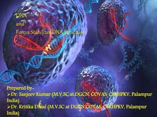
SlideShare une entreprise Scribd logo
1  sur  26
DNA
and
Forces Stabilizes DNA Structure
Prepared by-
Dr. Sanjeev Kumar (M.V.SC at DGCN, COVAS, CSKHPKV, Palampur
India)
Dr. Kritika Dhial (M.V.SC at DGCN,COVAS, CSKHPKV, Palampur
India)
Historical facts about DNA
DNA ?
• DNA Stands for “DeoxyriboNcleic Acid”.
• Term DNA was given by Zaccharis
• DNA is biopolymer consist of nucleotide as monomeric unit.
• DNA is double helical strcture in eukaryote and prokaryote, but in virus it
may be double stranded or single stranded and presented as monopartite
or multipartite.
• In eukaryotes, DNA is presented in nucleus surrounded by nuclear
membrane
• In prokaryotes, DNA is presented in nucleoid region of Cytoplasm without
nuclear membrane
• In virus, DNA is presented in the core of virus surrounded by Protein layer
(called as capsid).
DNA ?
• Cell perform various functions in specific way as per directed by DNA
contained by it.
• DNA contains all biological information of a cell/individual like
functioning, development, growth, life span etc. .
• The biological information transferred from one generation to the next
generation in the chemical form as DNA, so it is called as hereditary
material.
• DNA can be measured by the unit picogram
• DNA can be synthesized in vitro (in the laboratory).
Composition of DNA ?
• DNA is a biopolymer consist of repeated units called as nucleotides.
• Nucleotide= Nitrogen base + Sugar+ Phosphate group.
• Nitrogen bases are two types- 1. Purines, 2. Pyrimidine.
• Sugar present in DNA is 2-Deoxyribose.
• Purines are heterocyclic double ring molecules and contain nitogen
atoms at 1,3,7, and 9 positions.
• Purines are two types Adenine and Guanine.
• Pyrimidines are hterocyclic single ring molecule contain nitrogen
atoms at 1, and 3 positions.
• Pyrimidine are thee type thymine, cytosine, and uracil ( present in
RNA).
Composition of DNA ?
Watson and Crick model of DNA
In 1953, J.D. Watson (an American biologist) and
F.H.C. Crick (a British Physicist) proposed the
three-dimensional model of physiological DNA (i. e
B-DNA) on the basis of X-ray diffraction data of
DNA obtained by Franklin and Wilkins.
The important features of Watson – Crick Model
or double helix model of DNA are as follows:
1. The DNA molecule consists of two
polynucleotide chains or strands that spirally
twisted around each other and coiled around a
common axis to form a right-handed double-helix.
2. The two strands are antiparallel i.e. they ran in
opposite directions so that the 3′ end of one chain
facing the 5′ end of the other.
3. The sugar-phosphate backbones remain on the
outside, while the core of the helix contains the
purine and pyrimidine bases.
Watson and Crick model of DNA
4.The two strands are held together by hydrogen bonds between the
purine and pyrimidine bases of the opposite strands.
5. Adenine (A) always pairs with thymine (T) by two hydrogen bonds
and guanine (G) always pairs with cytosine (C) by three hydrogen
bonds. This complimentarily is known as the base pairing rule.
Thus, the two stands are complementary to one another.
6. The base sequence along a polynucleotide chain is variable and a
specific sequence of bases carries the genetic information.
7. The base compositions of DNA obey Chargaff s rules (E.E. Chargaff,
1950) according to which A=T and G=C; as a corollary ∑ purines
(A+G) = 2 pyrimidines (C+T); also (A+C) = (G+T). It also states that
ratio of (A+T) and (G+C) is constant for a species (range 0.4 to
1.9)
Watson and Crick model of DNA
8. The diameter of DNA is 2nm or 20
A. Adjacent bases are separated
0.34 nm or by 3.4 A along the
axis. The length of a complete
turn of helix is 3.4 nm or 34 A i.e.
there are 10bp per turn. (B- DNA-
Watson rick DNA)
9. The DNA helix has a shallow
groove called minor groove (-
1.2nm) and a deep groove called
major groove (- 2.2nm) across.
The conformation of a nucleotide unit is determined by the seven indicated torsion angles.
Forms of DNA
DNA is remarkably flexible molecule. Considerable
rotation is possible a number of bonds in the
sugar-phosphate backbone, and thermal
fluctuation can produce bending, stretching, and
unpairing of strands. Many significant deviations
from the Watson-Crick. DNA structure are found in
cellular DNA, some of which may play important
• A- DNA
• B- DNA
• Z-DNA
• Cruciform DNA and Hairpin DNA
• H- DNA or triplex
• G4- DNA
A- DNA B-DNA Z-DNA
Comparison Between different form of DNA
Cruciform and Hairpin DNA
H-DNA or Triplex DNA
G4- DNA
G4- DNA
• FIG. 8.Hypothetical mechanism of
formation and/or stabilization of G4 DNA
by Hop1 protein. In the model, bold and
light DNA molecules represent homologs
and closed circles denote guanine
residues. Once a DSB is produced at a hot
spot by Spo11 endonuclease, the free
ends are resected by exonuclease and/or
helicase activities to generate a 3′
overhang. Subsequently, Hop1 protein, by
binding to the guanine repeats in the 3′
overhang and to the G-rich strand in the
partially unwound homologous duplex
DNA, promotes the formation of
intermolecular G4 DNA. Alternatively
(right side), interstitial interactions
between chromatids could be mediated
by the formation of intermolecular G-G
pairing, regardless of whether the flanking
nucleotide sequences are homologous.
We suggest that such interactions might
either join homologs (as diagrammed
here) to facilitate interhomolog
recombination or join sister chromatids in
a manner that delimits sister chromatid
exchange.
Quaternary structure of the DNA molecule
• DNA is associated with proteins: histones and non histone
proteins, to form the chromatin.
•DNA as a whole is acidic (negatively charged) and binds to basic
(positively charged) proteins called histones
•There is 3 x 10 9 nucleotide pairs in the human haploid genome
representing about 30 000 genes dispersed over 23
chromosomes for a haploid set.
DNA Packaging
DNA Stability…..?
DNA double stranded helical structure is stabilize by
• Hydrogen bonding
• Base stacking interaction
• Hydrophobic force
• Ionic interaction
DNA Stability…..?
Hydrogen bonding-
 hydrogen bond b/w base pairs- G with C, and A
with T.
 It is important to note that three hydrogen bonds
can form between G and C, but only two bonds
can be found in A and T pairs.
 This is why it is more difficult to
separate DNA strands that contain more G-C pairs
than A-T pairs. On the other hand, A-T pairs seem
to destabilize the double helical structures. This
conclusion was made possible by a known fact that
in each species the G content is equal to that of C
content and the T content is equal to that of A
content.
 Although weak energy-wise, is able to stabilize the
helix because of the large number present
in DNA molecule
DNA Stability…..?
Base stacking interaction-
• also known as Van der Waals interactions between bases are weak, but the
large amounts of these interactions help to stabilize the overall structure of
the helix.
– Double helix is stabilized by hydrophobic effects by burying the bases in
the interior of the helix increases its stability; having the hydrophobic
bases clustered in the interior of the helix keeps it away from the
surrounding water, whereas the more polar surfaces, hence hydrophilic
heads are exposed and interaction with the exterior water
– Stacked base pairs also attract to one another through Van der Waals
forces the energy associated with a single van der Waals interaction has
small significant to the overall DNA structure however, the net effect
summed over the numerous atom pairs, results in substantial stability.
– Stacking also favors the conformations of rigid five-membered rings of the
sugars of backbone.
– Evidence of Stacking interactions: Compounds that interfere with
Hydrogen bonds (urea, formamide) don’t separate strands by themselves,
still requires heat.
DNA Stability…..?
Ionic interaction-
• Ion-ion repulsion of the negatively charged phosphate make DNA duplex
unstable.
• However the presence of Mg2+ and cationic proteins with abundant
Arginine and Lysine residues that stabilizes the double helix.
• Double-stranded helix structure thus, promoted by having phosphates on
outside, interact with H2O and counter ions (K+, Mg2+, etc.).
DNA Stability…..?
Hydrophobic force-
• The hydrophobic interactions
between the planar base pairs
stabilize the bases on the inside
of the helix, so these provide
stability to the structure but do
not contribute to the specificity.
• Hydrophobic Interactions are
important for the folding, stability
and biological activity.
References
• http://faculty.washington.edu/trawets/vc/theory/dna/DNA_big.jpg
• nptel.ac.in/courses/104103018/module4/lec3/2.html
• http://www.googleimage.com
• http://chem.libretexts.org/Core/Physical_and_Theoretical_Chemistry/Phy
sical_Properties_of_Matter/Atomic_and_Molecular_Properties/Intermole
cular_Forces/Hydrophobic_Interactions
• Text book of biology by True man
• bioinfosu.okstate.edu/um/napsweb/Lec.htmlfolder/836NAPS.ppt
• www.bio.brandeis.edu/classes/biochem104/DNA_lecture.pdf
• http://atlasgeneticsoncology.org/Educ/DNAEngID30001ES.html
• Principle of Biochemistry by Lehninger-forth edition
THANK YOU

Contenu connexe

Tendances

Tendances (20)

DNA structure and types
DNA structure and typesDNA structure and types
DNA structure and types
 
DNA as a Genetic Material
DNA as a Genetic MaterialDNA as a Genetic Material
DNA as a Genetic Material
 
DNA Supercoiling
DNA Supercoiling DNA Supercoiling
DNA Supercoiling
 
Maxam-Gilbert method of DNA sequencing
Maxam-Gilbert method of DNA sequencingMaxam-Gilbert method of DNA sequencing
Maxam-Gilbert method of DNA sequencing
 
Ramachandran plot
Ramachandran plotRamachandran plot
Ramachandran plot
 
Forces That Stabilize the DNA structure.pptx
Forces That Stabilize the DNA structure.pptxForces That Stabilize the DNA structure.pptx
Forces That Stabilize the DNA structure.pptx
 
MODELS OF REPLICATION
MODELS OF REPLICATIONMODELS OF REPLICATION
MODELS OF REPLICATION
 
tRNA structure and function
tRNA structure and functiontRNA structure and function
tRNA structure and function
 
repetitive and non repetitive dna.pptx
repetitive and non repetitive dna.pptxrepetitive and non repetitive dna.pptx
repetitive and non repetitive dna.pptx
 
Dna binding proteins
Dna binding proteinsDna binding proteins
Dna binding proteins
 
DNA Types
DNA TypesDNA Types
DNA Types
 
DNA Replication in eukaryotes and prokaryotes
DNA Replication in eukaryotes and prokaryotesDNA Replication in eukaryotes and prokaryotes
DNA Replication in eukaryotes and prokaryotes
 
watson and crick model of DNA(molecular biology)
watson and crick model of DNA(molecular biology) watson and crick model of DNA(molecular biology)
watson and crick model of DNA(molecular biology)
 
Griffith experiment
Griffith experimentGriffith experiment
Griffith experiment
 
Telomerase replication
Telomerase replicationTelomerase replication
Telomerase replication
 
Structure of DNA
Structure of DNAStructure of DNA
Structure of DNA
 
M. Meselson and F. Stahl experiment
M. Meselson and F. Stahl experimentM. Meselson and F. Stahl experiment
M. Meselson and F. Stahl experiment
 
Rna polymerase
Rna polymeraseRna polymerase
Rna polymerase
 
Dna replication in eukaryotes
Dna replication in eukaryotesDna replication in eukaryotes
Dna replication in eukaryotes
 
MODIFYING ENZYMES
MODIFYING ENZYMESMODIFYING ENZYMES
MODIFYING ENZYMES
 

En vedette

Partition chromatography 3
Partition chromatography 3Partition chromatography 3
Partition chromatography 3
MrSyedAmmar
 
ion exchange chromatography
ion exchange chromatographyion exchange chromatography
ion exchange chromatography
Shamili Kaparthi
 
DNA Structure PowerPoint
DNA Structure PowerPointDNA Structure PowerPoint
DNA Structure PowerPoint
BiologyIB
 

En vedette (18)

DNA structure, Functions and properties
DNA structure, Functions and propertiesDNA structure, Functions and properties
DNA structure, Functions and properties
 
Cloning3603
Cloning3603Cloning3603
Cloning3603
 
T/A CLONING
T/A CLONINGT/A CLONING
T/A CLONING
 
Secondary structural elements & ramachandran plot
Secondary structural elements & ramachandran plotSecondary structural elements & ramachandran plot
Secondary structural elements & ramachandran plot
 
Hydrogen bonding
Hydrogen bonding Hydrogen bonding
Hydrogen bonding
 
11.ramachandran plot
11.ramachandran plot11.ramachandran plot
11.ramachandran plot
 
Ramachandran plot
Ramachandran plotRamachandran plot
Ramachandran plot
 
Ion exchange Chromatography
Ion exchange ChromatographyIon exchange Chromatography
Ion exchange Chromatography
 
DNA structure, the bonds involved and it seperation
DNA structure, the bonds involved and it seperationDNA structure, the bonds involved and it seperation
DNA structure, the bonds involved and it seperation
 
Gene cloning
Gene cloningGene cloning
Gene cloning
 
x-ray-diffraction-technique
x-ray-diffraction-techniquex-ray-diffraction-technique
x-ray-diffraction-technique
 
POLYMERASE CHAIN REACTION
POLYMERASE CHAIN REACTIONPOLYMERASE CHAIN REACTION
POLYMERASE CHAIN REACTION
 
Agarose Gel Electrophoresis
Agarose Gel ElectrophoresisAgarose Gel Electrophoresis
Agarose Gel Electrophoresis
 
Partition chromatography 3
Partition chromatography 3Partition chromatography 3
Partition chromatography 3
 
DNA Cloning
DNA CloningDNA Cloning
DNA Cloning
 
Gene cloning
Gene cloningGene cloning
Gene cloning
 
ion exchange chromatography
ion exchange chromatographyion exchange chromatography
ion exchange chromatography
 
DNA Structure PowerPoint
DNA Structure PowerPointDNA Structure PowerPoint
DNA Structure PowerPoint
 

Similaire à DNA and Forces stabilizes dna structure

Nucleic acids med 2020-2021- l2&3-ayman-s
Nucleic acids  med 2020-2021- l2&3-ayman-sNucleic acids  med 2020-2021- l2&3-ayman-s
Nucleic acids med 2020-2021- l2&3-ayman-s
John Diggle
 
A592974226_23691_25_2019_Lecture11 onwards NUCLEIC ACIDS 2.pdf
A592974226_23691_25_2019_Lecture11 onwards NUCLEIC ACIDS 2.pdfA592974226_23691_25_2019_Lecture11 onwards NUCLEIC ACIDS 2.pdf
A592974226_23691_25_2019_Lecture11 onwards NUCLEIC ACIDS 2.pdf
RahulSharma123800
 

Similaire à DNA and Forces stabilizes dna structure (20)

DNA structure
DNA structure  DNA structure
DNA structure
 
IB Biology 2.6 & 7.1 Slides: DNA Structure
IB Biology 2.6 & 7.1 Slides: DNA StructureIB Biology 2.6 & 7.1 Slides: DNA Structure
IB Biology 2.6 & 7.1 Slides: DNA Structure
 
Deoxyribonucleic Acid (DNA)
Deoxyribonucleic Acid (DNA)Deoxyribonucleic Acid (DNA)
Deoxyribonucleic Acid (DNA)
 
Nucleic acids med 2020-2021- l2&3-ayman-s
Nucleic acids  med 2020-2021- l2&3-ayman-sNucleic acids  med 2020-2021- l2&3-ayman-s
Nucleic acids med 2020-2021- l2&3-ayman-s
 
STRUCTURE OF DNA.pptx
STRUCTURE OF DNA.pptxSTRUCTURE OF DNA.pptx
STRUCTURE OF DNA.pptx
 
Structure of dna
Structure of dnaStructure of dna
Structure of dna
 
Dna the thread of life watson crick model characteristics 2
Dna the thread of life watson crick model characteristics 2Dna the thread of life watson crick model characteristics 2
Dna the thread of life watson crick model characteristics 2
 
DNa Deoxyribonucleic acid- code of life
DNa Deoxyribonucleic acid-  code of lifeDNa Deoxyribonucleic acid-  code of life
DNa Deoxyribonucleic acid- code of life
 
molecular biology LECTURE 1 DNA & rna.pptx
molecular biology LECTURE 1 DNA & rna.pptxmolecular biology LECTURE 1 DNA & rna.pptx
molecular biology LECTURE 1 DNA & rna.pptx
 
Std 12 chapter 6
Std 12 chapter 6Std 12 chapter 6
Std 12 chapter 6
 
DNA STRUCTURE AND INTRODUCTION TO THE REPLICATION.pptx
DNA STRUCTURE AND INTRODUCTION TO THE REPLICATION.pptxDNA STRUCTURE AND INTRODUCTION TO THE REPLICATION.pptx
DNA STRUCTURE AND INTRODUCTION TO THE REPLICATION.pptx
 
Molecular organization of eukaryotic DNA
Molecular organization of eukaryotic DNAMolecular organization of eukaryotic DNA
Molecular organization of eukaryotic DNA
 
A592974226_23691_25_2019_Lecture11 onwards NUCLEIC ACIDS 2.pdf
A592974226_23691_25_2019_Lecture11 onwards NUCLEIC ACIDS 2.pdfA592974226_23691_25_2019_Lecture11 onwards NUCLEIC ACIDS 2.pdf
A592974226_23691_25_2019_Lecture11 onwards NUCLEIC ACIDS 2.pdf
 
Structure of dna
Structure  of  dnaStructure  of  dna
Structure of dna
 
Basics of DNA
Basics of DNA Basics of DNA
Basics of DNA
 
Structure of DNA
Structure of DNA Structure of DNA
Structure of DNA
 
DNA structure
DNA structureDNA structure
DNA structure
 
DNA structure
DNA structureDNA structure
DNA structure
 
Dna
DnaDna
Dna
 
Cell nucleus session 2
Cell nucleus session 2Cell nucleus session 2
Cell nucleus session 2
 

Dernier

Conjugation, transduction and transformation
Conjugation, transduction and transformationConjugation, transduction and transformation
Conjugation, transduction and transformation
Areesha Ahmad
 
GUIDELINES ON SIMILAR BIOLOGICS Regulatory Requirements for Marketing Authori...
GUIDELINES ON SIMILAR BIOLOGICS Regulatory Requirements for Marketing Authori...GUIDELINES ON SIMILAR BIOLOGICS Regulatory Requirements for Marketing Authori...
GUIDELINES ON SIMILAR BIOLOGICS Regulatory Requirements for Marketing Authori...
Lokesh Kothari
 
Discovery of an Accretion Streamer and a Slow Wide-angle Outflow around FUOri...
Discovery of an Accretion Streamer and a Slow Wide-angle Outflow around FUOri...Discovery of an Accretion Streamer and a Slow Wide-angle Outflow around FUOri...
Discovery of an Accretion Streamer and a Slow Wide-angle Outflow around FUOri...
Sérgio Sacani
 
Asymmetry in the atmosphere of the ultra-hot Jupiter WASP-76 b
Asymmetry in the atmosphere of the ultra-hot Jupiter WASP-76 bAsymmetry in the atmosphere of the ultra-hot Jupiter WASP-76 b
Asymmetry in the atmosphere of the ultra-hot Jupiter WASP-76 b
Sérgio Sacani
 
Presentation Vikram Lander by Vedansh Gupta.pptx
Presentation Vikram Lander by Vedansh Gupta.pptxPresentation Vikram Lander by Vedansh Gupta.pptx
Presentation Vikram Lander by Vedansh Gupta.pptx
gindu3009
 
Labelling Requirements and Label Claims for Dietary Supplements and Recommend...
Labelling Requirements and Label Claims for Dietary Supplements and Recommend...Labelling Requirements and Label Claims for Dietary Supplements and Recommend...
Labelling Requirements and Label Claims for Dietary Supplements and Recommend...
Lokesh Kothari
 
Seismic Method Estimate velocity from seismic data.pptx
Seismic Method Estimate velocity from seismic  data.pptxSeismic Method Estimate velocity from seismic  data.pptx
Seismic Method Estimate velocity from seismic data.pptx
AlMamun560346
 

Dernier (20)

module for grade 9 for distance learning
module for grade 9 for distance learningmodule for grade 9 for distance learning
module for grade 9 for distance learning
 
Feature-aligned N-BEATS with Sinkhorn divergence (ICLR '24)
Feature-aligned N-BEATS with Sinkhorn divergence (ICLR '24)Feature-aligned N-BEATS with Sinkhorn divergence (ICLR '24)
Feature-aligned N-BEATS with Sinkhorn divergence (ICLR '24)
 
Conjugation, transduction and transformation
Conjugation, transduction and transformationConjugation, transduction and transformation
Conjugation, transduction and transformation
 
Botany 4th semester file By Sumit Kumar yadav.pdf
Botany 4th semester file By Sumit Kumar yadav.pdfBotany 4th semester file By Sumit Kumar yadav.pdf
Botany 4th semester file By Sumit Kumar yadav.pdf
 
Factory Acceptance Test( FAT).pptx .
Factory Acceptance Test( FAT).pptx       .Factory Acceptance Test( FAT).pptx       .
Factory Acceptance Test( FAT).pptx .
 
GBSN - Biochemistry (Unit 1)
GBSN - Biochemistry (Unit 1)GBSN - Biochemistry (Unit 1)
GBSN - Biochemistry (Unit 1)
 
Connaught Place, Delhi Call girls :8448380779 Model Escorts | 100% verified
Connaught Place, Delhi Call girls :8448380779 Model Escorts | 100% verifiedConnaught Place, Delhi Call girls :8448380779 Model Escorts | 100% verified
Connaught Place, Delhi Call girls :8448380779 Model Escorts | 100% verified
 
GUIDELINES ON SIMILAR BIOLOGICS Regulatory Requirements for Marketing Authori...
GUIDELINES ON SIMILAR BIOLOGICS Regulatory Requirements for Marketing Authori...GUIDELINES ON SIMILAR BIOLOGICS Regulatory Requirements for Marketing Authori...
GUIDELINES ON SIMILAR BIOLOGICS Regulatory Requirements for Marketing Authori...
 
GBSN - Microbiology (Unit 1)
GBSN - Microbiology (Unit 1)GBSN - Microbiology (Unit 1)
GBSN - Microbiology (Unit 1)
 
Discovery of an Accretion Streamer and a Slow Wide-angle Outflow around FUOri...
Discovery of an Accretion Streamer and a Slow Wide-angle Outflow around FUOri...Discovery of an Accretion Streamer and a Slow Wide-angle Outflow around FUOri...
Discovery of an Accretion Streamer and a Slow Wide-angle Outflow around FUOri...
 
Asymmetry in the atmosphere of the ultra-hot Jupiter WASP-76 b
Asymmetry in the atmosphere of the ultra-hot Jupiter WASP-76 bAsymmetry in the atmosphere of the ultra-hot Jupiter WASP-76 b
Asymmetry in the atmosphere of the ultra-hot Jupiter WASP-76 b
 
Pulmonary drug delivery system M.pharm -2nd sem P'ceutics
Pulmonary drug delivery system M.pharm -2nd sem P'ceuticsPulmonary drug delivery system M.pharm -2nd sem P'ceutics
Pulmonary drug delivery system M.pharm -2nd sem P'ceutics
 
Presentation Vikram Lander by Vedansh Gupta.pptx
Presentation Vikram Lander by Vedansh Gupta.pptxPresentation Vikram Lander by Vedansh Gupta.pptx
Presentation Vikram Lander by Vedansh Gupta.pptx
 
Labelling Requirements and Label Claims for Dietary Supplements and Recommend...
Labelling Requirements and Label Claims for Dietary Supplements and Recommend...Labelling Requirements and Label Claims for Dietary Supplements and Recommend...
Labelling Requirements and Label Claims for Dietary Supplements and Recommend...
 
Zoology 4th semester series (krishna).pdf
Zoology 4th semester series (krishna).pdfZoology 4th semester series (krishna).pdf
Zoology 4th semester series (krishna).pdf
 
Site Acceptance Test .
Site Acceptance Test                    .Site Acceptance Test                    .
Site Acceptance Test .
 
American Type Culture Collection (ATCC).pptx
American Type Culture Collection (ATCC).pptxAmerican Type Culture Collection (ATCC).pptx
American Type Culture Collection (ATCC).pptx
 
Seismic Method Estimate velocity from seismic data.pptx
Seismic Method Estimate velocity from seismic  data.pptxSeismic Method Estimate velocity from seismic  data.pptx
Seismic Method Estimate velocity from seismic data.pptx
 
COST ESTIMATION FOR A RESEARCH PROJECT.pptx
COST ESTIMATION FOR A RESEARCH PROJECT.pptxCOST ESTIMATION FOR A RESEARCH PROJECT.pptx
COST ESTIMATION FOR A RESEARCH PROJECT.pptx
 
GBSN - Microbiology (Unit 3)
GBSN - Microbiology (Unit 3)GBSN - Microbiology (Unit 3)
GBSN - Microbiology (Unit 3)
 

DNA and Forces stabilizes dna structure

  • 1. DNA and Forces Stabilizes DNA Structure Prepared by- Dr. Sanjeev Kumar (M.V.SC at DGCN, COVAS, CSKHPKV, Palampur India) Dr. Kritika Dhial (M.V.SC at DGCN,COVAS, CSKHPKV, Palampur India)
  • 3. DNA ? • DNA Stands for “DeoxyriboNcleic Acid”. • Term DNA was given by Zaccharis • DNA is biopolymer consist of nucleotide as monomeric unit. • DNA is double helical strcture in eukaryote and prokaryote, but in virus it may be double stranded or single stranded and presented as monopartite or multipartite. • In eukaryotes, DNA is presented in nucleus surrounded by nuclear membrane • In prokaryotes, DNA is presented in nucleoid region of Cytoplasm without nuclear membrane • In virus, DNA is presented in the core of virus surrounded by Protein layer (called as capsid).
  • 4. DNA ? • Cell perform various functions in specific way as per directed by DNA contained by it. • DNA contains all biological information of a cell/individual like functioning, development, growth, life span etc. . • The biological information transferred from one generation to the next generation in the chemical form as DNA, so it is called as hereditary material. • DNA can be measured by the unit picogram • DNA can be synthesized in vitro (in the laboratory).
  • 5. Composition of DNA ? • DNA is a biopolymer consist of repeated units called as nucleotides. • Nucleotide= Nitrogen base + Sugar+ Phosphate group. • Nitrogen bases are two types- 1. Purines, 2. Pyrimidine. • Sugar present in DNA is 2-Deoxyribose. • Purines are heterocyclic double ring molecules and contain nitogen atoms at 1,3,7, and 9 positions. • Purines are two types Adenine and Guanine. • Pyrimidines are hterocyclic single ring molecule contain nitrogen atoms at 1, and 3 positions. • Pyrimidine are thee type thymine, cytosine, and uracil ( present in RNA).
  • 7.
  • 8. Watson and Crick model of DNA In 1953, J.D. Watson (an American biologist) and F.H.C. Crick (a British Physicist) proposed the three-dimensional model of physiological DNA (i. e B-DNA) on the basis of X-ray diffraction data of DNA obtained by Franklin and Wilkins. The important features of Watson – Crick Model or double helix model of DNA are as follows: 1. The DNA molecule consists of two polynucleotide chains or strands that spirally twisted around each other and coiled around a common axis to form a right-handed double-helix. 2. The two strands are antiparallel i.e. they ran in opposite directions so that the 3′ end of one chain facing the 5′ end of the other. 3. The sugar-phosphate backbones remain on the outside, while the core of the helix contains the purine and pyrimidine bases.
  • 9. Watson and Crick model of DNA 4.The two strands are held together by hydrogen bonds between the purine and pyrimidine bases of the opposite strands. 5. Adenine (A) always pairs with thymine (T) by two hydrogen bonds and guanine (G) always pairs with cytosine (C) by three hydrogen bonds. This complimentarily is known as the base pairing rule. Thus, the two stands are complementary to one another. 6. The base sequence along a polynucleotide chain is variable and a specific sequence of bases carries the genetic information. 7. The base compositions of DNA obey Chargaff s rules (E.E. Chargaff, 1950) according to which A=T and G=C; as a corollary ∑ purines (A+G) = 2 pyrimidines (C+T); also (A+C) = (G+T). It also states that ratio of (A+T) and (G+C) is constant for a species (range 0.4 to 1.9)
  • 10. Watson and Crick model of DNA 8. The diameter of DNA is 2nm or 20 A. Adjacent bases are separated 0.34 nm or by 3.4 A along the axis. The length of a complete turn of helix is 3.4 nm or 34 A i.e. there are 10bp per turn. (B- DNA- Watson rick DNA) 9. The DNA helix has a shallow groove called minor groove (- 1.2nm) and a deep groove called major groove (- 2.2nm) across.
  • 11. The conformation of a nucleotide unit is determined by the seven indicated torsion angles.
  • 12. Forms of DNA DNA is remarkably flexible molecule. Considerable rotation is possible a number of bonds in the sugar-phosphate backbone, and thermal fluctuation can produce bending, stretching, and unpairing of strands. Many significant deviations from the Watson-Crick. DNA structure are found in cellular DNA, some of which may play important • A- DNA • B- DNA • Z-DNA • Cruciform DNA and Hairpin DNA • H- DNA or triplex • G4- DNA A- DNA B-DNA Z-DNA
  • 17. G4- DNA • FIG. 8.Hypothetical mechanism of formation and/or stabilization of G4 DNA by Hop1 protein. In the model, bold and light DNA molecules represent homologs and closed circles denote guanine residues. Once a DSB is produced at a hot spot by Spo11 endonuclease, the free ends are resected by exonuclease and/or helicase activities to generate a 3′ overhang. Subsequently, Hop1 protein, by binding to the guanine repeats in the 3′ overhang and to the G-rich strand in the partially unwound homologous duplex DNA, promotes the formation of intermolecular G4 DNA. Alternatively (right side), interstitial interactions between chromatids could be mediated by the formation of intermolecular G-G pairing, regardless of whether the flanking nucleotide sequences are homologous. We suggest that such interactions might either join homologs (as diagrammed here) to facilitate interhomolog recombination or join sister chromatids in a manner that delimits sister chromatid exchange.
  • 18. Quaternary structure of the DNA molecule • DNA is associated with proteins: histones and non histone proteins, to form the chromatin. •DNA as a whole is acidic (negatively charged) and binds to basic (positively charged) proteins called histones •There is 3 x 10 9 nucleotide pairs in the human haploid genome representing about 30 000 genes dispersed over 23 chromosomes for a haploid set.
  • 20. DNA Stability…..? DNA double stranded helical structure is stabilize by • Hydrogen bonding • Base stacking interaction • Hydrophobic force • Ionic interaction
  • 21. DNA Stability…..? Hydrogen bonding-  hydrogen bond b/w base pairs- G with C, and A with T.  It is important to note that three hydrogen bonds can form between G and C, but only two bonds can be found in A and T pairs.  This is why it is more difficult to separate DNA strands that contain more G-C pairs than A-T pairs. On the other hand, A-T pairs seem to destabilize the double helical structures. This conclusion was made possible by a known fact that in each species the G content is equal to that of C content and the T content is equal to that of A content.  Although weak energy-wise, is able to stabilize the helix because of the large number present in DNA molecule
  • 22. DNA Stability…..? Base stacking interaction- • also known as Van der Waals interactions between bases are weak, but the large amounts of these interactions help to stabilize the overall structure of the helix. – Double helix is stabilized by hydrophobic effects by burying the bases in the interior of the helix increases its stability; having the hydrophobic bases clustered in the interior of the helix keeps it away from the surrounding water, whereas the more polar surfaces, hence hydrophilic heads are exposed and interaction with the exterior water – Stacked base pairs also attract to one another through Van der Waals forces the energy associated with a single van der Waals interaction has small significant to the overall DNA structure however, the net effect summed over the numerous atom pairs, results in substantial stability. – Stacking also favors the conformations of rigid five-membered rings of the sugars of backbone. – Evidence of Stacking interactions: Compounds that interfere with Hydrogen bonds (urea, formamide) don’t separate strands by themselves, still requires heat.
  • 23. DNA Stability…..? Ionic interaction- • Ion-ion repulsion of the negatively charged phosphate make DNA duplex unstable. • However the presence of Mg2+ and cationic proteins with abundant Arginine and Lysine residues that stabilizes the double helix. • Double-stranded helix structure thus, promoted by having phosphates on outside, interact with H2O and counter ions (K+, Mg2+, etc.).
  • 24. DNA Stability…..? Hydrophobic force- • The hydrophobic interactions between the planar base pairs stabilize the bases on the inside of the helix, so these provide stability to the structure but do not contribute to the specificity. • Hydrophobic Interactions are important for the folding, stability and biological activity.
  • 25. References • http://faculty.washington.edu/trawets/vc/theory/dna/DNA_big.jpg • nptel.ac.in/courses/104103018/module4/lec3/2.html • http://www.googleimage.com • http://chem.libretexts.org/Core/Physical_and_Theoretical_Chemistry/Phy sical_Properties_of_Matter/Atomic_and_Molecular_Properties/Intermole cular_Forces/Hydrophobic_Interactions • Text book of biology by True man • bioinfosu.okstate.edu/um/napsweb/Lec.htmlfolder/836NAPS.ppt • www.bio.brandeis.edu/classes/biochem104/DNA_lecture.pdf • http://atlasgeneticsoncology.org/Educ/DNAEngID30001ES.html • Principle of Biochemistry by Lehninger-forth edition