
Recommandé
Contenu connexe
Tendances
Tendances (20)
Similaire à Yersinia entero
Similaire à Yersinia entero (20)
Dernier
Dernier (20)
Yersinia entero
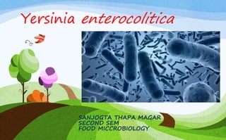
- 1. Yersinia enterocolitica SANJOGTA THAPA MAGAR SECOND SEM FOOD MICCROBIOLOGY
- 2. Yersinia enterocolitica is a member of the Enterobacteriaceae; an asporogenous, short (0.5-1.0 by 1-2 µm) Gram-negative rod which is facultatively anaerobic, catalase- positive and oxidase- negative Pigs are the major animal reservoir for the few strains of Y. enterocolitica that cause human illness, but rodents, rabbits, sheep, cattle, horses, dogs, and cats also can carry strains that cause human illness. Y. enterocolitica is psychrotrophic and has the ability to grow at temperatures below 4°C and can survive in frozen foods for extended periods. INTRODUCTION
- 3. 1. The doubling time, at 30°C, is 34 min; at 22°C, is 1 hr; and at 7°C, is 5 hrs. 2. The pH range for growth of Y. enterocolitica is about 4.6 to 9.6 3. It is sensitive to heat and acidity. 4. Y enterocolitica is destroyed in 1-3 minutes at 60° C. 5. It is motile at temperatures of 22–29°C, but becomes non motile at normal human body temperature. INTRODUCTION
- 5. Y. enterocolitica is a heterogeneous group of strains, which are traditionally classified by biotyping into six biogroups on the basis of phenotypic characteristics, and by serotyping into more than 57 O serogroups, on the basis of their O (lipopolysaccharide or LPS) surface antigen. Five of the six biogroups (1B and 2–5) are regarded as pathogens. The most commonly occurring Y enterocolitica serovars (serotypes) in human infections are 0:3, 0:5,27, 0:8, and 0:9. SEROTYPES
- 6. Virulence Factors • Y enterocolitica produces a heat-stable enterotoxin (ST) that survives 1000C for 20 minutes. • It is produced only at or below 300C,98 and its production is favored in the pH range 7-8 Incidence of Y. enterocolitica in Foods • Y. enterocolitica can grow easily at refrigeration temperature in vacuum-packed meat, boiled eggs, boiled fish, pasteurized liquid eggs, pasteurized whole milk, cottage cheese, and tofu. • Growth of the microorganism also occurs in refrigerated seafood – oysters, raw shrimp, and cooked crab meat
- 7. Incidence of Y. enterocolitica in Foods • However, the prevalence of this organism in soil, water, and animals, such as beavers, pigs, and squirrels, offers many opportunities for Yersinia to enter the food supply. • For example, poor sanitation and improper sterilization techniques by food handlers, including improper storage, may be a source of contamination. • Raw or undercooked pork products have drawn much attention as a source of Y. enterocolitica.
- 9. Pathogenesis The usual route of acquisition of this pathogen is through contaminated foods or water. The primary event of Y. enterocolitica pathogenesis is colonization of the intestinal tract, particularly the distal small intestine (terminal ileum) and proximal colon . Accordingly, most of the pathologic effects and, hence, clinical manifestations occur at this location There, virulent yersiniae must traverse the intestinal lumen, attach to and penetrate the mucus barrier overlying the mucosal epithelial cells, and eventually adhere to intestinal cells. Bacteria have shown to preferentially bind to and penetrate M cells of Peyer's patches (PP). Once internalized, the bacteria are transported across the epithelial barrier and expelled from the basolateral side of the M cell
- 10. Pathogenesis In the earliest steps of infection, phagocytes internalize the bacteria. Internalized bacteria reportedly replicate in native murine macrophages and hence they are assumed to be transported within migrating phagocytes to mesenteric lymph nodes (MLN), causing an inflammatory response that triggers abdominal pain. Furthermore, phagocytes that take up bacteria can disseminate via the bloodstream to the liver and spleen. Once located in PPs, MLNs, the spleen or liver, Y. enterocolitica replicate in an extracellular form within micro-abscesses Within these lesions the bacteria form microcolonies appear to be resistant to phagocytosis by macrophages and neutrophils
- 11. FIG : Pathogenesis model of Yersinia enterocolitica. (1) Yersinia cells traverse the intestinal epithelium via epithelial cells to the submucosa. (2) Submucosal macrophages phagocytose the pathogen and enter into the lymphatic system thereby reaching the MLN. (3) Alternatively, bacteria can be engulfed by M cells. (4) Once in the PP Yersinia forms microcolonies and starts replication. (5) Eventually, bacterial cells are located in the MLN and can equally form microcolonies to allow replication.
- 13. Disease: Yersiniosis refers to the illnesses caused by Y. enterocolitica Mortality: Fatalities are extremely rare. Infective dose: The medium infective dose for humans is not known, but is estimated to be between 104 to 106 organisms. The infective dose and clinical presentation of symptoms may depend on pathogen (strain-dependent) and host factors. For example, in some cases, in people with gastric hypoacidity, the infective dose may be lower. Onset: Incubation times from 1 to 11 days have been observed, but occasionally last for several months.
- 14. Illness / complications: • In some patients, complications arise due to the strain type causing the initial infection and specific human immunologic leukocyte antigen, HLA-B27. • These include reactive arthritis; glomerulonephritis; endocarditis; erythema nodosum (which occurs predominantly in women); uveitis; thyroid disorders, such as Graves’ disease; hyperthyroidism; nontoxic goiter; and Hashimoto’s thyroiditis. • Y. enterocolitica has been associated with reactive arthritis, which may occur even in the absence of obvious symptoms.
- 16. Illness / complications: • The frequency of such postenteritis arthritic conditions is about 2% to 3%. • Another complication is bacteremia, which raises the possibility of disease dissemination. However, this is rare. • Performance of unnecessary appendectomies also may be considered a major complication of yersiniosis, as one of the main symptoms of the disease is abdominal pain in the lower right quadrant. .
- 17. Symptoms: • Infection with Y. enterocolitica manifests as nonspecific, self- limiting diarrhea, but may cause a variety of autoimmune complications. • Most symptomatic infections occur in children younger than 5 years old. Yersiniosis in these children is frequently characterized as gastroenteritis, with diarrhea and/or vomiting; however, fever and abdominal pain are the hallmark symptoms. • A small proportion of children (less than 10%) produce bloody stools. Children usually complain of abdominal pain and headache and sore throat at the onset of the illness.
- 19. Symptoms: • Yersinia infections mimic appendicitis and mesenteric lymphadenitis, but the bacteria may also cause infection in other sites, such as wounds, joints, and the urinary tract Duration: The illness might last from a few days to 3 weeks, unless it becomes chronic enterocolitis, in which case it might continue for several months. Route of entry: Oral. Pathway: • As zoonotic pathogens, Y. enterocolitica enter the gastrointestinal tract after ingestion of contaminated food or water.
- 20. Pathway: • Gastric acid is a significant barrier to infection. • The infective dose might be lower among people with gastric hypoacidity. • This pathogens harbor plasmid (pYV)-encoded virulence genes that affect pathogenesis. • These include an outer-membrane protein, YadA (Yersinia adhesion A), and the genetic suite comprising the type III secretory system. • This process usually is facilitated by Yops proteins, which contribute to the ability of Y. enterocolitica cells to resist phagocytosis by causing disruption (cytotoxic changes) of mammalian (human)
- 21. Lab Diagnosis Sample specimen : Diagnosis of yersiniosis begins with isolation of the organism from the human host’s feces, blood, or vomit, and sometimes at the time of appendectomy Isolation and Identification : • The most commonly used enrichment media are phosphate buffered saline (PBS) or tryptone soya broth (TSB) most usually incubated at 4 °C for 21 days • Yersinia Enterocolitica are more alkali resistantso the pH of enrichment media is sometimes adjusted to 8.0-8.3 or cultures subjected to a short post-enrichment alkali treatment.
- 22. Lab Diagnosis Isolation and Identification : • The selective isolation of Yersinia Enterocolitica from foods and enrichment broths have been obtained with cefsulodin / irgasan / novobiocin (CIN) agar. • In addition to the antibiotics, the medium contains deoxycholate and crystal violet as selective agents and mannitol as a fermentable carbon source • After incubation at 28 °C for 24 h, typical colonies of Yersinia Enterocolitica appear with a dark- red centre surrounded by a transparent border. Isolates can be confirmed and bio-typed by biochemical tests.
- 23. Diagnosis • Several PCR primer sets are directed to either plasmid-borne genes, e.g., virF or yadA, or chromosomally located loci, such as ail. • Serology is used to identify the biotype (based on biochemical analysis) and serogroup (O-antigen). • Sera from acute or convalescent patients are titered against the suspect serotype of Yersinia spp. • Techniques using gene probes to detect the virulence-associated plasmid by a colony hybridization test have also been used with some success and offer the possibility of detecting potentially pathogenic strains in foods without the need for lengthy enrichment procedures.
- 24. Treatment • Treatment includes supportive care, since the gastroenteritis is self- limiting. If septicemia or other invasive diseases occur, antibiotic therapy with gentamicin or cefotaxime (doxycycline and ciprofloxacin) typically are administered.
- 25. PREVENTION : The first step in preventing Y. enterocolitica infection is to assume that all foods may cause foodborne illness. Follow steps can be done to prevent this infection: • Wash hands, food preparation surfaces and utensils thoroughly before and after handling raw foods to prevent recontamination of cooked foods. • Keep refrigerated foods below 40° F (5° C). However, this bacteria can survive and multiply at this temperature. • Serve hot foods immediately or keep them heated above 140° F (60° C). • Divide large volumes of food into small portions for rapid cooling in the refrigerator. Hot, bulky foods in the refrigerator can raise the temperature of foods already cooled. • Follow approved home-canning procedures. Heat canned foods thoroughly before tasting.When in doubt, throw it out. • Ensure that all pork is cooked at the correct temperature and cooking time. Do not eat undercooked pork.
- 26. PREVENTION : • Infants, young children, older persons and anyone with a compromised immune system are especially susceptible to foodborne illnesses. • These people should not consume inadequately cooked or raw meat products or milk or cheese products that have not been pasteurized.
- 28. Epidemiology • The populations most susceptible to Y. enterocolitica are small children, the elderly, and immuno-suppressed hosts. • 75% of patients with this disease are between the age of 5-15 years. • Most isolates of the disease are type O:3 and O:9 serotypes found in Canada and Europe. • In the US it is estimated that approximately 5% of enteric bacterial infections are in children under the age of 5. • The CDC reports 17,000 cases of infection in the United States annually. • This disease is much more common in Northern Europe, Scandinavia, and Japan.
- 29. REFERENCES • https://microbewiki.kenyon.edu/index.php/Y._enterocolitica • http://www.elsevier.es/es-revista-enfermedades-infecciosas-microbiologia- clinica-28-articulo-yersinia-enterocolitica-pathogenesis-virulence- antimicrobial-S0213005X11002655 • https://www.cdc.gov/yersinia/faq.html • Yeasmin Sabina, Atiqur Rahman, Ramesh Chandra Ray and Didier Montet,” Yersinia enterocolitica : Mode of Transmission, Molecular Insights of Virulence, and Pathogenesis of Infection’’ Journal of Pathogens Volume 2011 (2011), 10 pages • http://www.medic8.com/healthguide/food-poisoning/yersinia- enterocolitica.html • Anna Fàbrega, Jordi Vila,” Yersinia enterocolitica: Pathogenesis, virulence and antimicrobial resistance” Enferm Infecc Microbiol Clin 2012;30:24- 32 • https://secure.familyhealthtracker.com/deliver.aspx?s=sm&t=di&l=en&f=%7 B4AF57410-C58F-41C7-9E3F- D759B2FA5832%7D&key=26dd85c28d515a89bff6d8625ddf298a. • James M. Jay,” Modern Food Microbiology” Aspen Publishers, Inc,2000.pp no 556-559
- 30. REFERENCES • Shabbir Simjee,” Foodborne Diseases” Humana Press Inc, 2007,page no 79-90 • Bad Bug Book Handbook of Foodborne Pathogenic Microorganisms and Natural Toxins