1. > 1
Overview
A Single Photon Emission Computed Tomography
(SPECT) scan is a type of nuclear imaging test that
shows how blood flows to tissues and organs.
How does a SPECT scan work?
A SPECT scan integrates two technologies to view
your body: computed tomography (CT) and a
radioactive material (tracer). The tracer is what
allows doctors to see how blood flows to tissues and
organs.
Before the SPECT scan, you are injected with a
chemical that is radiolabled, meaning it emits
gamma rays that can be detected by the scanner.
The computer collects the information emitted by
the gamma rays and translates them into two-
dimensional cross-sections. These cross-sections
can be added back together to form a 3D image of
your brain.
The radioisotopes typically used in SPECT to label
tracers are iodine-123, technetium-99m, xenon-
133, thallium-201, and fluorine-18. These
radioactive forms of natural elements will pass
safely through your body and be detected by the
scanner. Various drugs and other chemicals can be
labeled with these isotopes.
The type of tracer used depends on what your
doctor wants to measure. For example, if your
doctor is looking at a tumor, he or she might use
radiolabled glucose (FDG) and watch how it is
metabolized by the tumor.
The test differs from a PET scan in that the tracer
stays in your blood stream rather than being
absorbed by surrounding tissues, thereby limiting
the images to areas where blood flows. SPECT
scans are cheaper and more readily available than
higher resolution PET scans.
What does a SPECT scan show?
A SPECT scan is primarily used to view how blood
flows through arteries and veins in the brain. Tests
have shown that it might be more sensitive to brain
injury than either MRI or CT scanning because it
can detect reduced blood flow to injured sites.
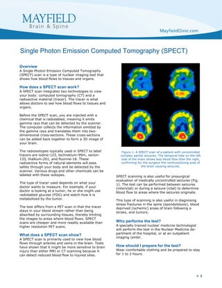
SPECT scanning is also useful for presurgical
evaluation of medically uncontrolled seizures (Fig.
1). The test can be performed between seizures
(interictal) or during a seizure (ictal) to determine
blood flow to areas where the seizures originate.
This type of scanning is also useful in diagnosing
stress fractures in the spine (spondylolysis), blood
deprived (ischemic) areas of brain following a
stroke, and tumors.
Who performs the test?
A specially trained nuclear medicine technologist
will perform the test in the Nuclear Medicine de-
partment of the hospital, or at an outpatient
imaging center.
How should I prepare for the test?
Wear comfortable clothing and be prepared to stay
for 1 to 2 hours.
Single Photon Emission Computed Tomography (SPECT)
Figure 1. A SPECT scan of a patient with uncontrolled
complex partial seizures. The temporal lobe on the left
side of the brain shows less blood flow than the right,
confirming for the surgeon the nonfunctioning area of
the brain causing seizures.