SlideShare une entreprise Scribd logo
1  sur  30
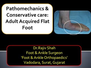
Dr.Rajiv Shah
Foot & Ankle Surgeon
‘Foot & Ankle Orthopaedics’
Vadodara, Surat, Gujarat
2
Tibialis Posterior
3
 14mm zone of
ischemia due to
lack of mesotenon
 Acute curve at
medial malleolus
 Shallow malleolar
groove
 Compression &
constriction under
Flexor retinaculum
Repetitive
micro trauma
Tendon &
sheath
inflammation
Tendon
elongation
Tendon
rupture
Ruptured
TP
Failed medial
restrains =
Flat foot
No locking ofTT joints +
unopposed pull of
peroneus brevis everts
heel = Heel valgus
The longitudinal axis of 1st metatarsal and
talus forms zero degree angle-Meary’s angle
Weight bearing biomechanics
On weight bearing talus plantarflexes and slides
distally on Calcaneum, which is restrained by spring
ligament
Weight bearing biomechanics
Calcaneum also plantarflexes and plantar fascia is
stretched to limit arch collapse
Weight bearing biomechanics
Navicular and cuneiform dorsiflex, evert & abduct
which is limited byTP
Weight bearing biomechanics
Metatarsals also dorsiflex and abduct
Weight bearing biomechanics
Final picture on weight bearing
Weight bearing biomechanics
Midfoot bones and metatarsals dorsiflex & abduct & flatfoot
results
Talus plantarflexes - moves distally and rotates medially
Calcaneum planterflexes & goes in valgus
Weak spring lig & ITCL fails to support
Clinical Stages
Stage 1
Tendinopathy
Normal tendon
length
No deformity
Stage 2
 Tendon
lengthening
 Flexible
deformity
Stage 3
 Tendon
lengthening
 Fixed
deformity
Stage 4
 Fixed
deformity
 Talus tilted in
ankle(ankle
involvement
 Dereymaeker:
Stage Zero
 Biomechanical
abnormality
 No symptoms
 Stage 2: 2a & 2b
 2a: Medial
symptoms
 2b: Lateral
symptoms
Clinical tests
Single Limb Heel RaiseTestToo many toes signHeelValgusTP function evaluation
Weight bearing X-rays
 LateralView: break inTalo-1st MT line
(Meary’s Line)
 Altered talar declination angle
Normal
Acquired Flatfoot
Radiological diagnosis
Normal Flat foot
 APView: talo-navicular uncoverage
 Forefoot abduction
Radiological diagnosis
Normal < 7 degree AAFD > 7 degree
 Less than 30% medial talar head
uncoverage (or no lateral
incongruence)
 No clinical forefoot abduction
 More than 30% medial talar head
uncoverage or lateral incongruence
 Significant clinical forefoot abduction
Congruent 2a Incongruent 2b
Arthritis of subtalar,TN & CC joints
Forefoot abduction
Heel valgus
Radiological diagnosis: Stage 3Radiological diagnosis: Stage 4
 Tendon
pathology, tear,
degeneration
 Spring ligament
visualization
 Usually not
necessary
 Magical effect
MRI???
 Stage 1: essentially
conservative
 Stage 2: conservative
care for at least 6
months or more
 Stage 3 & stage 4:
patients with co-
morbid conditions &
unfit for surgery
Conservative care
 Stage 1: prevent
tendon rupture by
giving rest to tendon
 Stage 2:prevent
progression of
deformity
 Stage 3 & stage 4:
accommodation of
deformity
NSAIDS
Conservative care: Modalities
Management of
systemic disease
Physical therapy
Strengthening
Theraband
Iontophoresis
Cryotherapy
Orthotics
Medial wedge
Medial column
post
Heel alterations
UCBL
Foot mold
BK cast
Boot
Activity modification
That’s all…
Thank you all..
 August 28th, 29th & 30th, 2015
 20 international faculties
 Day 1: Parekh family foundation
workshop (7 modules)
 Day2 & 3: Confenrence
A must attend meeting for 2015!

Contenu connexe

Tendances

Ankle fractures final
Ankle fractures finalAnkle fractures final
Ankle fractures finalRavi Malhotra
 
Techniques in primary total knee arthroplasty
Techniques in primary total knee arthroplastyTechniques in primary total knee arthroplasty
Techniques in primary total knee arthroplastyHBGMedical
 
Aseptic loosening total hip arthroplasty
Aseptic loosening total hip arthroplastyAseptic loosening total hip arthroplasty
Aseptic loosening total hip arthroplastyImran Ali
 
Medial Opening Wedge High Tibial Osteotomy
Medial Opening Wedge High Tibial Osteotomy Medial Opening Wedge High Tibial Osteotomy
Medial Opening Wedge High Tibial Osteotomy washingtonortho
 
Primary total knee arthroplasty
Primary total knee arthroplastyPrimary total knee arthroplasty
Primary total knee arthroplastyjatinder12345
 
Post polio residual paralysis of foot and ankle
Post polio residual paralysis of foot and anklePost polio residual paralysis of foot and ankle
Post polio residual paralysis of foot and ankleGIRIDHAR BOYAPATI
 
Periprosthetic fractures of knee
Periprosthetic fractures of kneePeriprosthetic fractures of knee
Periprosthetic fractures of kneeRukmangada Naidu
 
Periprosthetic fractures
Periprosthetic fracturesPeriprosthetic fractures
Periprosthetic fracturesPrajithVP2
 
Patellofemoral instability
Patellofemoral instabilityPatellofemoral instability
Patellofemoral instabilityGonzalo Samitier
 
Correcting Varus Deformity of the Knee in Total Knee Replacement
Correcting Varus Deformity of the Knee in Total Knee ReplacementCorrecting Varus Deformity of the Knee in Total Knee Replacement
Correcting Varus Deformity of the Knee in Total Knee ReplacementVaibhav Bagaria
 
Freedom pck revision knee system by meril life
Freedom pck revision knee system by meril lifeFreedom pck revision knee system by meril life
Freedom pck revision knee system by meril lifeRajRathore36
 
Principles of deformity correction
Principles of deformity correctionPrinciples of deformity correction
Principles of deformity correctionAbdulla Kamal
 
Algorithm to correct Varus Knee in a TKR
Algorithm to correct Varus Knee in a TKRAlgorithm to correct Varus Knee in a TKR
Algorithm to correct Varus Knee in a TKRVaibhav Bagaria
 
Bone defects in tkr
Bone defects in tkrBone defects in tkr
Bone defects in tkrankitjose
 
Periprosthetic fractures of knee
Periprosthetic fractures of kneePeriprosthetic fractures of knee
Periprosthetic fractures of kneeSupraja Avula
 
Implant Selection In Revision T.K.R
Implant Selection In Revision T.K.RImplant Selection In Revision T.K.R
Implant Selection In Revision T.K.RMurtuza Rassiwala
 
Proximal tibia fractures(Plateau, spine ,Tubercle and Epiphyseal )
Proximal tibia fractures(Plateau, spine ,Tubercle and Epiphyseal ) Proximal tibia fractures(Plateau, spine ,Tubercle and Epiphyseal )
Proximal tibia fractures(Plateau, spine ,Tubercle and Epiphyseal ) Prasanthmuddada
 
Patellar Instability
Patellar InstabilityPatellar Instability
Patellar InstabilityBijay Mehta
 
Periprosthetic fractures around the knee
Periprosthetic fractures around the kneePeriprosthetic fractures around the knee
Periprosthetic fractures around the kneeAhmed Azmy
 

Tendances (20)

Ankle fractures final
Ankle fractures finalAnkle fractures final
Ankle fractures final
 
Techniques in primary total knee arthroplasty
Techniques in primary total knee arthroplastyTechniques in primary total knee arthroplasty
Techniques in primary total knee arthroplasty
 
Aseptic loosening total hip arthroplasty
Aseptic loosening total hip arthroplastyAseptic loosening total hip arthroplasty
Aseptic loosening total hip arthroplasty
 
Medial Opening Wedge High Tibial Osteotomy
Medial Opening Wedge High Tibial Osteotomy Medial Opening Wedge High Tibial Osteotomy
Medial Opening Wedge High Tibial Osteotomy
 
Primary total knee arthroplasty
Primary total knee arthroplastyPrimary total knee arthroplasty
Primary total knee arthroplasty
 
Post polio residual paralysis of foot and ankle
Post polio residual paralysis of foot and anklePost polio residual paralysis of foot and ankle
Post polio residual paralysis of foot and ankle
 
Periprosthetic fractures of knee
Periprosthetic fractures of kneePeriprosthetic fractures of knee
Periprosthetic fractures of knee
 
Periprosthetic fractures
Periprosthetic fracturesPeriprosthetic fractures
Periprosthetic fractures
 
Patellofemoral instability
Patellofemoral instabilityPatellofemoral instability
Patellofemoral instability
 
Lecture 27 parekh pttd3 and 4
Lecture 27 parekh pttd3 and 4Lecture 27 parekh pttd3 and 4
Lecture 27 parekh pttd3 and 4
 
Correcting Varus Deformity of the Knee in Total Knee Replacement
Correcting Varus Deformity of the Knee in Total Knee ReplacementCorrecting Varus Deformity of the Knee in Total Knee Replacement
Correcting Varus Deformity of the Knee in Total Knee Replacement
 
Freedom pck revision knee system by meril life
Freedom pck revision knee system by meril lifeFreedom pck revision knee system by meril life
Freedom pck revision knee system by meril life
 
Principles of deformity correction
Principles of deformity correctionPrinciples of deformity correction
Principles of deformity correction
 
Algorithm to correct Varus Knee in a TKR
Algorithm to correct Varus Knee in a TKRAlgorithm to correct Varus Knee in a TKR
Algorithm to correct Varus Knee in a TKR
 
Bone defects in tkr
Bone defects in tkrBone defects in tkr
Bone defects in tkr
 
Periprosthetic fractures of knee
Periprosthetic fractures of kneePeriprosthetic fractures of knee
Periprosthetic fractures of knee
 
Implant Selection In Revision T.K.R
Implant Selection In Revision T.K.RImplant Selection In Revision T.K.R
Implant Selection In Revision T.K.R
 
Proximal tibia fractures(Plateau, spine ,Tubercle and Epiphyseal )
Proximal tibia fractures(Plateau, spine ,Tubercle and Epiphyseal ) Proximal tibia fractures(Plateau, spine ,Tubercle and Epiphyseal )
Proximal tibia fractures(Plateau, spine ,Tubercle and Epiphyseal )
 
Patellar Instability
Patellar InstabilityPatellar Instability
Patellar Instability
 
Periprosthetic fractures around the knee
Periprosthetic fractures around the kneePeriprosthetic fractures around the knee
Periprosthetic fractures around the knee
 

En vedette

Pes Planus by Dr. Mohammad Azhar ud din Darokhan
Pes Planus by Dr. Mohammad Azhar ud din DarokhanPes Planus by Dr. Mohammad Azhar ud din Darokhan
Pes Planus by Dr. Mohammad Azhar ud din Darokhanimazhardarokhan
 
Radiographic evaluation of shoulder
Radiographic evaluation of shoulderRadiographic evaluation of shoulder
Radiographic evaluation of shouldermrinal joshi
 
정재훈 Diabetic foot(웹게시용)v2
정재훈 Diabetic foot(웹게시용)v2정재훈 Diabetic foot(웹게시용)v2
정재훈 Diabetic foot(웹게시용)v2Woongap Chung
 
스마트 인솔 ( Smart in sole )
스마트 인솔 ( Smart in sole )스마트 인솔 ( Smart in sole )
스마트 인솔 ( Smart in sole )Ho Hyun Lee
 
Biomechanics and it’s economic advantages
Biomechanics and it’s economic advantagesBiomechanics and it’s economic advantages
Biomechanics and it’s economic advantageskhyatigupta
 
4 a adult acquired flat foot - Derek Park
4 a adult acquired flat foot - Derek Park4 a adult acquired flat foot - Derek Park
4 a adult acquired flat foot - Derek ParkDerek Park
 
Birmingham UK formthotics Seminar 2016
Birmingham UK  formthotics Seminar 2016 Birmingham UK  formthotics Seminar 2016
Birmingham UK formthotics Seminar 2016 Gerard Greene Physio
 

En vedette (20)

Pes Planus by Dr. Mohammad Azhar ud din Darokhan
Pes Planus by Dr. Mohammad Azhar ud din DarokhanPes Planus by Dr. Mohammad Azhar ud din Darokhan
Pes Planus by Dr. Mohammad Azhar ud din Darokhan
 
Lecture 42 shah calcaneal malunions
Lecture 42 shah calcaneal malunionsLecture 42 shah calcaneal malunions
Lecture 42 shah calcaneal malunions
 
Lecture 45 shah hallux rigidus
Lecture 45 shah hallux rigidusLecture 45 shah hallux rigidus
Lecture 45 shah hallux rigidus
 
Lecture 3 shah radiology in foot and ankle
Lecture 3 shah radiology in foot and ankleLecture 3 shah radiology in foot and ankle
Lecture 3 shah radiology in foot and ankle
 
Lecture 23 24 parekh peroneal pathology
Lecture 23 24 parekh peroneal pathologyLecture 23 24 parekh peroneal pathology
Lecture 23 24 parekh peroneal pathology
 
thefantasydoctorsStacked
thefantasydoctorsStackedthefantasydoctorsStacked
thefantasydoctorsStacked
 
Radiographic evaluation of shoulder
Radiographic evaluation of shoulderRadiographic evaluation of shoulder
Radiographic evaluation of shoulder
 
Flat foot
Flat footFlat foot
Flat foot
 
정재훈 Diabetic foot(웹게시용)v2
정재훈 Diabetic foot(웹게시용)v2정재훈 Diabetic foot(웹게시용)v2
정재훈 Diabetic foot(웹게시용)v2
 
Lecture 21 shah chronic achilles rupture
Lecture 21  shah chronic achilles ruptureLecture 21  shah chronic achilles rupture
Lecture 21 shah chronic achilles rupture
 
Foot tie : )
Foot tie : )Foot tie : )
Foot tie : )
 
스마트 인솔 ( Smart in sole )
스마트 인솔 ( Smart in sole )스마트 인솔 ( Smart in sole )
스마트 인솔 ( Smart in sole )
 
Upper limb
Upper limbUpper limb
Upper limb
 
Biomechanics and it’s economic advantages
Biomechanics and it’s economic advantagesBiomechanics and it’s economic advantages
Biomechanics and it’s economic advantages
 
4 a adult acquired flat foot - Derek Park
4 a adult acquired flat foot - Derek Park4 a adult acquired flat foot - Derek Park
4 a adult acquired flat foot - Derek Park
 
Lecture 12 shah orif calcaneal fractures
Lecture 12 shah orif calcaneal fracturesLecture 12 shah orif calcaneal fractures
Lecture 12 shah orif calcaneal fractures
 
Lecture 11 parekh pilon
Lecture 11 parekh pilonLecture 11 parekh pilon
Lecture 11 parekh pilon
 
Birmingham UK formthotics Seminar 2016
Birmingham UK  formthotics Seminar 2016 Birmingham UK  formthotics Seminar 2016
Birmingham UK formthotics Seminar 2016
 
Lecture 37 shah ttc fusion
Lecture 37 shah ttc fusionLecture 37 shah ttc fusion
Lecture 37 shah ttc fusion
 
Lecture 16 parekh jones
Lecture 16 parekh jonesLecture 16 parekh jones
Lecture 16 parekh jones
 

Similaire à Lecture 25 shah flat foot conservative

Approach to acute knee injuries (knee injury)
Approach to acute knee injuries (knee injury)Approach to acute knee injuries (knee injury)
Approach to acute knee injuries (knee injury)mahadev deuja
 
Distal Biceps Tendon Ruptures | Elbow Tendinopathies | South Windsor, Rocky H...
Distal Biceps Tendon Ruptures | Elbow Tendinopathies | South Windsor, Rocky H...Distal Biceps Tendon Ruptures | Elbow Tendinopathies | South Windsor, Rocky H...
Distal Biceps Tendon Ruptures | Elbow Tendinopathies | South Windsor, Rocky H...James Mazzara
 
Basal Joint Arthritis Of The Thumb
Basal Joint Arthritis Of The ThumbBasal Joint Arthritis Of The Thumb
Basal Joint Arthritis Of The ThumbChristian Veillette
 
Carpometacarpal ( C)
Carpometacarpal ( C)Carpometacarpal ( C)
Carpometacarpal ( C)drmomusa
 
hand injuries in sports
hand injuries in sportshand injuries in sports
hand injuries in sportsSumanta Ghosh
 
elbow sports injuries
elbow sports injurieselbow sports injuries
elbow sports injuriesmrinal joshi
 
osteoarthritis knee priyank
osteoarthritis knee priyankosteoarthritis knee priyank
osteoarthritis knee priyankDr Khushbu
 

Similaire à Lecture 25 shah flat foot conservative (20)

Hallux valgus UG lecture
Hallux valgus UG lectureHallux valgus UG lecture
Hallux valgus UG lecture
 
Hallux valgus
Hallux valgusHallux valgus
Hallux valgus
 
Patella Instability
Patella InstabilityPatella Instability
Patella Instability
 
Basal joint arthritis presentation
Basal joint arthritis presentationBasal joint arthritis presentation
Basal joint arthritis presentation
 
Medial ankle pain
Medial ankle pain Medial ankle pain
Medial ankle pain
 
Disorders Of The Hallux
Disorders Of The  HalluxDisorders Of The  Hallux
Disorders Of The Hallux
 
Disorders of the hallux
Disorders of the halluxDisorders of the hallux
Disorders of the hallux
 
Approach to acute knee injuries (knee injury)
Approach to acute knee injuries (knee injury)Approach to acute knee injuries (knee injury)
Approach to acute knee injuries (knee injury)
 
Distal Biceps Tendon Ruptures | Elbow Tendinopathies | South Windsor, Rocky H...
Distal Biceps Tendon Ruptures | Elbow Tendinopathies | South Windsor, Rocky H...Distal Biceps Tendon Ruptures | Elbow Tendinopathies | South Windsor, Rocky H...
Distal Biceps Tendon Ruptures | Elbow Tendinopathies | South Windsor, Rocky H...
 
Basal Joint Arthritis Of The Thumb
Basal Joint Arthritis Of The ThumbBasal Joint Arthritis Of The Thumb
Basal Joint Arthritis Of The Thumb
 
Case discussion 5
Case discussion 5Case discussion 5
Case discussion 5
 
All about pelvic
All about pelvicAll about pelvic
All about pelvic
 
Approach to Knee Pain I Dr.RAJAT JANGIR JAIPUR
Approach to Knee Pain  I Dr.RAJAT JANGIR JAIPURApproach to Knee Pain  I Dr.RAJAT JANGIR JAIPUR
Approach to Knee Pain I Dr.RAJAT JANGIR JAIPUR
 
2nd case discussion
2nd case discussion2nd case discussion
2nd case discussion
 
Carpometacarpal ( C)
Carpometacarpal ( C)Carpometacarpal ( C)
Carpometacarpal ( C)
 
hand injuries in sports
hand injuries in sportshand injuries in sports
hand injuries in sports
 
elbow sports injuries
elbow sports injurieselbow sports injuries
elbow sports injuries
 
5 pediatric #
5 pediatric #5 pediatric #
5 pediatric #
 
osteoarthritis knee priyank
osteoarthritis knee priyankosteoarthritis knee priyank
osteoarthritis knee priyank
 
8th case discussion
8th case discussion8th case discussion
8th case discussion
 

Plus de Selene G. Parekh, MD, MBA

Lecture 19 parekh non insertional and insertional achilles tears
Lecture 19 parekh non insertional and insertional achilles tearsLecture 19 parekh non insertional and insertional achilles tears
Lecture 19 parekh non insertional and insertional achilles tearsSelene G. Parekh, MD, MBA
 

Plus de Selene G. Parekh, MD, MBA (19)

Mauritius Course - Lecture 1
Mauritius Course - Lecture 1Mauritius Course - Lecture 1
Mauritius Course - Lecture 1
 
Lecture 50 shah morton neuroma
Lecture 50 shah morton neuromaLecture 50 shah morton neuroma
Lecture 50 shah morton neuroma
 
Lecture 47 parekh sports f&a
Lecture 47 parekh sports f&aLecture 47 parekh sports f&a
Lecture 47 parekh sports f&a
 
Lecture 46 parekh hr
Lecture 46 parekh hrLecture 46 parekh hr
Lecture 46 parekh hr
 
Lecture 44 shah delayed lisfranc
Lecture 44 shah delayed lisfrancLecture 44 shah delayed lisfranc
Lecture 44 shah delayed lisfranc
 
Lecture 41 parekh er f&a
Lecture 41 parekh er f&aLecture 41 parekh er f&a
Lecture 41 parekh er f&a
 
Lecture 40 parekh malunited ankle fracture
Lecture 40 parekh malunited ankle fractureLecture 40 parekh malunited ankle fracture
Lecture 40 parekh malunited ankle fracture
 
Lecture 39 parekh tar
Lecture 39 parekh tarLecture 39 parekh tar
Lecture 39 parekh tar
 
Lecture 35 shah subtalar fusion
Lecture 35 shah subtalar fusionLecture 35 shah subtalar fusion
Lecture 35 shah subtalar fusion
 
Lecture 33 34 parekh ankle arthritis
Lecture 33 34 parekh ankle arthritisLecture 33 34 parekh ankle arthritis
Lecture 33 34 parekh ankle arthritis
 
Lecture 31 parekh amputations
Lecture 31 parekh amputationsLecture 31 parekh amputations
Lecture 31 parekh amputations
 
Lecture 30 parekh charcot
Lecture 30 parekh charcotLecture 30 parekh charcot
Lecture 30 parekh charcot
 
Lecture 29 shah diabetic fractures copy
Lecture 29 shah diabetic fractures   copyLecture 29 shah diabetic fractures   copy
Lecture 29 shah diabetic fractures copy
 
Lecture 28 shah diabetic foot
Lecture 28 shah diabetic footLecture 28 shah diabetic foot
Lecture 28 shah diabetic foot
 
Lecture 19 parekh non insertional and insertional achilles tears
Lecture 19 parekh non insertional and insertional achilles tearsLecture 19 parekh non insertional and insertional achilles tears
Lecture 19 parekh non insertional and insertional achilles tears
 
Lecture 17 parekh achilles tears
Lecture 17 parekh achilles tearsLecture 17 parekh achilles tears
Lecture 17 parekh achilles tears
 
Lecture 15 parekh lisfranc
Lecture 15 parekh lisfrancLecture 15 parekh lisfranc
Lecture 15 parekh lisfranc
 
Lecture 14 shah fracture talus
Lecture 14 shah fracture talusLecture 14 shah fracture talus
Lecture 14 shah fracture talus
 
Lecture 9 shah ankle fractures
Lecture 9 shah ankle fracturesLecture 9 shah ankle fractures
Lecture 9 shah ankle fractures
 

Lecture 25 shah flat foot conservative

  • 1. Dr.Rajiv Shah Foot & Ankle Surgeon ‘Foot & Ankle Orthopaedics’ Vadodara, Surat, Gujarat
  • 3. 3  14mm zone of ischemia due to lack of mesotenon  Acute curve at medial malleolus  Shallow malleolar groove  Compression & constriction under Flexor retinaculum
  • 5. Ruptured TP Failed medial restrains = Flat foot No locking ofTT joints + unopposed pull of peroneus brevis everts heel = Heel valgus
  • 6. The longitudinal axis of 1st metatarsal and talus forms zero degree angle-Meary’s angle Weight bearing biomechanics
  • 7. On weight bearing talus plantarflexes and slides distally on Calcaneum, which is restrained by spring ligament Weight bearing biomechanics
  • 8. Calcaneum also plantarflexes and plantar fascia is stretched to limit arch collapse Weight bearing biomechanics
  • 9. Navicular and cuneiform dorsiflex, evert & abduct which is limited byTP Weight bearing biomechanics
  • 10. Metatarsals also dorsiflex and abduct Weight bearing biomechanics
  • 11. Final picture on weight bearing Weight bearing biomechanics
  • 12. Midfoot bones and metatarsals dorsiflex & abduct & flatfoot results Talus plantarflexes - moves distally and rotates medially Calcaneum planterflexes & goes in valgus Weak spring lig & ITCL fails to support
  • 13. Clinical Stages Stage 1 Tendinopathy Normal tendon length No deformity Stage 2  Tendon lengthening  Flexible deformity Stage 3  Tendon lengthening  Fixed deformity Stage 4  Fixed deformity  Talus tilted in ankle(ankle involvement  Dereymaeker: Stage Zero  Biomechanical abnormality  No symptoms  Stage 2: 2a & 2b  2a: Medial symptoms  2b: Lateral symptoms
  • 14. Clinical tests Single Limb Heel RaiseTestToo many toes signHeelValgusTP function evaluation
  • 15. Weight bearing X-rays  LateralView: break inTalo-1st MT line (Meary’s Line)  Altered talar declination angle Normal Acquired Flatfoot Radiological diagnosis Normal Flat foot
  • 16.  APView: talo-navicular uncoverage  Forefoot abduction Radiological diagnosis Normal < 7 degree AAFD > 7 degree
  • 17.  Less than 30% medial talar head uncoverage (or no lateral incongruence)  No clinical forefoot abduction
  • 18.  More than 30% medial talar head uncoverage or lateral incongruence  Significant clinical forefoot abduction
  • 20. Arthritis of subtalar,TN & CC joints Forefoot abduction Heel valgus Radiological diagnosis: Stage 3Radiological diagnosis: Stage 4
  • 21.  Tendon pathology, tear, degeneration  Spring ligament visualization  Usually not necessary  Magical effect MRI???
  • 22.  Stage 1: essentially conservative  Stage 2: conservative care for at least 6 months or more  Stage 3 & stage 4: patients with co- morbid conditions & unfit for surgery Conservative care  Stage 1: prevent tendon rupture by giving rest to tendon  Stage 2:prevent progression of deformity  Stage 3 & stage 4: accommodation of deformity
  • 23. NSAIDS Conservative care: Modalities Management of systemic disease Physical therapy Strengthening Theraband Iontophoresis Cryotherapy Orthotics Medial wedge Medial column post Heel alterations UCBL Foot mold BK cast Boot Activity modification
  • 24.
  • 25.
  • 26.
  • 27.
  • 28.
  • 30.  August 28th, 29th & 30th, 2015  20 international faculties  Day 1: Parekh family foundation workshop (7 modules)  Day2 & 3: Confenrence A must attend meeting for 2015!