SlideShare une entreprise Scribd logo
1  sur  15
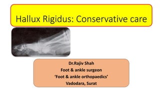
Hallux Rigidus: Conservative care
Dr.Rajiv Shah
Foot & ankle surgeon
‘Foot & ankle orthopaedics’
Vadodara, Surat
• Reported in 1887 by Davies-Colley - Hallux Flexus
• Cotterill coined the term Hallux Rigidus
• DuVries in 1959 and Moberg - hallux rigidus is the most common
condition to affect the first MTP joint
• Also called as
- hallux limitus
- dorsal bunion
- hallux dolorosus
- hallux malleus
- metatarsus primus elevatus (MPE)
• Painful condition of
great toe MTP joint
• Characterized by
- Restricted motion
(mainly
dorsiflexion)
- Proliferative periarticular
bone formation
Trauma
Intra articular fracture
Single episode Repetitive micro traumas
Crush Injury
Acute chondral/
osteochondral injury
Forced hyperextension/
plantar flexion injury
Congenital flattened/squared
metatrsal head
Short first metatarsal
Long first metatrsal
Pes planus
Tight intrinsic muscles
Congruent MTP joint
Hindfoot pronation
Metatarsus primus
elevatus
Dorsal and
dorsolateral
exostosis
Bony ledge
Increased bulk
around the joint
Constricting
footwear Dorsal
exostosis
enlargement
Limitation of
dorsiflexion
Pain Swelling MTP synovitis
Late
Stage
Initial
Stage
• Swollen metatarsophalangeal
(MTP) joint
• Everted gait
• Numbness develops over medial
sensory nerve to the hallux
• Callus develops on lateral heel
because of everted gait
• Hyperextension of hallux
interphalangeal joint
• Initial stages
- Nonuniform joint space
narrowing
- Widening and flattening of first
metatarsal head
• Advanced stages
- Subchondral cysts and sclerosis
of metatarsal head
- Widening of base of proximal
phalanx
- Hypertrophy of sesamoids
• Clinical
- Only stiffness
- Loss of passive motion
• ROM
- Dorsiflexion 40-60
• Radiograph
- Normal
• Clinical
- Occasional pain
- Pain at extremes of dorsiflexion
• ROM
- Dorsiflexion 30-40 degrees
• Radiograph
- Dorsal spur
- Min joint narrowing
- Min periarticular sclerosis
- Minimal flattening of metatarsal head
• Clinical
- Nearly constant subjective pain
substantial stiffness pain through out ROM
(but not at mid ROM)
• ROM
- 10 degrees or less Dorsiflexion
• Radiograph
- Grade 2+ substantial narrowing, periarticular
changes,>25% joint space involved no dorsal
side,sesamoids enlarged
• Clinical
- Moderate to severe pain and stiffness
- Pain just before maximal dorsiflexion
• ROM
- Dorsiflexion 10-30 degrees
• Radiograph
- Osteophytes
- <25%dorsal joint space involved
- Mild to moderate joint narrowing and
sclerosis
• Clinical
- nearly constant subjective pain
andsubstantial stiffness
- pain through out ROM+definite oain at mid
ROM
• Radiograph
-Grade 2+ substantial
- narrowing,periarticular changes,>25% joint
space involved no dorsal side,sesamoids
enlarged
• ROM 10
- degrees or less Dorsiflexion
Management
Conservative
Surgical
•Cheilectomy
•Phalangeal osteotomy
•Plantar flexion osteotomy
•Arthroplasty
•Arthrodesis
NSAIDs: reduces inflammation
& pain due to synovitis
Modification of activities
Neurotropics: if neuritic pain Steroid injections
•Morton’s extension
•Carbon fibre plate insert
•Commercially available
inserts
•Custom mould inserts
Tapping
Footwear modifications
Orthotics:
To stiffen the fore foot
To offload forefoot
To reduce the forefoot/MTP
joint motion
•Stiff insoles
•Rocker bottom shoe
•Still medial shank
•Wide/deep toe box
•Low heels
Joint manipulation
That’s all…
Thank you all…

Contenu connexe

Tendances

Tendances (20)

Ctev symposium 2015
Ctev symposium 2015Ctev symposium 2015
Ctev symposium 2015
 
Hallux valgus UG lecture
Hallux valgus UG lectureHallux valgus UG lecture
Hallux valgus UG lecture
 
Hip biomechanics
Hip biomechanicsHip biomechanics
Hip biomechanics
 
Pes planus / Flat Foot
Pes planus / Flat Foot Pes planus / Flat Foot
Pes planus / Flat Foot
 
High Tibial Osteotomy_UTSAV
High Tibial Osteotomy_UTSAVHigh Tibial Osteotomy_UTSAV
High Tibial Osteotomy_UTSAV
 
Hallux valgus Deformity
Hallux valgus DeformityHallux valgus Deformity
Hallux valgus Deformity
 
Hallux valgus.pptx
Hallux valgus.pptxHallux valgus.pptx
Hallux valgus.pptx
 
Acromio clavicular joint injury
Acromio clavicular joint injuryAcromio clavicular joint injury
Acromio clavicular joint injury
 
CORA (center of rotation of angulation)
CORA (center of rotation of angulation)CORA (center of rotation of angulation)
CORA (center of rotation of angulation)
 
Recurrent shoulder dislocation
Recurrent shoulder dislocationRecurrent shoulder dislocation
Recurrent shoulder dislocation
 
Shoulder dislocation with physiotherapy management
Shoulder dislocation with physiotherapy managementShoulder dislocation with physiotherapy management
Shoulder dislocation with physiotherapy management
 
Shoulder Rehabilitation
Shoulder RehabilitationShoulder Rehabilitation
Shoulder Rehabilitation
 
Osteotomy
OsteotomyOsteotomy
Osteotomy
 
Hallux valgus
Hallux valgusHallux valgus
Hallux valgus
 
Hallux rigidus
Hallux rigidus Hallux rigidus
Hallux rigidus
 
ILIZAROV EXTERNAL FIXATOR
ILIZAROV  EXTERNAL FIXATORILIZAROV  EXTERNAL FIXATOR
ILIZAROV EXTERNAL FIXATOR
 
Ankle sprain
Ankle sprainAnkle sprain
Ankle sprain
 
Physiotherapy for ankle & foot deformities
Physiotherapy for ankle & foot deformitiesPhysiotherapy for ankle & foot deformities
Physiotherapy for ankle & foot deformities
 
Recurrent Dislocation of patella -PAWAN
Recurrent Dislocation of patella -PAWANRecurrent Dislocation of patella -PAWAN
Recurrent Dislocation of patella -PAWAN
 
ACL rehabilitation
ACL rehabilitationACL rehabilitation
ACL rehabilitation
 

En vedette

Hallux Rigidus
Hallux RigidusHallux Rigidus
Hallux Rigidus
Pablo Vollmar
 
Alluce valgo
Alluce valgoAlluce valgo
Alluce valgo
Anslasax
 
Sure Orthotics Guide & Catalogue 2015
Sure Orthotics Guide & Catalogue 2015Sure Orthotics Guide & Catalogue 2015
Sure Orthotics Guide & Catalogue 2015
David Myatt
 

En vedette (20)

Lecture 46 parekh hr
Lecture 46 parekh hrLecture 46 parekh hr
Lecture 46 parekh hr
 
Lecture 42 shah calcaneal malunions
Lecture 42 shah calcaneal malunionsLecture 42 shah calcaneal malunions
Lecture 42 shah calcaneal malunions
 
hallux valgus & hallux rigidus
hallux valgus & hallux rigidushallux valgus & hallux rigidus
hallux valgus & hallux rigidus
 
Hallux rigidus
Hallux rigidusHallux rigidus
Hallux rigidus
 
Lecture 50 shah morton neuroma
Lecture 50 shah morton neuromaLecture 50 shah morton neuroma
Lecture 50 shah morton neuroma
 
Lecture 37 shah ttc fusion
Lecture 37 shah ttc fusionLecture 37 shah ttc fusion
Lecture 37 shah ttc fusion
 
Mauritius Course - Lecture 1
Mauritius Course - Lecture 1Mauritius Course - Lecture 1
Mauritius Course - Lecture 1
 
Lecture 44 shah delayed lisfranc
Lecture 44 shah delayed lisfrancLecture 44 shah delayed lisfranc
Lecture 44 shah delayed lisfranc
 
Lecture 35 shah subtalar fusion
Lecture 35 shah subtalar fusionLecture 35 shah subtalar fusion
Lecture 35 shah subtalar fusion
 
Lecture 27 parekh pttd3 and 4
Lecture 27 parekh pttd3 and 4Lecture 27 parekh pttd3 and 4
Lecture 27 parekh pttd3 and 4
 
Lecture 25 shah flat foot conservative
Lecture 25 shah flat foot conservativeLecture 25 shah flat foot conservative
Lecture 25 shah flat foot conservative
 
Lecture 47 parekh sports f&a
Lecture 47 parekh sports f&aLecture 47 parekh sports f&a
Lecture 47 parekh sports f&a
 
thefantasydoctorsStacked
thefantasydoctorsStackedthefantasydoctorsStacked
thefantasydoctorsStacked
 
Disorders of the hallux
Disorders of the halluxDisorders of the hallux
Disorders of the hallux
 
Hammer toe
Hammer toeHammer toe
Hammer toe
 
Hallux Rigidus
Hallux RigidusHallux Rigidus
Hallux Rigidus
 
Richard Schuster: Life Lessons and Legacy
Richard Schuster: Life Lessons and LegacyRichard Schuster: Life Lessons and Legacy
Richard Schuster: Life Lessons and Legacy
 
Alluce valgo
Alluce valgoAlluce valgo
Alluce valgo
 
Sure Orthotics Guide & Catalogue 2015
Sure Orthotics Guide & Catalogue 2015Sure Orthotics Guide & Catalogue 2015
Sure Orthotics Guide & Catalogue 2015
 
Il piede
Il piedeIl piede
Il piede
 

Similaire à Lecture 45 shah hallux rigidus

Anatomy and Injuries to the Wrist and Hand.ppt
Anatomy and Injuries to the Wrist and Hand.pptAnatomy and Injuries to the Wrist and Hand.ppt
Anatomy and Injuries to the Wrist and Hand.ppt
Rajveer71
 
Fracture calcaneum and talus by dr ashutosh
Fracture calcaneum and talus by dr ashutoshFracture calcaneum and talus by dr ashutosh
Fracture calcaneum and talus by dr ashutosh
Ashutosh Kumar
 
anatomy and deformities of Elbow Joint.pptx
anatomy and deformities of Elbow Joint.pptxanatomy and deformities of Elbow Joint.pptx
anatomy and deformities of Elbow Joint.pptx
HariraLatif1
 
Handand wristexam
Handand wristexamHandand wristexam
Handand wristexam
Ireland64
 
Kin 191 B – Wrist, Hand And Finger Evaluation And Pathologies
Kin 191 B – Wrist, Hand And Finger Evaluation And PathologiesKin 191 B – Wrist, Hand And Finger Evaluation And Pathologies
Kin 191 B – Wrist, Hand And Finger Evaluation And Pathologies
JLS10
 

Similaire à Lecture 45 shah hallux rigidus (20)

hallux valgus.pptx
hallux valgus.pptxhallux valgus.pptx
hallux valgus.pptx
 
Role of ultrasound in clinical evaluation of shoulder Dr. Muhammad Bin Zulfiqar
Role of ultrasound in clinical evaluation of shoulder Dr. Muhammad Bin ZulfiqarRole of ultrasound in clinical evaluation of shoulder Dr. Muhammad Bin Zulfiqar
Role of ultrasound in clinical evaluation of shoulder Dr. Muhammad Bin Zulfiqar
 
Calcaneal fractures
Calcaneal fracturesCalcaneal fractures
Calcaneal fractures
 
Anatomy and Injuries to the Wrist and Hand.ppt
Anatomy and Injuries to the Wrist and Hand.pptAnatomy and Injuries to the Wrist and Hand.ppt
Anatomy and Injuries to the Wrist and Hand.ppt
 
Shoulder Disorders
Shoulder DisordersShoulder Disorders
Shoulder Disorders
 
Ankle ppt
Ankle pptAnkle ppt
Ankle ppt
 
Common Fractures
Common FracturesCommon Fractures
Common Fractures
 
Knee joint anatomy, biomechanics, pathomechanics and assessment
Knee joint anatomy, biomechanics, pathomechanics and assessmentKnee joint anatomy, biomechanics, pathomechanics and assessment
Knee joint anatomy, biomechanics, pathomechanics and assessment
 
Fracture calcaneum and talus by dr ashutosh
Fracture calcaneum and talus by dr ashutoshFracture calcaneum and talus by dr ashutosh
Fracture calcaneum and talus by dr ashutosh
 
Common sports injuries.2560
Common sports injuries.2560Common sports injuries.2560
Common sports injuries.2560
 
MSK Part 2 LE.pdf
MSK Part 2 LE.pdfMSK Part 2 LE.pdf
MSK Part 2 LE.pdf
 
anatomy and deformities of Elbow Joint.pptx
anatomy and deformities of Elbow Joint.pptxanatomy and deformities of Elbow Joint.pptx
anatomy and deformities of Elbow Joint.pptx
 
Proximal tibia fractures(Plateau, spine ,Tubercle and Epiphyseal )
Proximal tibia fractures(Plateau, spine ,Tubercle and Epiphyseal ) Proximal tibia fractures(Plateau, spine ,Tubercle and Epiphyseal )
Proximal tibia fractures(Plateau, spine ,Tubercle and Epiphyseal )
 
Humerus Fractures
Humerus FracturesHumerus Fractures
Humerus Fractures
 
Handand wristexam
Handand wristexamHandand wristexam
Handand wristexam
 
Skeletal tuberculosis
Skeletal tuberculosisSkeletal tuberculosis
Skeletal tuberculosis
 
Lower limb deformities.pptx
Lower limb deformities.pptxLower limb deformities.pptx
Lower limb deformities.pptx
 
Tarsal tunnel syndrome
Tarsal tunnel syndromeTarsal tunnel syndrome
Tarsal tunnel syndrome
 
Wrist Joint
Wrist JointWrist Joint
Wrist Joint
 
Kin 191 B – Wrist, Hand And Finger Evaluation And Pathologies
Kin 191 B – Wrist, Hand And Finger Evaluation And PathologiesKin 191 B – Wrist, Hand And Finger Evaluation And Pathologies
Kin 191 B – Wrist, Hand And Finger Evaluation And Pathologies
 

Plus de Selene G. Parekh, MD, MBA

Lecture 19 parekh non insertional and insertional achilles tears
Lecture 19 parekh non insertional and insertional achilles tearsLecture 19 parekh non insertional and insertional achilles tears
Lecture 19 parekh non insertional and insertional achilles tears
Selene G. Parekh, MD, MBA
 

Plus de Selene G. Parekh, MD, MBA (19)

Lecture 41 parekh er f&a
Lecture 41 parekh er f&aLecture 41 parekh er f&a
Lecture 41 parekh er f&a
 
Lecture 40 parekh malunited ankle fracture
Lecture 40 parekh malunited ankle fractureLecture 40 parekh malunited ankle fracture
Lecture 40 parekh malunited ankle fracture
 
Lecture 39 parekh tar
Lecture 39 parekh tarLecture 39 parekh tar
Lecture 39 parekh tar
 
Lecture 33 34 parekh ankle arthritis
Lecture 33 34 parekh ankle arthritisLecture 33 34 parekh ankle arthritis
Lecture 33 34 parekh ankle arthritis
 
Lecture 31 parekh amputations
Lecture 31 parekh amputationsLecture 31 parekh amputations
Lecture 31 parekh amputations
 
Lecture 30 parekh charcot
Lecture 30 parekh charcotLecture 30 parekh charcot
Lecture 30 parekh charcot
 
Lecture 29 shah diabetic fractures copy
Lecture 29 shah diabetic fractures   copyLecture 29 shah diabetic fractures   copy
Lecture 29 shah diabetic fractures copy
 
Lecture 28 shah diabetic foot
Lecture 28 shah diabetic footLecture 28 shah diabetic foot
Lecture 28 shah diabetic foot
 
Lecture 26 parekh pttd2
Lecture 26 parekh pttd2Lecture 26 parekh pttd2
Lecture 26 parekh pttd2
 
Lecture 23 24 parekh peroneal pathology
Lecture 23 24 parekh peroneal pathologyLecture 23 24 parekh peroneal pathology
Lecture 23 24 parekh peroneal pathology
 
Lecture 21 shah chronic achilles rupture
Lecture 21  shah chronic achilles ruptureLecture 21  shah chronic achilles rupture
Lecture 21 shah chronic achilles rupture
 
Lecture 19 parekh non insertional and insertional achilles tears
Lecture 19 parekh non insertional and insertional achilles tearsLecture 19 parekh non insertional and insertional achilles tears
Lecture 19 parekh non insertional and insertional achilles tears
 
Lecture 17 parekh achilles tears
Lecture 17 parekh achilles tearsLecture 17 parekh achilles tears
Lecture 17 parekh achilles tears
 
Lecture 16 parekh jones
Lecture 16 parekh jonesLecture 16 parekh jones
Lecture 16 parekh jones
 
Lecture 15 parekh lisfranc
Lecture 15 parekh lisfrancLecture 15 parekh lisfranc
Lecture 15 parekh lisfranc
 
Lecture 14 shah fracture talus
Lecture 14 shah fracture talusLecture 14 shah fracture talus
Lecture 14 shah fracture talus
 
Lecture 12 shah orif calcaneal fractures
Lecture 12 shah orif calcaneal fracturesLecture 12 shah orif calcaneal fractures
Lecture 12 shah orif calcaneal fractures
 
Lecture 11 parekh pilon
Lecture 11 parekh pilonLecture 11 parekh pilon
Lecture 11 parekh pilon
 
Lecture 9 shah ankle fractures
Lecture 9 shah ankle fracturesLecture 9 shah ankle fractures
Lecture 9 shah ankle fractures
 

Lecture 45 shah hallux rigidus

  • 1. Hallux Rigidus: Conservative care Dr.Rajiv Shah Foot & ankle surgeon ‘Foot & ankle orthopaedics’ Vadodara, Surat
  • 2. • Reported in 1887 by Davies-Colley - Hallux Flexus • Cotterill coined the term Hallux Rigidus • DuVries in 1959 and Moberg - hallux rigidus is the most common condition to affect the first MTP joint • Also called as - hallux limitus - dorsal bunion - hallux dolorosus - hallux malleus - metatarsus primus elevatus (MPE)
  • 3. • Painful condition of great toe MTP joint • Characterized by - Restricted motion (mainly dorsiflexion) - Proliferative periarticular bone formation
  • 4. Trauma Intra articular fracture Single episode Repetitive micro traumas Crush Injury Acute chondral/ osteochondral injury Forced hyperextension/ plantar flexion injury
  • 5. Congenital flattened/squared metatrsal head Short first metatarsal Long first metatrsal Pes planus Tight intrinsic muscles Congruent MTP joint Hindfoot pronation Metatarsus primus elevatus
  • 6. Dorsal and dorsolateral exostosis Bony ledge Increased bulk around the joint Constricting footwear Dorsal exostosis enlargement Limitation of dorsiflexion Pain Swelling MTP synovitis Late Stage Initial Stage
  • 7. • Swollen metatarsophalangeal (MTP) joint • Everted gait • Numbness develops over medial sensory nerve to the hallux • Callus develops on lateral heel because of everted gait • Hyperextension of hallux interphalangeal joint
  • 8. • Initial stages - Nonuniform joint space narrowing - Widening and flattening of first metatarsal head • Advanced stages - Subchondral cysts and sclerosis of metatarsal head - Widening of base of proximal phalanx - Hypertrophy of sesamoids
  • 9. • Clinical - Only stiffness - Loss of passive motion • ROM - Dorsiflexion 40-60 • Radiograph - Normal • Clinical - Occasional pain - Pain at extremes of dorsiflexion • ROM - Dorsiflexion 30-40 degrees • Radiograph - Dorsal spur - Min joint narrowing - Min periarticular sclerosis - Minimal flattening of metatarsal head
  • 10. • Clinical - Nearly constant subjective pain substantial stiffness pain through out ROM (but not at mid ROM) • ROM - 10 degrees or less Dorsiflexion • Radiograph - Grade 2+ substantial narrowing, periarticular changes,>25% joint space involved no dorsal side,sesamoids enlarged • Clinical - Moderate to severe pain and stiffness - Pain just before maximal dorsiflexion • ROM - Dorsiflexion 10-30 degrees • Radiograph - Osteophytes - <25%dorsal joint space involved - Mild to moderate joint narrowing and sclerosis
  • 11. • Clinical - nearly constant subjective pain andsubstantial stiffness - pain through out ROM+definite oain at mid ROM • Radiograph -Grade 2+ substantial - narrowing,periarticular changes,>25% joint space involved no dorsal side,sesamoids enlarged • ROM 10 - degrees or less Dorsiflexion
  • 13. NSAIDs: reduces inflammation & pain due to synovitis Modification of activities Neurotropics: if neuritic pain Steroid injections
  • 14. •Morton’s extension •Carbon fibre plate insert •Commercially available inserts •Custom mould inserts Tapping Footwear modifications Orthotics: To stiffen the fore foot To offload forefoot To reduce the forefoot/MTP joint motion •Stiff insoles •Rocker bottom shoe •Still medial shank •Wide/deep toe box •Low heels Joint manipulation