SlideShare une entreprise Scribd logo
1  sur  37
Télécharger pour lire hors ligne
A Novel Stem Cell Delivery Device
Final Report
Project Sponsor: Dr. Luis Garza M.D., Ph.D., Department of Dermatology, Johns Hopkins Hospital
Design Team 8: Michael Clark (Team Leader), Angelica Herrera, Arianne Papa, Seung Jung, Michael Mow,
Annabeth Rodriguez, Jose Solis, Prateek Gowda
Contents
1 Abstract 3
2 Introduction 4
2.1 Clinical Problem . . . . . . . . . . . . . . . . . . . . . . . . . . . . . . . . . . . . . . . . . . . 4
2.2 Clinical Need . . . . . . . . . . . . . . . . . . . . . . . . . . . . . . . . . . . . . . . . . . . . . 4
2.3 Solution and Design Specifications . . . . . . . . . . . . . . . . . . . . . . . . . . . . . . . . . 4
3 Design 6
3.1 Design Process . . . . . . . . . . . . . . . . . . . . . . . . . . . . . . . . . . . . . . . . . . . . 6
3.2 Device Overview . . . . . . . . . . . . . . . . . . . . . . . . . . . . . . . . . . . . . . . . . . . 6
3.3 Base Station . . . . . . . . . . . . . . . . . . . . . . . . . . . . . . . . . . . . . . . . . . . . . 7
3.4 Cell Thawing Subsystem . . . . . . . . . . . . . . . . . . . . . . . . . . . . . . . . . . . . . . . 7
3.5 Peristaltic Pump Subsystem . . . . . . . . . . . . . . . . . . . . . . . . . . . . . . . . . . . . . 8
3.6 Closed-Loop Cell Pathway . . . . . . . . . . . . . . . . . . . . . . . . . . . . . . . . . . . . . . 8
3.7 Cryobag Freezing Mold . . . . . . . . . . . . . . . . . . . . . . . . . . . . . . . . . . . . . . . 8
3.8 Power Systems . . . . . . . . . . . . . . . . . . . . . . . . . . . . . . . . . . . . . . . . . . . . 9
3.9 Angle and Depth Guards . . . . . . . . . . . . . . . . . . . . . . . . . . . . . . . . . . . . . . 9
4 Results 10
4.1 Post-Injection Cell Viability Testing . . . . . . . . . . . . . . . . . . . . . . . . . . . . . . . . 10
4.1.1 Methods . . . . . . . . . . . . . . . . . . . . . . . . . . . . . . . . . . . . . . . . . . . . 10
4.1.2 Testing and Results . . . . . . . . . . . . . . . . . . . . . . . . . . . . . . . . . . . . . 10
4.2 Cell Thawing System Testing . . . . . . . . . . . . . . . . . . . . . . . . . . . . . . . . . . . . 11
Appendix A Materials 12
Appendix B Prototype Budget 13
Appendix C Figures, Photos, and Sketches 14
C.1 Injector . . . . . . . . . . . . . . . . . . . . . . . . . . . . . . . . . . . . . . . . . . . . . . . . 14
C.2 Base Station . . . . . . . . . . . . . . . . . . . . . . . . . . . . . . . . . . . . . . . . . . . . . 14
C.3 Motor and Driver . . . . . . . . . . . . . . . . . . . . . . . . . . . . . . . . . . . . . . . . . . . 15
C.4 Onboard Computer . . . . . . . . . . . . . . . . . . . . . . . . . . . . . . . . . . . . . . . . . . 15
C.5 Microcontroller . . . . . . . . . . . . . . . . . . . . . . . . . . . . . . . . . . . . . . . . . . . . 15
C.6 Thermocouple . . . . . . . . . . . . . . . . . . . . . . . . . . . . . . . . . . . . . . . . . . . . . 16
C.7 Cryobag Freezing Mold . . . . . . . . . . . . . . . . . . . . . . . . . . . . . . . . . . . . . . . 16
C.8 Cryobag . . . . . . . . . . . . . . . . . . . . . . . . . . . . . . . . . . . . . . . . . . . . . . . . 16
C.9 Luer Lock Connector . . . . . . . . . . . . . . . . . . . . . . . . . . . . . . . . . . . . . . . . . 17
C.10 Needle . . . . . . . . . . . . . . . . . . . . . . . . . . . . . . . . . . . . . . . . . . . . . . . . . 17
C.11 Power Delivery Schematic . . . . . . . . . . . . . . . . . . . . . . . . . . . . . . . . . . . . . . 17
C.12 Microcontroller Code . . . . . . . . . . . . . . . . . . . . . . . . . . . . . . . . . . . . . . . . . 18
C.13 Heating System Performance Data . . . . . . . . . . . . . . . . . . . . . . . . . . . . . . . . . 19
C.14 Viability Testing Data . . . . . . . . . . . . . . . . . . . . . . . . . . . . . . . . . . . . . . . . 19
Appendix D Calculations 20
D.1 Volume Resolution Calculation . . . . . . . . . . . . . . . . . . . . . . . . . . . . . . . . . . . 20
D.2 Tubing Dead Space Calculation . . . . . . . . . . . . . . . . . . . . . . . . . . . . . . . . . . . 20
Appendix E References 21
Appendix F Proposal 22
2
1 Abstract
Recent advancements in stem cell therapies have shown the potential to revolutionize the treatment of many
conditions. However, there is still a need to consistently and accurately deliver stem cells to target regions
in the solid organs of the body–particularly the skin. Skin stem cell therapies currently under investigation
have the potential to reverse hair loss, heal wounds, and alter the phenotype of the epidermis. We have
designed a device to deliver stem cells to the skin at adjustable volumes with minimal viability loss or risk
of contamination. The device makes use of a peristaltic pump to provide precise control over injection rate,
volume, and the resulting shearing forces. Cell viability is also improved through an automated heating
system that thaws the cells at an expeditious but controlled rate immediately prior to injection. The entire
cell pathway is a closed loop, which minimizes the risk of contamination. Cryobags were utilized to increase
the volume of cells available to the physician administering the therapy. Interchangeable angle and depth
guards were designed to improve the consistency and repeatably of injections. In preparation for ex vivo
testing, an exploratory in vitro test was performed on the injection mechanism using fibroblast cells. The
thermal performance of the heating system was also evaluated experimentally.
3
2 Introduction
2.1 Clinical Problem
Stem cell therapies are an expanding market in regenerative medicine. The global stem cell industry has
grown 13.6% annually from $5.6 billion in the year 2013 [1]. Currently, 4,681 clinical trials involving stem
cell therapies are being carried out in the United States with over 3,000 in early phases of testing [2]. The
NIH estimates spending approximately $2.77 billion on stem cell research in 2015 [3]. Public funding from
individual states will total over $4.1 billion by 2018 [4]. As thousands of therapies advance past initial stages
of testing, there will be an increased demand for a safe and effective way to deliver the stem cells to patients.
Current stem cell therapies for the skin involve altering the characteristics of dermal cells to treat wounds,
rashes, and burns. In the United States, the treatment of wounds and associated complications exceeds $20
billion annually [5]. Existing treatments for these conditions are prohibitively expensive; skin allograft
therapies typically cost $500,000 per patient [6]. Skin stem cell therapies also have cosmetic applications
such as regenerating hair follicles, repairing scar tissue, and changing the phenotypic expression of the skin.
Today, physicians have difficulty delivering stem cells to the skin in a consistent manner. Providers rely
on tactile feedback and previous experience to deliver the cells to the desired location. Subjectivity and
variance are inherent in the current treatment administration procedure. In order to generate useful clinical
trial data, the stem cell delivery procedure needs to be optimized to be as consistent and repeatable as
possible.
A primary concern is ensuring post-injection cell viability. If cells are injected too quickly, shearing
forces in the needle can tear them apart. Another concern is contamination: in the current procedure, cells
are exposed to air when they are transferred from the freezing vessel to the syringe. This can lead to cell
contamination and an increased risk of post-treatment infection. As a result of these complications, many
existing dermal injectors cannot be adapted to this highly specialized application.
2.2 Clinical Need
No devices on the market are specifically designed to mitigate the difficulties associated with delivering stem
cell therapies to the skin. Consequently, there is a need for a device that allows physicians to deliver stem
cells to target dermal regions at adjustable volumes with minimal risk of contamination or viability loss.
2.3 Solution and Design Specifications
The team’s dermal injector addresses the biological hurdles associated with stem cell delivery by providing
adjustable injection rates, integrated cell thawing capabilities, and a closed-loop delivery system that reduces
the risk of contamination. Stem cells are commonly stored and frozen in cryogenic vials, which typically
hold a one to two milliliter volume. The team will make use of cryogenic bags, which can hold volumes
ranging from five to ten milliliters. This reduces the number of times the physician has to reload the device
– effectively decreasing procedure time and air exposure, thus reducing the risk of contamination. The
cryobag will be connected to a cartridge-tubing system and a needle to form the closed-loop system. The
physicians will place the cartridge onto a peristaltic pump, which will accurately output desired volumes with
an accuracy of ±1 µl and physician-specified rates for each injection averaged at one minute per injection.
To further enhance cell viability, the team spoke with Dr. Luis Garza, M.D., Ph.D., who is the sponsor
and medical adviser for the team, and found that a heating system should be included as well.
Several other possibilities why a new device might be better than a syringe might be the problem
of temperature, where we know that cells will likely be shipped to a consumer frozen. But
frozen cells lose viability or thaw too slowly. If they are thawed more quickly, then viability is
maintained better, so a device that eliminates user variability in terms of quick thawing rate
could also improve final outcome for cellular therapies.
4
An integrated cell thawing system was designed that heats frozen stem cells at a consistent rate, which
dramatically improves cell viability [7]. In order to achieve this, the frozen stem cells must be heated from
−196 ◦
C, the temperature of the liquid nitrogen, to 37 ◦
C, the average temperature of the human body, in
under two minutes per 1.0 ml thawed, which is the current standard established from a traditional water
bath.
The dermal injector will be kept in its base station when not in use. This docking station will charge
the device and will be used to adjust device settings. Every system in the dermal injector will be controlled
electronically. The physician will only have to input his or her desired injection volume and injection rate,
and then start the thawing process using a touch screen user interface on the base station. This LCD touch
screen will indicate when cell thawing is complete and alert the physician when the device is ready to use.
This simplified procedure is to ensure that this method is no more difficult than the current standard of care.
5
3 Design
3.1 Design Process
The team conducted months of extensive research to gain a thorough understanding of the market. An
intellectual property search was conducted, which revealed ample room to innovate as there were no other
devices that delivered stem cells to the dermis. This meant that the team had no competitors, but also had
no predicate devices upon which to improve their design. Therefore, the group decided to expand the search
to include devices that delivered stem cells to other organs of the body. However this also revealed a lack
of devices and the team switched to drug delivery methods instead. As a result, the team developed four
promising devices that could have been adapted to deliver stem cells.
The first idea was a direct stem cell insertion. This method was considered as it was the simplest way
to get stem cells from point A to point B. The stem cells would be cultured in a laboratory setting into a
cell matrix. The method would then require an operation where the patient’s skin was cut and pulled back,
the cell matrix inserted into place, and then skin sutured back on. Although this method was the most
direct treatment, it was also the most invasive. The team thought it best to avoid operations that required
a lengthy recovery time and continued to seek less invasive methods of delivery.
The next idea was an injection gun. This was by far the fastest way the team found to deliver a treatment.
This device penetrated the skin with the aid of pressurized gas, which evenly distributed the drug. However,
this method raised concerns as the velocity of injection was deemed detrimental to the viability of the cells.
In addition, the location that the stem cells were delivered also raised concerns as the cells traveled too deep
into the dermis and passed its optimal location. From this idea, the team determined that a slow consistent
rate of injection was a vital part of the device as it ensured that the cells maintained a <40% reduction in
viability.
The use of a microneedle array was one of the best methods to deliver drugs at the optimal location. This
device consisted of a series of microneedles that were able to deliver drugs at consistent rates. However, the
team found that a small needle gauge would damage the cells and lower cell viability as it passed through
the needle. Though this method had a consistent flow rate, the team determined that the correct needle
gauge was just as important. After an extensive literature search, the team pursued a 23 gauge needle,
which was found to be the most optimal size for their objective as it was big enough for the cells to travel
the needle without much shearing, but small enough to penetrate the skin without the pain associated with
bigger needles.8
The final idea was an insulin pen injector. Similar to a needle and syringe, this device simplified the
delivery process. The patient would set the volume he or she needed and then press a button to deliver the
treatment. Using this method as a springboard, the team adapted this simple design and incorporated all
the features that the team determined was vital from the previous ideas: minimal invasiveness, consistent
flow rate, use of a 23 gauge needle, and adjustable volume concentration from the pen injector.
From there, the team generated ideas and selected designs that yielded the best treatment outcomes and
per-dollar performance. After soliciting feedback from experts, the team modified the device to include the
elements described in §3.2.
3.2 Device Overview
The device is a hand-held electronic cell injection system (Appendix C.1). The injector itself is cordless
– it docks in a powered base station, which allows the device to be charged and programmed by the user
(Appendix C.2). The injector has four discrete subsystems: an automated cell thawing system with a
temperature feedback mechanism, a peristaltic pump system that offers precise control over injection rate
and volume, a closed-loop cell pathway, and a power delivery system. A sealed septum separates the electronic
systems from the “wet” cellular pathway, which effectively mitigates the risk of electrical shock in the case
of a breach in the closed-loop system.
The injector device is designed to be held like a computer mouse, with the pointer finger resting on the
injection button. This position provides a great deal of stability to the physician administering the therapy.
6
The long tip of the device allows clinicians to achieve very shallow injection angles. The tip also accepts
interchangeable angle and depth guards.
The base station houses an onboard computer (Raspberry Pi Model B+) and a touch-screen user interface.
A bag freezing mold was also developed that facilitates increased heat transfer from the heating pads to the
cryobag.
3.3 Base Station
The base station (Appendix C.2) was developed as a means of improving physician usability by utilizing a
large full color capacitive LCD touchscreen with a user-friendly graphic user interface (GUI), which allows the
physician to set the parameters of the injection easily. After the physician attaches the cell cryobag cartridge
to the device, the program switches on and prompts the physician to enter the desired injection volume of
stem cells After the physician inputs the information, the data is sent to an internal computer inside the base
station. The Raspberry Pi was utilized as the internal computer (Appendix C.4) due to its compatibility with
touch screen interfaces and its ability to communicate with our specific microcontroller, the Arduino Micro
(Appendix C.5). The internal computer carries out calculations from the selected volume of injection in
order to determine the time necessary for cell thawing, while automatically setting the injection rate. Inside
the handheld device, The data computed by the internal computer is programmed onto the microcontroller
in the device via contacts between the base station and the resting device. The Arduino carries out the
heating process and signals when the process is complete. The screen then prompts the physician to remove
the device from the base station and begin the injection.
3.4 Cell Thawing Subsystem
The heating system functions to thaw the stem cells quickly, but in a controlled manner that prevents the
risk of destroying the cells due to overheating. Thawing cells quickly helps reduce the risk that the cells
will be damaged by ice crystals that are present during the thawing process. Literature also suggests that
rapid thawing reduces protein damage [8]. The heating process takes place while the device is resting in the
base station. The physician places the cryobag cartridge into the device and selects the heating option on
the base station user interface. The cells are thawed by two heating pads that envelope the cryobag. Due
to the large surface area of the cryobag and thin distribution of the cells in the bag itself, the heating is
uniform along the cells in the bag. The heating pads maintain a constant temperature of 37.0 ◦
C. This is
the ideal temperature of the cells inside the human body. It is important the heating pads do not exceed
this temperature, as it leads to potential cell death and an overall loss in cell viability. To achieve constant
temperature control, the system utilizes a negative feedback loop and control systems to provide safe heating
of the cells.
The heating subsystem is comprised of two thin parallel heating pads constructed using a mesh of polyester
filament and conductive fiber folded into a protective Polyimide Film. The fact that these are low power,
flexible and draw little power makes them ideal for things like hand-warmers and other heated garments. The
current source for these heating pads is controlled and obtained from a regulated DC power supply provided
by a grounded wall outlet. The circuit is regulated by an NPN transistor switch that can be opened and closed
based on feedback from data obtained by a thermocouple (Appendix C.6). The thermocouple concurrently
sends the temperature of the heating pads as digital data to an Arduino Micro (Appendix C.5) that then
regulates the switching of the transistor (See code in Appendix C.12). This transistor was used as a switching
mechanism over other options, such as mechanical relays for example, because of its solid state design. This
allows the team to utilize power with modulation techniques to rapidly switch the current to more safely
control the heating of the pads. Using this method, the pads will heat rapidly at lower temperatures and
slowly as the temperature approaches the maximum of 37.0 ◦
C. Once the cells have thawed, the heating has
been completed, at which time the physician will be signaled to remove the device from the base station and
prepare for injection.
7
3.5 Peristaltic Pump Subsystem
The pump system is designed to control the stem cell injection rates and reduce viability losses due to
shearing forces in the needle. The user interface for the pumping process is comprised of two mechanical
buttons on the device, a clear-air button and an injection button. The purpose of the clear-air button is
to remove excess air in the tubing and replace it with the cell from the cryobag. This is performed at the
discretion of the physician and is analogous to clearing air bubbles in the common needle and syringe. The
clear-air function pumps at a higher rate than the injection button. The injection button, when toggled
by the physician, pumps the stem cell solution out at a slow, constant rate to maximize cell viability and
reduce shearing forces. Testing will be carried out to determine the ideal rate of injection. To perform the
injection, the physician inserts the needle into the patient and holds the injection button.
The pump system is comprised of a peristaltic pump head that is rotated by a small four-wire unipolar
stepper motor (Appendix C.3). Peristaltic pumps work by pushing and collapsing the walls of a flexible
tubing material to create a vacuum and a source of suction, that when rotating along the tubing walls,
draws out the cells from the cryobag into the tubing. This method prevents contact between the cells
and any external pumping mechanism, preventing contamination during the injection. A stepper motor is
utilized to turn the peristaltic pump and control the rotation without using a separate mechanical or optical
encoder. Removing the encoder prevents bulky attachments, reduces the weight and controls space within
the device. Instead, stepper motors work by moving in steps that are designated by the Arduino Micro,
which eliminates a need for a negative feedback loop.The motor is geared to afford a resolution of 1024 turns
per revolution, which gives a pumping resolution of 0.0455 µl based on the size of the tubing and pump head
radius (Appendix D.1). This high resolution and controlled pump system ensures that even small volumes
of stem cell solution can be injected without the device.
3.6 Closed-Loop Cell Pathway
The closed-loop cell pathway is comprised of four components: the cryobag, tubing, needle, and Luer-lock
connectors. Cryobags were chosen over more traditional cryovials because they are able to contain a larger
(and more clinically appropriate) volume of cells - this decreases the frequency with which the physician has
to halt the procedure to load more cells into the device. OriGen Biomedicals PermaLife Cell Culture Bag
(Appendix C.8) was selected for the device. The bag is made from biologically-inert Fluorinated ethylene
propylene (FEP), which offers an operational temperature range of −196 ◦
C to 137 ◦
C permitting the bag to
be frozen in liquid nitrogen and sterilized by autoclave.
Unlike the cryobag, the tubing will not be subjected to extreme temperatures. Clear silicone tubing
made from FDA-compliant resins (McMaster-Carr 5236K501) was selected for its plyability and ability to
be sterilized by autoclave. The inner diameter (ID) and length of the tubing was minimized (794 µm) in
order to reduce dead space losses, i.e. the volume of cells required to fill the tubing that become unavailable
for injection. In the current design, less than 76 µl of dead space exists in the tubing (Appendix D.2).
Hypodermic needles are widely available in health care settings, so the needle selection was guided by
industry standards. Becton, Dickinson, and Company (BD) has a significant share of the hypodermic needle
market and is therefore a suitable supplier, although a wide variety of manufactures sell needles that can be
used with the device. The device will accept any needle with a Luer-lock connector - various gauges, lengths,
and bevels are available to suit a variety of procedures. A 23 gauge needle (Appendix C.10) was utilized for
testing purposes since it is commonly used in intradermal injection procedures.
All interfaces between the cryobag, tubing, and needle feature a Luer-lock type connector (Appendix
C.9). Luer-lock is a commonly used and ISO-standard fitting that will be familiar to physicians. Luer-lock
connectors were chosen over Luer-slip connectors due to their ability to withstand higher injection pressures.
3.7 Cryobag Freezing Mold
Maximum heat transfer is achieved when the contact area between the heating pad and cryobag is maxi-
mized. In order to increase the contact area, freezing the cryobag at a uniform (flat) thickness works to
8
the physician’s advantage. A cryobag freezing mold (Appendix C.7, isometric drawing) was developed as a
means to that end. The mold allows two 10 ml cryobags to be frozen flat inside of a standard 135 mm by
135 mm cryobox. Polylactide (PLA) was used to prototype the mold, but the final product will implement a
low-temperature polymer capable of thermocycling from room temperature to liquid nitrogen temperatures
(e.g. polypropylene). The cryobag sits in between the two halves of the mold. Stainless steel springs will be
employed to apply pressure on both sides, gently compressing the bag in the middle.
3.8 Power Systems
Two systems provide power to the device subsystems (Appendix C.11). A power supply in the base station
transforms, rectifies, filters and regulates 120V AC current to 5.1V DC at 2.1A. The 5.1V rail is used to
power the onboard computer in the base station, charge the 12V battery in the injector, and operate a NPN
transistor along with the Arduino. The power supply is controlled by a switch located on the back of the
base station. The second power source is the 12V battery in the injector, which allows the device to be
operated wirelessly when undocked from the base station. The battery supplies the power necessary to run
the onboard microcontroller that regulates the pump motor and thermocouple feedback system.
3.9 Angle and Depth Guards
A selection of nine angle and depth guards were designed. The guards clip on to the tip of the device.
Three clinically relevant angles (90◦
, 45◦
, and 15◦
) and three physiologically relevant depths (intradermal,
subdermal, and intramuscular) were represented. The guards are made from clear plastic so that physicians
can visualize the injection site while they administer the cellular therapy.
9
4 Results
4.1 Post-Injection Cell Viability Testing
4.1.1 Methods
After finalizing each component of the stem cell injector, preliminary testing for the two major subsystems
of the device was performed. Both the closed-loop cell injection system and automated cell thawing system
(and feedback mechanism) were examined. Literature suggests that cell death occurs when cells are exposed
to high levels of pressure or shear force, specifically above 1.0 Pascal [9]. It has been shown that viability
losses up to forty percent can occur when cells are injected through a needle (size) and syringe(rate, cell
density, media (PBS in this case)) due to shearing forces [10].
Preliminary tests were designed to correlate post-injection cell viability to injection rate through a 23
gauge needle. A 23 gauge needle was chosen since this is the most commonly used needle size for cell
viability testing and has clinical relevance. A needle of this size is optimal for the balance between a low
pain level for the patient and a large enough width to maintain cell viability [11]. Also, a small diameter
allows precise regions, such as the dermoepidermal junction, to be reached. Injection rates of 6.0 mL/min,
3.0 mL/min, 1.0 mL/min and 0.5 mL/min were chosen based on physician feedback and literature13. The
fastest injection rate (6.0 mL/min) was used to simulate high shear forces through the needle. An injection
rate of 3.0 mL/min mimicked a physicians typical injection speed. Slower injection rates (1.0 mL/min) were
used in other viability studies and used to simulate the lowest amount of shear forces through the needle
(0.5mL/min).13
4.1.2 Testing and Results
Initial viability testing was performed with mouse spleen cells due to their relative availability. Cells were
obtained from Dr. Luis Garzas laboratory at Johns Hopkins Hospital.
Mouse spleen cells were harvested, filtered with PBS through a mesh netting to isolate the cells, and
centrifuged at 1000 RPM for 5 minutes. The cells were then resuspended in Phosphate-buffered saline
(PBS). In order to determine cell density, 10 uL of the cell solution was mixed with 10 uL of trypan blue
and placed on a hemocytometer slide. Using a Countess Automated Cell Counter, the number of live cells
was approximated. However, the countess did not provide clear estimates for cell density. This could be due
to the fact that red blood cells were not separated from the spleen cells. In future tests, the red blood cells
can be lysed and then washed away using ACK Lysing Buffer (Life Technologies, A10492-01).
Additional testing was performed with volar fibroblasts biopsied from a patients sole. Fibroblasts were
acquired from Dr. Luis Garzas laboratory at Johns Hopkins Hospital (IRB NA 00068684) and cultured
in Dulbecco’s Modified Eagle Medium (DMEM) for one week before experimentation. Due to the short
expansion time, cells were only used at a density of 3.0 × 10⁵
cells/mL.
Injection testing was performed using a 23 gauge needle and syringe. For the 6.0 ml/min rate, the injection
was performed manually to model the accuracy and repeatability of a physician. For the slower rates (3.0,
1.0, and 0.5 ml/min) a syringe infusion pump was used to provide consistent injection rates. In each trial,
300 µl of the cell solution were loaded into the 1.0 ml syringe by hand. Using the infusion pump (or by hand
in the 6.0 ml/min trial), 100 µl of the solution were injected into a 1.0 ml vial at a predetermined controlled
rate. The ejected cells were then placed on ice. Three trials were conducted for every injection rate. To
serve as a control, 100 µl of cell solution were drawn up manually into the syringe and injected into a 1.0 ml
without using a needle. After completing all injections, 10 µl from each 1.0 ml vial were drawn up with a
pipette and placed in a new 1.0 mL vial with 10 µl of trypan blue. After mixing, 10 µl of the solution were
pipetted onto a hemocytometer slide and imaged.
No losses in viability were seen during this experiment; all visible cells appeared viable with intact
membranes and no dark stains from the trypan blue (Appendix C.14). However, there was much variation
between cell densities in each hemocytometer image. Across different injection rates, there did not seem to be
a constantly increasing or decreasing trend in the number of cells present. Additionally, between injections
of the the same rate, results would vary from no visible cells to large inordinate amounts of cells. As a
10
result, standard deviation values from these injection rates tend to be greater than the average of observable
cells within that rate. For example, the trials for the 1.0 ml/min injection rate were: 0 cells visible, 1
cell, and 61 cells visible. This results in an average of 20 cells visible in this injection rate and a standard
deviation of 35 cells. These high standard deviation numbers made any quantifying results inconclusive or
at least statistically invalid. In the future, cell solutions will be mixed thoroughly before pipetting onto the
hemocytometer slide to ensure homogeneity throughout the solution. A 24 hour time point will be used as
well to see if viability losses are seen over time and not immediately after injection. By using trypan blue,
cell death will only be seen once the cell membrane is lysed. This may not occur immediately since cells
may only initially be damaged.
Revised testing is currently underway at Johns Hopkins University Homewood campus. Mouse fibrob-
last (L929, P3 11334) cells were acquired from Dr. Elizabeth Logsdon. The cells, which were originally
frozen at ninety percent confluence were thawed quickly and cultured in DMEM (10% FBS with Penicillin-
Streptomycin (Sigma-Aldrich P4333, 5:500) for four days prior to testing.
Before experimentation, cell density was determined using standard cell counting procedures and a hemo-
cytometer slide. Dimethyl sulfoxide (DMSO) was added to the cell solution to mimic the conditions in Dr.
Garzas clinical trial. It is expected that DMSO will make the cells more susceptible to shearing forces and
viability losses [12]. Many studies have demonstrated reduced cell viability in a dose-dependent manner.
Although DMSO is used to protect cells while frozen, they may induce some cytotoxicity when they are
thawed due to permeability [13]. The previous procedure using fibroblasts was slightly modified for this
experimentation. Injections were performed directly into 24 well plates for easier imaging. An identical
second well plate was used for the 24 hour time point. Injections were repeated three times, either by hand
or with the infusion pump, per injection rate. Trypan blue was added to the wells before imaging.
4.2 Cell Thawing System Testing
The automated thawing system and feedback loop were thoroughly tested and examined. Before use in the
clinical setting, stem cells are frozen in liquid nitrogen and stored. When needed, the physician removes and
thaws these vials before injection. Cells can withstand a maximum temperature of 37 ◦
C, any higher and
they face the potential of cell death [14]. To maintain a consistent thawing procedure, the in device heating
capabilities were examined. Testing the heating component of the dermal injector was vital to the device.
To ensure a maximum temperature of 37 ◦
C, the heating pads were connected to a thermocouple, recording
the temperature during the experiment. Along the heating circuit, these pads were controlled by a switching
system and power supply. The thermocouple created a negative feedback loop, allowing us to reach a
maximum temperature of 37 ◦
C. The arduino read the temperature from the thermocouple and controlled
the switch along the heating circuit.
The test was carried out with a supply voltage of 1.1V to the collector input of the transistor. The
feedback program on the microprocessor utilized power width modulation techniques in three stages to
regulate current flow to the heating pads. Rapid heating occurred when the temperature of heating pads
were below 30 ◦
C. The temperature then increased slowly between the period of 30 ◦
C-37 ◦
C. Once the pads
had reached 37 ◦
C, it began to oscillate along this target temperature, with maximum at 37.6 ◦
C (Appendix
C.13).
The thermocouple feedback system, along with the PWM technique used in the programming, demon-
strated the ability to control temperature with 1.0 ◦
C precision from the 37 ◦
C target temperature. Im-
provements can be made in the software by increasing the reading rate of the program. In this test, the
temperature was taken at a rate of one reading every 250 milliseconds. Increasing this rate would increase
the number of times that the temperature is managed, resulting in smaller fluctuations along the target
temperature and a reduction in time between heating and cooling cycles.
11
A Materials
System Component Description Source Item Number
Base Station Outer Shell PLA Makerbot Indus. White PLA
Computer Raspberry Pi Model B+ Raspberry Pi Model B+
Power Button SPST (Round) Sparkfun COM-11138
LCD Screen 2.8-Inch, TFT, 320x240 Adafruit Indus. 1601
Power Cord 18AWG, 72”, Black Digi-Key Elec. 221001-01
Contacts Universal 1.8MM SMD Digi-Key Elec. 1003-1010-2-ND
USB/Data Cable Adafruit 70
Device Outer Shell PLA Makerbot Indus. White PLA
Microcontroller Arduino Micro Arduino A000053
Buttons Tactile Button Assortment Sparkfun COM-10302
LEDs Blue LED Digi-Key Elec. 160-1602-ND
Pins Stainless Steel McMaster-Carr 6517K65
Springs Compression, Steel Conical McMaster-Carr 1692K11
Stepper Motor 12V Adafruit 918
Motor Driver Lighted Texas Instruments ULN2003ADR
Heating Pads Flexible Sparkfun COM-11289
Battery 12V Enegizer A23
Transistor General Purpose Transistor ON Semiconductor PN2222
Thermocouple K-type Adafruit Indus. 270
Transducer For K-type Thermocouple Adafruit Indus. 269
Resistors 10KΩ, 1KΩ Digi-Key Elec. CF14JT10K0,
CF14JT1K50
Cell Pathway Tubing For Peristaltic Pumps McMaster-Carr 5236K501
Needle 23 Gauge (for testing) Becton & Dickenson 305148
Cryobag 10 ml OriGen PL07
Leur Lock 0.8mm ID Barb Qosina 11106
Freezing Mold Mold PLA Makerbot Indus. Black PLA
Springs Conical, Stainless Steel McMaster-Carr 1692K12
Angle Guards Guard Clear, Polycarbonate McMaster-Carr 8585K51
12
B Prototype Budget
13
C Figures, Photos, and Sketches
C.1 Injector
C.2 Base Station
14
C.3 Motor and Driver
C.4 Onboard Computer
C.5 Microcontroller
15
C.6 Thermocouple
C.7 Cryobag Freezing Mold
C.8 Cryobag
16
C.9 Luer Lock Connector
C.10 Needle
C.11 Power Delivery Schematic
17
C.12 Microcontroller Code
18
C.13 Heating System Performance Data
C.14 Viability Testing Data
Figure 1: Cell viability testing using sole fibroblasts from Dr. Luis Garzas laboratory. Variability in cell
density is seen between experimental and control groups when injected onto a hemocytometer slide and
imaged a) Injection rate of 1.0 mL per minute and b) control group injected by hand without a needle.
19
D Calculations
D.1 Volume Resolution Calculation
Stem cell therapies are often carried out with very small injection volumes, <10 ml. It is important, therefore,
that the device is able to provide pumping resolutions small enough and with enough precision for any stem
cell therapy that the physician might carry out. Volume resolution is characterized as the smallest volume
that the device can inject with one motion of the pump.
In our calculations, volume resolution was calculated as a function of the radius of the peristaltic pump
head, the step resolution of the peristaltic pump, and the interior diameter of the tubing.
The manufacturer’s specification sheet shows an interior tube diameter of 0.0794 cm, while the peristaltic
pump head is designed to be a half circle of a 1.5 cm radius. Therefore in one full rotation of the pump head,
we calculate an output of 0.0466 ml of solution. The 1/64th geared stepper motor has a full 1024 steps per
rotation.
Therefore there is a calculated volume resolution of 0.0455 µl.
D.2 Tubing Dead Space Calculation
As stated in Appendix D.1, the cross-sectional area of the tubing is:
A = π×r2
= π×
.0794
2
2
= .0049514cm2
Approximately 15.24 cm of tubing is used in the device. The tubing dead space is therefore:
V = A×l = .0049514 ∗ 15.24 = .0754cm3
The total dead space is therefore 75.46 µl.
20
E References
[1] “Global Market for Stem Cells to Reach $10.6 Billion in 2018.” The Global Market for Stem Cells. BCC
Research, July 2014. Web. 19 Jan. 2015.
[2] “Search Results.” Search Of: Stem+cell. ClinicalTrials.gov, n.d. Web. 19 Jan. 2015.
[3] “Estimates of Funding for Various Research, Condition, and Disease Categories (RCDC).” NIH Cate-
gorical Spending. NIH, 7 Mar. 2014. Web. 19 Jan. 2015.
[4] “Embryonic Stem Cell Research by the Numbers.” Center for American Progress, 17 Apr. 2007. Web.
19 Jan. 2015.
[5] Chen, Ming, Melissa Przyborowski, and Francois Berthiaume. Stem Cells for Skin Tissue Engineering
and Wound Healing. Critical reviews in biomedical engineering 37.4-5 (2009): 399-421. Print.
[6] Clark, R.A. Ghosh, K. Tonnesen, M.G. (2007). Tissue engineering for cutaneous wounds. J Invest
Dermatology. 127, 1018-1029.10.1038/sj.jid.5700715
[7] Finucane, M. L., & Williams, A. E. (2011). Psychosocial and cultural factors affecting judgments and
decisions about translational stem-cell research. In Translational stem cell research: Issues beyond the debate
on the moral status of the human embryo (pp. 391-398). Totowa, NJ: Humana Press.
[8] Cao, E., Chen, Y., Cui, Z. and Foster, P. R. (2003), Effect of freezing and thawing rates on denaturation
of proteins in aqueous solutions. Biotechnol. Bioeng., 82: 684690.
[9] Pagn R, Mackey B. Relationship between Membrane Damage and Cell Death in Pressure-Treated Es-
cherichia coli Cells: Differences between Exponential- and Stationary-Phase Cells and Variation among
Strains. Applied and Environmental Microbiology. 2000;66(7):2829-2834.
[10] Aguado BA1, Mulyasasmita W, Su J, Lampe KJ, Heilshorn SC. Improving viability of stem cells during
syringe needle flow through the design of hydrogel cell carriers. Tissue Eng Part A. 2012 Apr;18(7-8):806-15.
doi: 10.1089/ten.TEA.2011.0391. Epub 2011 Dec 20
[11] Walker PA, Jimenez F, Gerber MH, Aroom KR, Shah SK, Harting MT, Gill BS, Savitz SI, Cox CS.,
Jr Effect of needle diameter and flow rate on rat and human mesenchymal stromal cell characterization and
viability. Tissue Eng Part C Methods. 2010;16:989997.
[12] Aguado BA1, Mulyasasmita W, Su J, Lampe KJ, Heilshorn SC. Improving viability of stem cells during
syringe needle flow through the design of hydrogel cell carriers. Tissue Eng Part A. 2012 Apr;18(7-8):806-15.
Epub 2011 Dec 20
[13] Chen, X., & Thibeault, S. (2013). Effect of DMSO Concentration, Cell Density and Needle Gauge on the
Viability of Cryopreserved Cells in Three Dimensional Hyaluronan Hydrogel. Conference Proceedings: An-
nual International Conference of the IEEE Engineering in Medicine and Biology Society. IEEE Engineering
in Medicine and Biology Society. Conference, 2013, 62286231.
[14] Finucane, M. L., & Williams, A. E. (2011). Psychosocial and cultural factors affecting judgments and
decisions about translational stem-cell research. In Translational stem cell research: Issues beyond the debate
on the moral status of the human embryo (pp. 391-398). Totowa, NJ: Humana Press.
21
F Proposal
Document begins on the next page.
22
Design Team 8 Project Proposal
A Novel Stem Cell Delivery Device
Michael Clark, Angelica Herrera, Arianne Papa, Michael Mow, and Jack Jung
Project Sponsor: Dr. Luis Garza∗
Revision: September 30, 2014
An intradermal injection. Source: Novosanis.
Abstract
To facilitate consistent and accurate placement of skin stem cells at the dermoepidermal junction, we
plan to design, develop, and test a novel intradermal stem cell delivery device. Although this delivery
device has a wide range of applications in the field of intradermal cellular therapies, the development
of the device will be examined within the context of an existing clinical trial taking place at Johns
Hopkins Hospital: an investigational stem cell treatment for amputees that aims to initiate growth of
palmoplantar skin on amputees’ stumps. The device will be designed to deliver stem cells at adjustable
depths and volumes as determined by the physician administering the therapy. The nature of the clinical
need requires the device to effectively deliver cells at a wide variety of locations. The device will deliver
the cells in such a way as to maximize cell viability and treatment sterility. Patient comfort and device
cost will also be considered and optimized. The device will be tested in vitro in a variety of skin models,
and eventually in vivo as part of the aforementioned clinical trial. Valuable subjective feedback will also
be obtained through the use of several IRB-approved surveys.
∗Assistant Professor, Johns Hopkins Medicine Department of Dermatology, lag@jhmi.edu
Contents
1 Clinical Background 3
2 Clinical Trial 4
3 Standard of Care 4
4 Clinical Problem 5
5 Clinical Need 5
6 Project Goals and Design Constraints 5
6.1 Project Goals . . . . . . . . . . . . . . . . . . . . . . . . . . . . . . . . . . . . . . . . . . . 6
6.2 Design Constraints . . . . . . . . . . . . . . . . . . . . . . . . . . . . . . . . . . . . . . . . 7
7 Existing Solution Landscape 8
7.1 Technologies . . . . . . . . . . . . . . . . . . . . . . . . . . . . . . . . . . . . . . . . . . . . 8
7.2 Patents . . . . . . . . . . . . . . . . . . . . . . . . . . . . . . . . . . . . . . . . . . . . . . 10
References 11
A Microneedle Design Specifications 12
B Current Eight-Week Plan 14
2
1 Clinical Background
Human skin is broken up into three main layers – the epidermis, the dermis, and the subcutaneous tissue
(NCI, 2014). Figure 1 depicts the basic anatomy of the skin. The epidermis is the outermost layer, and
its thickness varies by location. It is only 0.05mm thick on the eyelids, and is 1.5mm thick on the palms
and the soles of the feet. Keratinocytes are cells found predominantly in the epidermis and primarily
function as a barrier against the exterior environment.
Figure 1: Basic anatomy of the skin. Image source:
SEER, National Cancer Institute.
In humans, the palmoplantar epidermis is a highly specialized tissue found on the palms and soles
that expresses a wide range of keratins. This palmoplantar (“volar”) skin has different cellular properties
compared to nonpalmoplantar skin. Figure 2 highlights these differences. This palmoplantar tissue is
subjected to the highest degree of mechanical stress that the body is exposed to from extrinsic factors
(Fu et al., 2013)
Figure 2: Differences in cellular properties between
volar/palmoplantar skin and non-palmoplantar skin.
3
Keratin 9 (KRT9) is a protein that is found only in the palmoplantar epidermis of palms and soles. It
has been found that KRT9 is required for terminal differentiation as well as maintaining the mechanical
integrity and structural resilience of the palmoplantar epidermis. Since KRT9 is exclusively expressed in
the palmoplantar epidermis, it can be used as a differentiation marker of palms and soles (Yamaguchi et
al., 1999).
Directly below the epidermis is the dermal skin layer, the thickest of the three layers of the skin (1.5
to 4.0mm). The dermis makes up approximately ninety percent of the total thickness of the skin. It
consists of dermal fibroblasts that produce collagen and extracellular matrix components. These cells
also generate connective tissue and allow the skin to recover from injury by aiding in wound healing.
Derived from mesenchymal stem cells within the body, dermal fibroblasts can be further differentiated.
Epithelial-mesenchymal interactions play a key role in the growth and differentiation of keratinocytes.
Dermal fibroblasts are directly involved in regulating and controlling epithelial (keratinocyte) function by
secreting diffusible growth factors (Coulomb, Lebreton, & Dubertret, 1989). It has been determined that
KRT9 can be regulated by extrinsic signals from dermal fibroblasts (Yamaguchi et al., 1999). Since the
dermal layer determines the phenotypic expression of the epidermal layer, there is great potential to use
these cellular interactions to control the characteristics of the epidermal cells. In order to use epithelial-
mesenchymal interactions to differentiate epidermal cells, it is necessary to have a full understanding of
effective delivery techniques of stem cells to a target skin region for use in stem cell therapy.
2 Clinical Trial
Dr. Luis Garza, a dermatologist at The Johns Hopkins Hospital, has been investigating the use of site-
specific autologous fibroblasts to alter skin identity. It has been shown that skin identity can be altered
by obtaining tissue samples from the desired skin type on the patient, culturing the tissue to expand the
fibroblasts, and transplanting these cells into the target region on the patient (see Garza, et al. in Section
7.2). Garza’s new procedure for injecting fibroblast cells into the skin begins by culturing autologous
fibroblast cells with keratinocytes obtained from the epidermis of human foreskin in vitro and freezing
the cells in a solution of DMSO and hetastarch. In order to adhere to FDA regulations and to minimize
contamination, the cells are then injected into the skin in the same solution in which they are frozen
(Garza, 2014).
Currently, injections are performed with a needle and syringe. The ideal injection location is thought
to be at the dermoepidermal junction. Cells must remain sterile, viable, and as concentrated as possible
within the tissue. Clinical trials of this procedure will begin in Fall 2014 (Garza, 2014).
Garza’s treatment has the potential to be used for scars, discolored skin, rashes, ulcers, and alopecia.
Currently, Garza is working on a clinical trial to inject fibroblast cells into the stump sites of amputees
who wear prosthetic devices. The study aims to change the phenotype of the stump site skin from
nonvolar to volar. This transformation will prevent skin degradation and provide a better skin-device
interface.
3 Standard of Care
In the United States, an estimated 185,000 people undergo an upper or lower limb amputation each
year. Studies show that about 54% of these amputations arise from dysvascular disease, 45% due to
trauma accounts, and less than 2% due to cancer (Ziegler-Graham, MacKenzie, Ephraim, Travison, &
Brookmeyer, 2008).
Around 50% of all amputees who wear a lower limb prosthesis report some type of skin degradation at
their stump site due to their prosthetic device. In a study of 247 Vietnam War veterans with amputations,
48.2% of patients reported skin breakdown, 39% reported pressure ulcers, 25% reported infection, 25%
of patients reported scars and wounds, 21.8% reported rashes, and 21% reported abrasion of the skin
4
(Meulenbelt, Geertzen, Jonkman, & Dijkstra, 2011). These skin conditions are believed to be caused
primarily by friction and mechanical trauma from the prosthesis. Many patients report that these skin
complications lead to substantial pain and discomfort. Examples of these skin complications are depicted
in Figure 3. The current standard of care for this problem involves using bandages on the stump site, or
altering or replacing the prosthesis. When those treatments fail, prosthetic abandonment can occur.
Figure 3: Common complications arising from pros-
thetic use. Image source: Procellera.
Common interventions include the use of antibiotics for superinfection, emollients and topical corti-
costeroids for contact dermatitis, and surgery for epidermal inclusion cysts. Various companies, such as
Procellera, provide wound care for amputees, especially athletes who demand the highest performance
from their prosthetic device. Procellera provides a wound dressing that takes advantage of microcurrent
technology to prevent the growth of harmful bacteria in the stump dressing and enhance the rate of
wound healing. These treatments only alleviate the problem for the patient. However, Garza’s novel
treatment to inject stem cells into the stump site to create durable, volar skin will prevent the occurrence
of skin degradation (Yamaguchi et al., 1999).
4 Clinical Problem
Currently, physicians have difficulty delivering stem cells to the dermoepidermal junction at consistent
depths. Providers rely on tactile feedback and previous experience to deliver the cell suspension to the
desired location. As a result, variance is inherent in the treatment administration procedure. In order to
standardize the clinical trial data, the stem cell delivery procedure must be as objective and repeatable
as possible. A novel stem cell delivery device capable of delivering specific volumes of a cell suspension
at precise, repeatable depths would be very useful.
5 Clinical Need
In patient populations receiving skin stem cell therapies, there is a need for a device that allows physicians
to deliver stem cells to target intradermal regions at adjustable depths and volumes with minimal risk
of contamination or damage to the stem cells.
6 Project Goals and Design Constraints
The need specifications for the novel stem cell delivery device are reported below as project goals and de-
sign constraints. Project goals reflect desirable project outcomes. Design constraints describe prototype
specifications and testing procedures that will shape the final design.
5
6.1 Project Goals
Goal Reasoning Assessment
Increase Patient Comfort Each year, 11% of Americans who
receive an amputation abandon
their prosthetic device. Prosthetic
abandonment is a direct
consequence of patient discomfort
secondary to prosthetic use.
Increasing patient comfort will
reduce prosthetic abandonment.
IRB-approved survey to assess
patient comfort after receiving the
skin stem cell therapy. Assessing
this project goal is the primary
responsibility of Dr. Garza, who
oversees the biological and
therapeutic aspect of the project.
Decrease Procedure Pain Cell delivery via hypodermic
needle (the current standard of
care) can be painful to patients.
Reducing the pain of the
procedure will increase patient
satisfaction and lead to more
positive patient outcomes.
IRB-approved survey to assess
patient pain during and after the
stem cell delivery takes place.
Procedure observation to
determine patients immediate
reaction to the delivery procedure.
Increase Cell Viability Stem cells can burst if subjected to
high shearing forces, such as those
experienced when cells are forced
through a small needle. Any
considerable strain, such as high
pressure, placed on the cells will
result in a reduction in cell
viability. For this reason,
hypodermic needles smaller than
twenty-five gauge are thought to
be damaging to the stem cells.
A physical skin model will be
developed to test cell viability. A
mock delivery procedure will be
performed on the model, and cell
(or cell analogue) growth will be
monitored. Within the context of
the clinical trial, post-injection
patient follow-up will allow volar
skin growth (and thus cell
viability) to be observed.
Increase Treatment Sterility The risk of cells becoming
contaminated increases as they are
transferred from one container to
another. Ultimately, the completed
device should require a minimum
number of container transfers.
A physical skin model will be
developed, and a mock cell (or cell
analogue) delivery procedure will
take place. Cell growth and skin
contamination can then be
monitored using basic wet lab
techniques. Within the context of
the clinical trial, post-delivery
follow-up with patients will help
determine treatment sterility.
Decrease Device Cost The stem cell therapy needs to be
an accessible, viable option for
patients. Reducing the cost to
providers and heath care systems
will encourage them to utilize the
technology.
The cost of the novel delivery
device will be compared to the
cost of the current standard of
care. The device cost will also be
compared to the cost of the stem
cell therapy, which is estimated to
be between $1000 and $5000 USD.
Health care providers and
administrations will be surveyed to
determine if costs are prohibitive,
competitive, and/or incentivising.
6
6.2 Design Constraints
Constraint Reasoning Assessment
A Repeatable, Consistent
Delivery Procedure
The specifics of the stem cell
delivery procedure is currently
under the discretion of the doctor
performing the injection. Changes
in the procedure may arise from
variation from patient to patient
variation as well as variation from
doctor to doctor. The delivery
device must perform consistently
and repeatably despite these
variations.
An IRB-approved survey will be
employed to obtain feedback from
physicians. A physical skin model
will be sourced and/or developed,
which will allow physicians to try
the device and offer feedback. The
model will also be used to verify
the correct placement of the stem
cells. Post-delivery follow-up with
patients treated with the novel
delivery technology will be
completed in order to compare
their outcomes with “current
standard of care” patient
outcomes.
Adjustable Depths Different locations on the body
have different epidural thicknesses.
Epidural thickness may also vary
from patient to patient. The
device will need to adjust to these
varying depths (50-150
micrometers), with a resolution of
at least 1 micrometer.
An IRB-approved survey will be
used to assess physicians’ sense of
depth control. Physical skin
models of different epidural
thicknesses will be employed to
test the accuracy and precision of
the device. Post-delivery follow-up
with patients will be conducted to
evaluate stem cell placement.
Adjustable Volumes The novel device must be able to
compete with the existing syringe
method used in clinical trials,
which has the ability to deliver
specific volumes of the stem cell
treatment. The final device will
need to be capable of delivering
37 × 106
cells in 750µL of
cryopreservation fluid.
An IRB-approved survey will be
used to assess physicians’ sense of
volume control. Physical skin
models will be employed to assess
the volume of cell suspension
delivered to the epidermis-dermis
interface.
Reusability Device cost is an important
consideration, and can be reduced
if all or part of the device is
reusable.
An IRB-approved survey can be
conducted to assess physicians’
thoughts on reusable components.
Cost analyses and a continuing
survey of existing technologies will
guide decisions relating to device
reusability.
Delivery Locations The final device must be able to
target different stump locations on
the body of multiple sizes,
contours, and underlying
structures. If the device is to be
used outside Garza’s clinical trial,
the device will need to work in
locations other than stump skin.
Various skin models will be used
to evaluate the efficacy of the
device when confronted with a
variety of epidural thicknesses,
surface contours, and underlying
structures.
7
7 Existing Solution Landscape
7.1 Technologies
Stem cell therapy is an emerging field in medicine. Despite its rapid growth, there has been little devel-
opment in the area of intradermal stem cell delivery technologies in recent years. Instead, doctors must
depend on traditional drug delivery methods to administer cellular therapies. Fortunately, traditional
devices for general transdermal and intradermal drug delivery are plentiful. Research has been con-
ducted to determine how these existing drug delivery technologies can be modified and adapted to suit
the emerging challenge of intradermal stem cell delivery. Examples of these existing technologies include
hypodermic needles, microneedle arrays, and liquid jet injectors. A summary of these technologies is
displayed in Figure 4.
Figure 4: A visual survey of different injection technolo-
gies.
Macromolecular drug delivery across the skin is primarily accomplished using a hypodermic needle
and the Mantoux technique. Not only is this the cheapest device on the market but also the most widely
available around the world. Hypodermic needles come in various sizes ranging from 6 (I.D. = 4.39 mm)
to 34 gauge (I.D. = 0.08 mm) . The smallest gauge that can be feasibly implemented is 25 (I.D. = 0.26
mm) as any larger gauge would increase the shear stress on the cells during extensional flow. Hypo-
dermic needles possess major drawbacks, such as acute cell death due to shear stress, inconsistencies in
delivered dosage, and safety concerns (such as accidental needle sticks) (Kis, Winter, & Myschik, 2012).
Providers typically utilize standard luer-lock syringes. Garza has developed a novel syringe that allows
the stem cell suspension to be injected directly from a cryogenic storage tube. This device has not been
manufactured or used to deliver any clinical therapies.
Microneedle arrays are micron-scale needles used for transdermal drug delivery. Typically, micronee-
dles are fabricated as an array of up to hundreds of microneedles over a base substrate. There are four
main classifications and designs, listed here in the order they appear in Figure 5: solid microneedles that
pierce the skin to make it more permeable, solid microneedles coated with dry powder drugs or vaccines
for dissolution in the skin, microneedles prepared from polymer with encapsulated vaccine for rapid or
controlled release in the skin, and hollow microneedles for injections (Arora, Prausnitz, & Mitragotri,
2008). Due to the relatively large size of the stem cells, microneedles will be difficult to adapt for stem
cell injection. Though clinical trials are ongoing, there has so far been no significant adverse reactions
to microneedles other than minor pain and mild skin irritation, which occur in most manual injection
methods. A complete list of materials used and design specifications is attached in the appendix.
8
Figure 5: A visual summary of microneedle technologies.
Image source: Arora et al., 2008.
Liquid jet injections employ a high-speed jet of liquid that punctures the skin and delivers drugs
without the use of a needle. The basic design of commercial liquid jet injectors consists of a power
source, piston, drug-loaded compartment and a nozzle with an orifice size typically ranging between
150 and 300 micrometers. Drug delivery via jet injection takes place in two phases. Upon triggering
the actuation mechanism, the power source, either a spring or compressed gas, pushes the piston which
impacts the drug-loaded compartment, leading to a quick increase in pressure. This forces the drug
solution through the nozzle as a liquid jet with a velocity ranging between 100 to 200 meters per second
(Baxter & Mitragotri, 2005). The jet punctures through the skin and initiates hole formation as shown
in Figure 6. The second phase then begins with a multi-directional jet dispersion from the end point of
penetration. However, concerns regarding cell viability and the forces involved with the expulsion of the
drug solution at high pressures make this approach infeasible in its current state (Aguado, Mulyasasmita,
Su, Lampe, & Heilshorn, 2012).
Figure 6: A visual summary of liquid jet injector tech-
nology. Image source: Arora et al., 2008.
9
7.2 Patents
A summary of relevant patents and devices is presented below. Due to length concerns, the full text of
the patents are omitted from this document.
Arora, et al. — Allergan, Inc.
Soft tissue augmentation by needle-free injection
Granted Patent, US 8021323 B2
The invention relates to needle-free apparatus that can be used to augment soft tissue. More specifically,
the needle-free injectors of the present invention allow injection of more viscous materials such as col-
lagen, hyaluronic acid, and other polymers that are useful as dermal fillers. The needle-free injectors of
the present invention allow injection of such materials to fill the undesired lines, wrinkles, and folds of a
patient. The present invention also relates to kits comprising such needle-free injectors and a quantity of
dermal filling material. In addition, the present invention relates to methods of augmenting soft tissue
using needle-free apparatus.
Mudd, et al. — Allergan Inc
Modular Injection Device
Granted Patent, US 8480630 B2, EP 2571550 B1
A modular injection device for administration of dermal filler compositions is provided. The injection
device may include a handheld injector unit including a drive unit, the drive unit configured to apply
an extrusion force to a fluid; a control unit remote from the injector unit, the control unit configured to
control the drive unit; and a cable configured to connect the control unit to the injector unit.
Sheldon et al, — Antares Pharma Inc.
Single Use Disposable Jet Injector
Granted Patent, EP 1265663 B1
The present invention is directed to a device for delivery of medicament, and in particular to a single use
disposable jet injector.
Heneveld, et al. — Aesthetic Sciences Corp. Apparatus And Methods For Injecting High Viscos-
ity Dermal Fillers
Patent Applications, US 2009/0124996 A, AU 2008/283868 A1, WO 2009/021020 A1
A method includes inserting a distal end portion of a needle of a medical injector into a skin of a body. An
energy source operatively coupled to the medical injector is actuated such that a dermal filler is conveyed
from the medical injector into the skin through the distal end portion of the needle. The distal end portion
of the needle is moved within the skin during the actuating.
Garza, et al. — The Johns Hopkins University
Methods For Using Autologous Fibroblasts To Alter Skin Identity
Patent Applications, WO 2013/166045 Al
The present invention relates to the field of autologous fibroblasts. More specifically, the present inven-
tion provides methods and compositions comprising autologous fibroblasts and uses thereof to alter skin
identity. In certain embodiments, volar fibroblasts can be expanded for the ability to induce volar skin
at the stump site in amputees. In other embodiments, fibroblasts from haired scalp can be expanded to
ameliorate alopecias.
Replicel Life Sciences, Inc.
RCl-02: Dermatology injector device
No Patent Found
10
References
Aguado, B., Mulyasasmita, W., Su, J., Lampe, K., & Heilshorn, S. (2012). Improving viability
of stem cells during syringe needle flow through the design of hydrogel cell carriers. Tissue
Enginering. Part A., 18(7–8), 806-815.
Arora, A., Prausnitz, M. R., & Mitragotri, S. (2008). Micro-scale devices for transdermal drug
delivery. International Journal of Pharmaceutics, 364(2), 227–236.
Baxter, J., & Mitragotri, S. (2005). Jet-induced skin puncture and its impact on needle-free
jet injections: experimental studies and a predictive model. Journal of Controlled Release,
106(3), 361–373.
Coulomb, B., Lebreton, C., & Dubertret, L. (1989). Influence of human dermal fibroblasts on
epidermalization. Journal of Investigative Dermatology, 92(1), 122–125.
Fu, D. J., Thomson, C., Lunny, D. P., Dopping-Hepenstal, P. J., McGrath, J. A., Smith, F. J.,
. . . Pedrioli, D. M. L. (2013). Keratin 9 is required for the structural integrity and terminal
differentiation of the palmoplantar epidermis. Journal of Investigative Dermatology.
Garza, L. (2014). Design team 8 interivew..
Kis, E. E., Winter, G., & Myschik, J. (2012). Devices for intradermal vaccination. Vaccine,
30(3), 523–538.
Meulenbelt, H. E., Geertzen, J. H., Jonkman, M. F., & Dijkstra, P. U. (2011). Skin problems of
the stump in lower limb amputees: 1. a clinical study. Acta dermato-venereologica, 91(2),
173–177.
NCI. (2014). Seer training modules: Layers of the skin. Online.
Yamaguchi, Y., Itami, S., Tarutani, M., Hosokawa, K., Miura, H., & Yoshikawa, K. (1999). Reg-
ulation of keratin 9 in nonpalmoplantar keratinocytes by palmoplantar fibroblasts through
epithelial–mesenchymal interactions. Journal of Investigative Dermatology, 112(4), 483–
488.
Ziegler-Graham, K., MacKenzie, E. J., Ephraim, P. L., Travison, T. G., & Brookmeyer, R.
(2008). Estimating the prevalence of limb loss in the united states: 2005 to 2050. Archives
of physical medicine and rehabilitation, 89(3), 422–429.
11
A Microneedle Design Specifications
12
13
B Current Eight-Week Plan
(Document begins on next page)
14
15

Contenu connexe

Tendances (11)

PCrompton_510(k)
PCrompton_510(k)PCrompton_510(k)
PCrompton_510(k)
 
xMAP Cookbook 2nd Edition
xMAP Cookbook 2nd EditionxMAP Cookbook 2nd Edition
xMAP Cookbook 2nd Edition
 
Mechanical simulations in tissue engineering
Mechanical simulations in tissue engineeringMechanical simulations in tissue engineering
Mechanical simulations in tissue engineering
 
Cd003408
Cd003408Cd003408
Cd003408
 
Darbepoetin cochrane review
Darbepoetin cochrane reviewDarbepoetin cochrane review
Darbepoetin cochrane review
 
Senior Capstone - Nasogastruc Intubation Training
Senior Capstone - Nasogastruc Intubation TrainingSenior Capstone - Nasogastruc Intubation Training
Senior Capstone - Nasogastruc Intubation Training
 
Scoping insight final publication (1)
Scoping insight final publication (1)Scoping insight final publication (1)
Scoping insight final publication (1)
 
X map.cookbook.3rdedition.wr
X map.cookbook.3rdedition.wrX map.cookbook.3rdedition.wr
X map.cookbook.3rdedition.wr
 
Speech Therapy Intern Study Gudie
Speech Therapy Intern Study Gudie Speech Therapy Intern Study Gudie
Speech Therapy Intern Study Gudie
 
Who blood products
Who blood productsWho blood products
Who blood products
 
Eddy Current Evaluation 3-1
Eddy Current Evaluation 3-1Eddy Current Evaluation 3-1
Eddy Current Evaluation 3-1
 

Similaire à Soterias Medical Final Report

60969_Orsted2003-Morten Høgholm Pedersen-New Digital Techniques in Medical Ul...
60969_Orsted2003-Morten Høgholm Pedersen-New Digital Techniques in Medical Ul...60969_Orsted2003-Morten Høgholm Pedersen-New Digital Techniques in Medical Ul...
60969_Orsted2003-Morten Høgholm Pedersen-New Digital Techniques in Medical Ul...
Morten Høgholm Pedersen
 
Opinion Formation about Childhood Immunization and Disease Spread on Networks
Opinion Formation about Childhood Immunization and Disease Spread on NetworksOpinion Formation about Childhood Immunization and Disease Spread on Networks
Opinion Formation about Childhood Immunization and Disease Spread on Networks
Zhao Shanshan
 
Classification of squamous cell cervical cytology
Classification of squamous cell cervical cytologyClassification of squamous cell cervical cytology
Classification of squamous cell cervical cytology
karthigailakshmi
 
Tesi master Giulia Lanza
Tesi master Giulia LanzaTesi master Giulia Lanza
Tesi master Giulia Lanza
thinfilmsworkshop
 
The Design and Build of Biodigester Toilet
The Design and Build of Biodigester Toilet The Design and Build of Biodigester Toilet
The Design and Build of Biodigester Toilet
Natasha Rayan
 
Masters Thesis - Ankit_Kukreja
Masters Thesis - Ankit_KukrejaMasters Thesis - Ankit_Kukreja
Masters Thesis - Ankit_Kukreja
ANKIT KUKREJA
 

Similaire à Soterias Medical Final Report (20)

Thesispdf
ThesispdfThesispdf
Thesispdf
 
thesis
thesisthesis
thesis
 
Cmc2
Cmc2Cmc2
Cmc2
 
60969_Orsted2003-Morten Høgholm Pedersen-New Digital Techniques in Medical Ul...
60969_Orsted2003-Morten Høgholm Pedersen-New Digital Techniques in Medical Ul...60969_Orsted2003-Morten Høgholm Pedersen-New Digital Techniques in Medical Ul...
60969_Orsted2003-Morten Høgholm Pedersen-New Digital Techniques in Medical Ul...
 
cescon2013phd
cescon2013phdcescon2013phd
cescon2013phd
 
Thesis
ThesisThesis
Thesis
 
Isl1408681688437
Isl1408681688437Isl1408681688437
Isl1408681688437
 
Opinion Formation about Childhood Immunization and Disease Spread on Networks
Opinion Formation about Childhood Immunization and Disease Spread on NetworksOpinion Formation about Childhood Immunization and Disease Spread on Networks
Opinion Formation about Childhood Immunization and Disease Spread on Networks
 
DNV GL Integrated Multiscale Modeling of Materials
DNV GL Integrated Multiscale Modeling of MaterialsDNV GL Integrated Multiscale Modeling of Materials
DNV GL Integrated Multiscale Modeling of Materials
 
Classification of squamous cell cervical cytology
Classification of squamous cell cervical cytologyClassification of squamous cell cervical cytology
Classification of squamous cell cervical cytology
 
Tesi master Giulia Lanza
Tesi master Giulia LanzaTesi master Giulia Lanza
Tesi master Giulia Lanza
 
ThesisB
ThesisBThesisB
ThesisB
 
FULLTEXT01 (1).pdf
FULLTEXT01 (1).pdfFULLTEXT01 (1).pdf
FULLTEXT01 (1).pdf
 
The Design and Build of Biodigester Toilet
The Design and Build of Biodigester Toilet The Design and Build of Biodigester Toilet
The Design and Build of Biodigester Toilet
 
CDP FINAL REPORT
CDP FINAL REPORTCDP FINAL REPORT
CDP FINAL REPORT
 
Automated Image Detection Of Retinal Pathology.pdf
Automated Image Detection Of Retinal Pathology.pdfAutomated Image Detection Of Retinal Pathology.pdf
Automated Image Detection Of Retinal Pathology.pdf
 
Masters Thesis - Ankit_Kukreja
Masters Thesis - Ankit_KukrejaMasters Thesis - Ankit_Kukreja
Masters Thesis - Ankit_Kukreja
 
Micro robotic cholesteatoma surgery
Micro robotic cholesteatoma surgeryMicro robotic cholesteatoma surgery
Micro robotic cholesteatoma surgery
 
Integrating IoT Sensory Inputs For Cloud Manufacturing Based Paradigm
Integrating IoT Sensory Inputs For Cloud Manufacturing Based ParadigmIntegrating IoT Sensory Inputs For Cloud Manufacturing Based Paradigm
Integrating IoT Sensory Inputs For Cloud Manufacturing Based Paradigm
 
JPMthesis
JPMthesisJPMthesis
JPMthesis
 

Soterias Medical Final Report

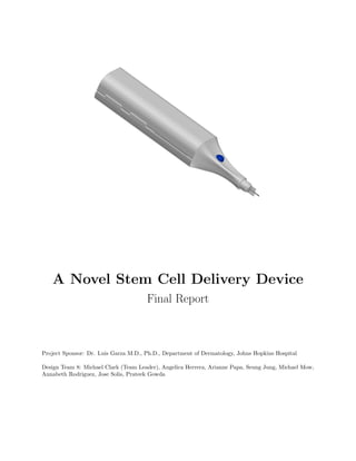
  • 1. A Novel Stem Cell Delivery Device Final Report Project Sponsor: Dr. Luis Garza M.D., Ph.D., Department of Dermatology, Johns Hopkins Hospital Design Team 8: Michael Clark (Team Leader), Angelica Herrera, Arianne Papa, Seung Jung, Michael Mow, Annabeth Rodriguez, Jose Solis, Prateek Gowda
  • 2. Contents 1 Abstract 3 2 Introduction 4 2.1 Clinical Problem . . . . . . . . . . . . . . . . . . . . . . . . . . . . . . . . . . . . . . . . . . . 4 2.2 Clinical Need . . . . . . . . . . . . . . . . . . . . . . . . . . . . . . . . . . . . . . . . . . . . . 4 2.3 Solution and Design Specifications . . . . . . . . . . . . . . . . . . . . . . . . . . . . . . . . . 4 3 Design 6 3.1 Design Process . . . . . . . . . . . . . . . . . . . . . . . . . . . . . . . . . . . . . . . . . . . . 6 3.2 Device Overview . . . . . . . . . . . . . . . . . . . . . . . . . . . . . . . . . . . . . . . . . . . 6 3.3 Base Station . . . . . . . . . . . . . . . . . . . . . . . . . . . . . . . . . . . . . . . . . . . . . 7 3.4 Cell Thawing Subsystem . . . . . . . . . . . . . . . . . . . . . . . . . . . . . . . . . . . . . . . 7 3.5 Peristaltic Pump Subsystem . . . . . . . . . . . . . . . . . . . . . . . . . . . . . . . . . . . . . 8 3.6 Closed-Loop Cell Pathway . . . . . . . . . . . . . . . . . . . . . . . . . . . . . . . . . . . . . . 8 3.7 Cryobag Freezing Mold . . . . . . . . . . . . . . . . . . . . . . . . . . . . . . . . . . . . . . . 8 3.8 Power Systems . . . . . . . . . . . . . . . . . . . . . . . . . . . . . . . . . . . . . . . . . . . . 9 3.9 Angle and Depth Guards . . . . . . . . . . . . . . . . . . . . . . . . . . . . . . . . . . . . . . 9 4 Results 10 4.1 Post-Injection Cell Viability Testing . . . . . . . . . . . . . . . . . . . . . . . . . . . . . . . . 10 4.1.1 Methods . . . . . . . . . . . . . . . . . . . . . . . . . . . . . . . . . . . . . . . . . . . . 10 4.1.2 Testing and Results . . . . . . . . . . . . . . . . . . . . . . . . . . . . . . . . . . . . . 10 4.2 Cell Thawing System Testing . . . . . . . . . . . . . . . . . . . . . . . . . . . . . . . . . . . . 11 Appendix A Materials 12 Appendix B Prototype Budget 13 Appendix C Figures, Photos, and Sketches 14 C.1 Injector . . . . . . . . . . . . . . . . . . . . . . . . . . . . . . . . . . . . . . . . . . . . . . . . 14 C.2 Base Station . . . . . . . . . . . . . . . . . . . . . . . . . . . . . . . . . . . . . . . . . . . . . 14 C.3 Motor and Driver . . . . . . . . . . . . . . . . . . . . . . . . . . . . . . . . . . . . . . . . . . . 15 C.4 Onboard Computer . . . . . . . . . . . . . . . . . . . . . . . . . . . . . . . . . . . . . . . . . . 15 C.5 Microcontroller . . . . . . . . . . . . . . . . . . . . . . . . . . . . . . . . . . . . . . . . . . . . 15 C.6 Thermocouple . . . . . . . . . . . . . . . . . . . . . . . . . . . . . . . . . . . . . . . . . . . . . 16 C.7 Cryobag Freezing Mold . . . . . . . . . . . . . . . . . . . . . . . . . . . . . . . . . . . . . . . 16 C.8 Cryobag . . . . . . . . . . . . . . . . . . . . . . . . . . . . . . . . . . . . . . . . . . . . . . . . 16 C.9 Luer Lock Connector . . . . . . . . . . . . . . . . . . . . . . . . . . . . . . . . . . . . . . . . . 17 C.10 Needle . . . . . . . . . . . . . . . . . . . . . . . . . . . . . . . . . . . . . . . . . . . . . . . . . 17 C.11 Power Delivery Schematic . . . . . . . . . . . . . . . . . . . . . . . . . . . . . . . . . . . . . . 17 C.12 Microcontroller Code . . . . . . . . . . . . . . . . . . . . . . . . . . . . . . . . . . . . . . . . . 18 C.13 Heating System Performance Data . . . . . . . . . . . . . . . . . . . . . . . . . . . . . . . . . 19 C.14 Viability Testing Data . . . . . . . . . . . . . . . . . . . . . . . . . . . . . . . . . . . . . . . . 19 Appendix D Calculations 20 D.1 Volume Resolution Calculation . . . . . . . . . . . . . . . . . . . . . . . . . . . . . . . . . . . 20 D.2 Tubing Dead Space Calculation . . . . . . . . . . . . . . . . . . . . . . . . . . . . . . . . . . . 20 Appendix E References 21 Appendix F Proposal 22 2
  • 3. 1 Abstract Recent advancements in stem cell therapies have shown the potential to revolutionize the treatment of many conditions. However, there is still a need to consistently and accurately deliver stem cells to target regions in the solid organs of the body–particularly the skin. Skin stem cell therapies currently under investigation have the potential to reverse hair loss, heal wounds, and alter the phenotype of the epidermis. We have designed a device to deliver stem cells to the skin at adjustable volumes with minimal viability loss or risk of contamination. The device makes use of a peristaltic pump to provide precise control over injection rate, volume, and the resulting shearing forces. Cell viability is also improved through an automated heating system that thaws the cells at an expeditious but controlled rate immediately prior to injection. The entire cell pathway is a closed loop, which minimizes the risk of contamination. Cryobags were utilized to increase the volume of cells available to the physician administering the therapy. Interchangeable angle and depth guards were designed to improve the consistency and repeatably of injections. In preparation for ex vivo testing, an exploratory in vitro test was performed on the injection mechanism using fibroblast cells. The thermal performance of the heating system was also evaluated experimentally. 3
  • 4. 2 Introduction 2.1 Clinical Problem Stem cell therapies are an expanding market in regenerative medicine. The global stem cell industry has grown 13.6% annually from $5.6 billion in the year 2013 [1]. Currently, 4,681 clinical trials involving stem cell therapies are being carried out in the United States with over 3,000 in early phases of testing [2]. The NIH estimates spending approximately $2.77 billion on stem cell research in 2015 [3]. Public funding from individual states will total over $4.1 billion by 2018 [4]. As thousands of therapies advance past initial stages of testing, there will be an increased demand for a safe and effective way to deliver the stem cells to patients. Current stem cell therapies for the skin involve altering the characteristics of dermal cells to treat wounds, rashes, and burns. In the United States, the treatment of wounds and associated complications exceeds $20 billion annually [5]. Existing treatments for these conditions are prohibitively expensive; skin allograft therapies typically cost $500,000 per patient [6]. Skin stem cell therapies also have cosmetic applications such as regenerating hair follicles, repairing scar tissue, and changing the phenotypic expression of the skin. Today, physicians have difficulty delivering stem cells to the skin in a consistent manner. Providers rely on tactile feedback and previous experience to deliver the cells to the desired location. Subjectivity and variance are inherent in the current treatment administration procedure. In order to generate useful clinical trial data, the stem cell delivery procedure needs to be optimized to be as consistent and repeatable as possible. A primary concern is ensuring post-injection cell viability. If cells are injected too quickly, shearing forces in the needle can tear them apart. Another concern is contamination: in the current procedure, cells are exposed to air when they are transferred from the freezing vessel to the syringe. This can lead to cell contamination and an increased risk of post-treatment infection. As a result of these complications, many existing dermal injectors cannot be adapted to this highly specialized application. 2.2 Clinical Need No devices on the market are specifically designed to mitigate the difficulties associated with delivering stem cell therapies to the skin. Consequently, there is a need for a device that allows physicians to deliver stem cells to target dermal regions at adjustable volumes with minimal risk of contamination or viability loss. 2.3 Solution and Design Specifications The team’s dermal injector addresses the biological hurdles associated with stem cell delivery by providing adjustable injection rates, integrated cell thawing capabilities, and a closed-loop delivery system that reduces the risk of contamination. Stem cells are commonly stored and frozen in cryogenic vials, which typically hold a one to two milliliter volume. The team will make use of cryogenic bags, which can hold volumes ranging from five to ten milliliters. This reduces the number of times the physician has to reload the device – effectively decreasing procedure time and air exposure, thus reducing the risk of contamination. The cryobag will be connected to a cartridge-tubing system and a needle to form the closed-loop system. The physicians will place the cartridge onto a peristaltic pump, which will accurately output desired volumes with an accuracy of ±1 µl and physician-specified rates for each injection averaged at one minute per injection. To further enhance cell viability, the team spoke with Dr. Luis Garza, M.D., Ph.D., who is the sponsor and medical adviser for the team, and found that a heating system should be included as well. Several other possibilities why a new device might be better than a syringe might be the problem of temperature, where we know that cells will likely be shipped to a consumer frozen. But frozen cells lose viability or thaw too slowly. If they are thawed more quickly, then viability is maintained better, so a device that eliminates user variability in terms of quick thawing rate could also improve final outcome for cellular therapies. 4
  • 5. An integrated cell thawing system was designed that heats frozen stem cells at a consistent rate, which dramatically improves cell viability [7]. In order to achieve this, the frozen stem cells must be heated from −196 ◦ C, the temperature of the liquid nitrogen, to 37 ◦ C, the average temperature of the human body, in under two minutes per 1.0 ml thawed, which is the current standard established from a traditional water bath. The dermal injector will be kept in its base station when not in use. This docking station will charge the device and will be used to adjust device settings. Every system in the dermal injector will be controlled electronically. The physician will only have to input his or her desired injection volume and injection rate, and then start the thawing process using a touch screen user interface on the base station. This LCD touch screen will indicate when cell thawing is complete and alert the physician when the device is ready to use. This simplified procedure is to ensure that this method is no more difficult than the current standard of care. 5
  • 6. 3 Design 3.1 Design Process The team conducted months of extensive research to gain a thorough understanding of the market. An intellectual property search was conducted, which revealed ample room to innovate as there were no other devices that delivered stem cells to the dermis. This meant that the team had no competitors, but also had no predicate devices upon which to improve their design. Therefore, the group decided to expand the search to include devices that delivered stem cells to other organs of the body. However this also revealed a lack of devices and the team switched to drug delivery methods instead. As a result, the team developed four promising devices that could have been adapted to deliver stem cells. The first idea was a direct stem cell insertion. This method was considered as it was the simplest way to get stem cells from point A to point B. The stem cells would be cultured in a laboratory setting into a cell matrix. The method would then require an operation where the patient’s skin was cut and pulled back, the cell matrix inserted into place, and then skin sutured back on. Although this method was the most direct treatment, it was also the most invasive. The team thought it best to avoid operations that required a lengthy recovery time and continued to seek less invasive methods of delivery. The next idea was an injection gun. This was by far the fastest way the team found to deliver a treatment. This device penetrated the skin with the aid of pressurized gas, which evenly distributed the drug. However, this method raised concerns as the velocity of injection was deemed detrimental to the viability of the cells. In addition, the location that the stem cells were delivered also raised concerns as the cells traveled too deep into the dermis and passed its optimal location. From this idea, the team determined that a slow consistent rate of injection was a vital part of the device as it ensured that the cells maintained a <40% reduction in viability. The use of a microneedle array was one of the best methods to deliver drugs at the optimal location. This device consisted of a series of microneedles that were able to deliver drugs at consistent rates. However, the team found that a small needle gauge would damage the cells and lower cell viability as it passed through the needle. Though this method had a consistent flow rate, the team determined that the correct needle gauge was just as important. After an extensive literature search, the team pursued a 23 gauge needle, which was found to be the most optimal size for their objective as it was big enough for the cells to travel the needle without much shearing, but small enough to penetrate the skin without the pain associated with bigger needles.8 The final idea was an insulin pen injector. Similar to a needle and syringe, this device simplified the delivery process. The patient would set the volume he or she needed and then press a button to deliver the treatment. Using this method as a springboard, the team adapted this simple design and incorporated all the features that the team determined was vital from the previous ideas: minimal invasiveness, consistent flow rate, use of a 23 gauge needle, and adjustable volume concentration from the pen injector. From there, the team generated ideas and selected designs that yielded the best treatment outcomes and per-dollar performance. After soliciting feedback from experts, the team modified the device to include the elements described in §3.2. 3.2 Device Overview The device is a hand-held electronic cell injection system (Appendix C.1). The injector itself is cordless – it docks in a powered base station, which allows the device to be charged and programmed by the user (Appendix C.2). The injector has four discrete subsystems: an automated cell thawing system with a temperature feedback mechanism, a peristaltic pump system that offers precise control over injection rate and volume, a closed-loop cell pathway, and a power delivery system. A sealed septum separates the electronic systems from the “wet” cellular pathway, which effectively mitigates the risk of electrical shock in the case of a breach in the closed-loop system. The injector device is designed to be held like a computer mouse, with the pointer finger resting on the injection button. This position provides a great deal of stability to the physician administering the therapy. 6
  • 7. The long tip of the device allows clinicians to achieve very shallow injection angles. The tip also accepts interchangeable angle and depth guards. The base station houses an onboard computer (Raspberry Pi Model B+) and a touch-screen user interface. A bag freezing mold was also developed that facilitates increased heat transfer from the heating pads to the cryobag. 3.3 Base Station The base station (Appendix C.2) was developed as a means of improving physician usability by utilizing a large full color capacitive LCD touchscreen with a user-friendly graphic user interface (GUI), which allows the physician to set the parameters of the injection easily. After the physician attaches the cell cryobag cartridge to the device, the program switches on and prompts the physician to enter the desired injection volume of stem cells After the physician inputs the information, the data is sent to an internal computer inside the base station. The Raspberry Pi was utilized as the internal computer (Appendix C.4) due to its compatibility with touch screen interfaces and its ability to communicate with our specific microcontroller, the Arduino Micro (Appendix C.5). The internal computer carries out calculations from the selected volume of injection in order to determine the time necessary for cell thawing, while automatically setting the injection rate. Inside the handheld device, The data computed by the internal computer is programmed onto the microcontroller in the device via contacts between the base station and the resting device. The Arduino carries out the heating process and signals when the process is complete. The screen then prompts the physician to remove the device from the base station and begin the injection. 3.4 Cell Thawing Subsystem The heating system functions to thaw the stem cells quickly, but in a controlled manner that prevents the risk of destroying the cells due to overheating. Thawing cells quickly helps reduce the risk that the cells will be damaged by ice crystals that are present during the thawing process. Literature also suggests that rapid thawing reduces protein damage [8]. The heating process takes place while the device is resting in the base station. The physician places the cryobag cartridge into the device and selects the heating option on the base station user interface. The cells are thawed by two heating pads that envelope the cryobag. Due to the large surface area of the cryobag and thin distribution of the cells in the bag itself, the heating is uniform along the cells in the bag. The heating pads maintain a constant temperature of 37.0 ◦ C. This is the ideal temperature of the cells inside the human body. It is important the heating pads do not exceed this temperature, as it leads to potential cell death and an overall loss in cell viability. To achieve constant temperature control, the system utilizes a negative feedback loop and control systems to provide safe heating of the cells. The heating subsystem is comprised of two thin parallel heating pads constructed using a mesh of polyester filament and conductive fiber folded into a protective Polyimide Film. The fact that these are low power, flexible and draw little power makes them ideal for things like hand-warmers and other heated garments. The current source for these heating pads is controlled and obtained from a regulated DC power supply provided by a grounded wall outlet. The circuit is regulated by an NPN transistor switch that can be opened and closed based on feedback from data obtained by a thermocouple (Appendix C.6). The thermocouple concurrently sends the temperature of the heating pads as digital data to an Arduino Micro (Appendix C.5) that then regulates the switching of the transistor (See code in Appendix C.12). This transistor was used as a switching mechanism over other options, such as mechanical relays for example, because of its solid state design. This allows the team to utilize power with modulation techniques to rapidly switch the current to more safely control the heating of the pads. Using this method, the pads will heat rapidly at lower temperatures and slowly as the temperature approaches the maximum of 37.0 ◦ C. Once the cells have thawed, the heating has been completed, at which time the physician will be signaled to remove the device from the base station and prepare for injection. 7
  • 8. 3.5 Peristaltic Pump Subsystem The pump system is designed to control the stem cell injection rates and reduce viability losses due to shearing forces in the needle. The user interface for the pumping process is comprised of two mechanical buttons on the device, a clear-air button and an injection button. The purpose of the clear-air button is to remove excess air in the tubing and replace it with the cell from the cryobag. This is performed at the discretion of the physician and is analogous to clearing air bubbles in the common needle and syringe. The clear-air function pumps at a higher rate than the injection button. The injection button, when toggled by the physician, pumps the stem cell solution out at a slow, constant rate to maximize cell viability and reduce shearing forces. Testing will be carried out to determine the ideal rate of injection. To perform the injection, the physician inserts the needle into the patient and holds the injection button. The pump system is comprised of a peristaltic pump head that is rotated by a small four-wire unipolar stepper motor (Appendix C.3). Peristaltic pumps work by pushing and collapsing the walls of a flexible tubing material to create a vacuum and a source of suction, that when rotating along the tubing walls, draws out the cells from the cryobag into the tubing. This method prevents contact between the cells and any external pumping mechanism, preventing contamination during the injection. A stepper motor is utilized to turn the peristaltic pump and control the rotation without using a separate mechanical or optical encoder. Removing the encoder prevents bulky attachments, reduces the weight and controls space within the device. Instead, stepper motors work by moving in steps that are designated by the Arduino Micro, which eliminates a need for a negative feedback loop.The motor is geared to afford a resolution of 1024 turns per revolution, which gives a pumping resolution of 0.0455 µl based on the size of the tubing and pump head radius (Appendix D.1). This high resolution and controlled pump system ensures that even small volumes of stem cell solution can be injected without the device. 3.6 Closed-Loop Cell Pathway The closed-loop cell pathway is comprised of four components: the cryobag, tubing, needle, and Luer-lock connectors. Cryobags were chosen over more traditional cryovials because they are able to contain a larger (and more clinically appropriate) volume of cells - this decreases the frequency with which the physician has to halt the procedure to load more cells into the device. OriGen Biomedicals PermaLife Cell Culture Bag (Appendix C.8) was selected for the device. The bag is made from biologically-inert Fluorinated ethylene propylene (FEP), which offers an operational temperature range of −196 ◦ C to 137 ◦ C permitting the bag to be frozen in liquid nitrogen and sterilized by autoclave. Unlike the cryobag, the tubing will not be subjected to extreme temperatures. Clear silicone tubing made from FDA-compliant resins (McMaster-Carr 5236K501) was selected for its plyability and ability to be sterilized by autoclave. The inner diameter (ID) and length of the tubing was minimized (794 µm) in order to reduce dead space losses, i.e. the volume of cells required to fill the tubing that become unavailable for injection. In the current design, less than 76 µl of dead space exists in the tubing (Appendix D.2). Hypodermic needles are widely available in health care settings, so the needle selection was guided by industry standards. Becton, Dickinson, and Company (BD) has a significant share of the hypodermic needle market and is therefore a suitable supplier, although a wide variety of manufactures sell needles that can be used with the device. The device will accept any needle with a Luer-lock connector - various gauges, lengths, and bevels are available to suit a variety of procedures. A 23 gauge needle (Appendix C.10) was utilized for testing purposes since it is commonly used in intradermal injection procedures. All interfaces between the cryobag, tubing, and needle feature a Luer-lock type connector (Appendix C.9). Luer-lock is a commonly used and ISO-standard fitting that will be familiar to physicians. Luer-lock connectors were chosen over Luer-slip connectors due to their ability to withstand higher injection pressures. 3.7 Cryobag Freezing Mold Maximum heat transfer is achieved when the contact area between the heating pad and cryobag is maxi- mized. In order to increase the contact area, freezing the cryobag at a uniform (flat) thickness works to 8
  • 9. the physician’s advantage. A cryobag freezing mold (Appendix C.7, isometric drawing) was developed as a means to that end. The mold allows two 10 ml cryobags to be frozen flat inside of a standard 135 mm by 135 mm cryobox. Polylactide (PLA) was used to prototype the mold, but the final product will implement a low-temperature polymer capable of thermocycling from room temperature to liquid nitrogen temperatures (e.g. polypropylene). The cryobag sits in between the two halves of the mold. Stainless steel springs will be employed to apply pressure on both sides, gently compressing the bag in the middle. 3.8 Power Systems Two systems provide power to the device subsystems (Appendix C.11). A power supply in the base station transforms, rectifies, filters and regulates 120V AC current to 5.1V DC at 2.1A. The 5.1V rail is used to power the onboard computer in the base station, charge the 12V battery in the injector, and operate a NPN transistor along with the Arduino. The power supply is controlled by a switch located on the back of the base station. The second power source is the 12V battery in the injector, which allows the device to be operated wirelessly when undocked from the base station. The battery supplies the power necessary to run the onboard microcontroller that regulates the pump motor and thermocouple feedback system. 3.9 Angle and Depth Guards A selection of nine angle and depth guards were designed. The guards clip on to the tip of the device. Three clinically relevant angles (90◦ , 45◦ , and 15◦ ) and three physiologically relevant depths (intradermal, subdermal, and intramuscular) were represented. The guards are made from clear plastic so that physicians can visualize the injection site while they administer the cellular therapy. 9
  • 10. 4 Results 4.1 Post-Injection Cell Viability Testing 4.1.1 Methods After finalizing each component of the stem cell injector, preliminary testing for the two major subsystems of the device was performed. Both the closed-loop cell injection system and automated cell thawing system (and feedback mechanism) were examined. Literature suggests that cell death occurs when cells are exposed to high levels of pressure or shear force, specifically above 1.0 Pascal [9]. It has been shown that viability losses up to forty percent can occur when cells are injected through a needle (size) and syringe(rate, cell density, media (PBS in this case)) due to shearing forces [10]. Preliminary tests were designed to correlate post-injection cell viability to injection rate through a 23 gauge needle. A 23 gauge needle was chosen since this is the most commonly used needle size for cell viability testing and has clinical relevance. A needle of this size is optimal for the balance between a low pain level for the patient and a large enough width to maintain cell viability [11]. Also, a small diameter allows precise regions, such as the dermoepidermal junction, to be reached. Injection rates of 6.0 mL/min, 3.0 mL/min, 1.0 mL/min and 0.5 mL/min were chosen based on physician feedback and literature13. The fastest injection rate (6.0 mL/min) was used to simulate high shear forces through the needle. An injection rate of 3.0 mL/min mimicked a physicians typical injection speed. Slower injection rates (1.0 mL/min) were used in other viability studies and used to simulate the lowest amount of shear forces through the needle (0.5mL/min).13 4.1.2 Testing and Results Initial viability testing was performed with mouse spleen cells due to their relative availability. Cells were obtained from Dr. Luis Garzas laboratory at Johns Hopkins Hospital. Mouse spleen cells were harvested, filtered with PBS through a mesh netting to isolate the cells, and centrifuged at 1000 RPM for 5 minutes. The cells were then resuspended in Phosphate-buffered saline (PBS). In order to determine cell density, 10 uL of the cell solution was mixed with 10 uL of trypan blue and placed on a hemocytometer slide. Using a Countess Automated Cell Counter, the number of live cells was approximated. However, the countess did not provide clear estimates for cell density. This could be due to the fact that red blood cells were not separated from the spleen cells. In future tests, the red blood cells can be lysed and then washed away using ACK Lysing Buffer (Life Technologies, A10492-01). Additional testing was performed with volar fibroblasts biopsied from a patients sole. Fibroblasts were acquired from Dr. Luis Garzas laboratory at Johns Hopkins Hospital (IRB NA 00068684) and cultured in Dulbecco’s Modified Eagle Medium (DMEM) for one week before experimentation. Due to the short expansion time, cells were only used at a density of 3.0 × 10⁵ cells/mL. Injection testing was performed using a 23 gauge needle and syringe. For the 6.0 ml/min rate, the injection was performed manually to model the accuracy and repeatability of a physician. For the slower rates (3.0, 1.0, and 0.5 ml/min) a syringe infusion pump was used to provide consistent injection rates. In each trial, 300 µl of the cell solution were loaded into the 1.0 ml syringe by hand. Using the infusion pump (or by hand in the 6.0 ml/min trial), 100 µl of the solution were injected into a 1.0 ml vial at a predetermined controlled rate. The ejected cells were then placed on ice. Three trials were conducted for every injection rate. To serve as a control, 100 µl of cell solution were drawn up manually into the syringe and injected into a 1.0 ml without using a needle. After completing all injections, 10 µl from each 1.0 ml vial were drawn up with a pipette and placed in a new 1.0 mL vial with 10 µl of trypan blue. After mixing, 10 µl of the solution were pipetted onto a hemocytometer slide and imaged. No losses in viability were seen during this experiment; all visible cells appeared viable with intact membranes and no dark stains from the trypan blue (Appendix C.14). However, there was much variation between cell densities in each hemocytometer image. Across different injection rates, there did not seem to be a constantly increasing or decreasing trend in the number of cells present. Additionally, between injections of the the same rate, results would vary from no visible cells to large inordinate amounts of cells. As a 10
  • 11. result, standard deviation values from these injection rates tend to be greater than the average of observable cells within that rate. For example, the trials for the 1.0 ml/min injection rate were: 0 cells visible, 1 cell, and 61 cells visible. This results in an average of 20 cells visible in this injection rate and a standard deviation of 35 cells. These high standard deviation numbers made any quantifying results inconclusive or at least statistically invalid. In the future, cell solutions will be mixed thoroughly before pipetting onto the hemocytometer slide to ensure homogeneity throughout the solution. A 24 hour time point will be used as well to see if viability losses are seen over time and not immediately after injection. By using trypan blue, cell death will only be seen once the cell membrane is lysed. This may not occur immediately since cells may only initially be damaged. Revised testing is currently underway at Johns Hopkins University Homewood campus. Mouse fibrob- last (L929, P3 11334) cells were acquired from Dr. Elizabeth Logsdon. The cells, which were originally frozen at ninety percent confluence were thawed quickly and cultured in DMEM (10% FBS with Penicillin- Streptomycin (Sigma-Aldrich P4333, 5:500) for four days prior to testing. Before experimentation, cell density was determined using standard cell counting procedures and a hemo- cytometer slide. Dimethyl sulfoxide (DMSO) was added to the cell solution to mimic the conditions in Dr. Garzas clinical trial. It is expected that DMSO will make the cells more susceptible to shearing forces and viability losses [12]. Many studies have demonstrated reduced cell viability in a dose-dependent manner. Although DMSO is used to protect cells while frozen, they may induce some cytotoxicity when they are thawed due to permeability [13]. The previous procedure using fibroblasts was slightly modified for this experimentation. Injections were performed directly into 24 well plates for easier imaging. An identical second well plate was used for the 24 hour time point. Injections were repeated three times, either by hand or with the infusion pump, per injection rate. Trypan blue was added to the wells before imaging. 4.2 Cell Thawing System Testing The automated thawing system and feedback loop were thoroughly tested and examined. Before use in the clinical setting, stem cells are frozen in liquid nitrogen and stored. When needed, the physician removes and thaws these vials before injection. Cells can withstand a maximum temperature of 37 ◦ C, any higher and they face the potential of cell death [14]. To maintain a consistent thawing procedure, the in device heating capabilities were examined. Testing the heating component of the dermal injector was vital to the device. To ensure a maximum temperature of 37 ◦ C, the heating pads were connected to a thermocouple, recording the temperature during the experiment. Along the heating circuit, these pads were controlled by a switching system and power supply. The thermocouple created a negative feedback loop, allowing us to reach a maximum temperature of 37 ◦ C. The arduino read the temperature from the thermocouple and controlled the switch along the heating circuit. The test was carried out with a supply voltage of 1.1V to the collector input of the transistor. The feedback program on the microprocessor utilized power width modulation techniques in three stages to regulate current flow to the heating pads. Rapid heating occurred when the temperature of heating pads were below 30 ◦ C. The temperature then increased slowly between the period of 30 ◦ C-37 ◦ C. Once the pads had reached 37 ◦ C, it began to oscillate along this target temperature, with maximum at 37.6 ◦ C (Appendix C.13). The thermocouple feedback system, along with the PWM technique used in the programming, demon- strated the ability to control temperature with 1.0 ◦ C precision from the 37 ◦ C target temperature. Im- provements can be made in the software by increasing the reading rate of the program. In this test, the temperature was taken at a rate of one reading every 250 milliseconds. Increasing this rate would increase the number of times that the temperature is managed, resulting in smaller fluctuations along the target temperature and a reduction in time between heating and cooling cycles. 11
  • 12. A Materials System Component Description Source Item Number Base Station Outer Shell PLA Makerbot Indus. White PLA Computer Raspberry Pi Model B+ Raspberry Pi Model B+ Power Button SPST (Round) Sparkfun COM-11138 LCD Screen 2.8-Inch, TFT, 320x240 Adafruit Indus. 1601 Power Cord 18AWG, 72”, Black Digi-Key Elec. 221001-01 Contacts Universal 1.8MM SMD Digi-Key Elec. 1003-1010-2-ND USB/Data Cable Adafruit 70 Device Outer Shell PLA Makerbot Indus. White PLA Microcontroller Arduino Micro Arduino A000053 Buttons Tactile Button Assortment Sparkfun COM-10302 LEDs Blue LED Digi-Key Elec. 160-1602-ND Pins Stainless Steel McMaster-Carr 6517K65 Springs Compression, Steel Conical McMaster-Carr 1692K11 Stepper Motor 12V Adafruit 918 Motor Driver Lighted Texas Instruments ULN2003ADR Heating Pads Flexible Sparkfun COM-11289 Battery 12V Enegizer A23 Transistor General Purpose Transistor ON Semiconductor PN2222 Thermocouple K-type Adafruit Indus. 270 Transducer For K-type Thermocouple Adafruit Indus. 269 Resistors 10KΩ, 1KΩ Digi-Key Elec. CF14JT10K0, CF14JT1K50 Cell Pathway Tubing For Peristaltic Pumps McMaster-Carr 5236K501 Needle 23 Gauge (for testing) Becton & Dickenson 305148 Cryobag 10 ml OriGen PL07 Leur Lock 0.8mm ID Barb Qosina 11106 Freezing Mold Mold PLA Makerbot Indus. Black PLA Springs Conical, Stainless Steel McMaster-Carr 1692K12 Angle Guards Guard Clear, Polycarbonate McMaster-Carr 8585K51 12
  • 14. C Figures, Photos, and Sketches C.1 Injector C.2 Base Station 14
  • 15. C.3 Motor and Driver C.4 Onboard Computer C.5 Microcontroller 15
  • 16. C.6 Thermocouple C.7 Cryobag Freezing Mold C.8 Cryobag 16
  • 17. C.9 Luer Lock Connector C.10 Needle C.11 Power Delivery Schematic 17
  • 19. C.13 Heating System Performance Data C.14 Viability Testing Data Figure 1: Cell viability testing using sole fibroblasts from Dr. Luis Garzas laboratory. Variability in cell density is seen between experimental and control groups when injected onto a hemocytometer slide and imaged a) Injection rate of 1.0 mL per minute and b) control group injected by hand without a needle. 19
  • 20. D Calculations D.1 Volume Resolution Calculation Stem cell therapies are often carried out with very small injection volumes, <10 ml. It is important, therefore, that the device is able to provide pumping resolutions small enough and with enough precision for any stem cell therapy that the physician might carry out. Volume resolution is characterized as the smallest volume that the device can inject with one motion of the pump. In our calculations, volume resolution was calculated as a function of the radius of the peristaltic pump head, the step resolution of the peristaltic pump, and the interior diameter of the tubing. The manufacturer’s specification sheet shows an interior tube diameter of 0.0794 cm, while the peristaltic pump head is designed to be a half circle of a 1.5 cm radius. Therefore in one full rotation of the pump head, we calculate an output of 0.0466 ml of solution. The 1/64th geared stepper motor has a full 1024 steps per rotation. Therefore there is a calculated volume resolution of 0.0455 µl. D.2 Tubing Dead Space Calculation As stated in Appendix D.1, the cross-sectional area of the tubing is: A = π×r2 = π× .0794 2 2 = .0049514cm2 Approximately 15.24 cm of tubing is used in the device. The tubing dead space is therefore: V = A×l = .0049514 ∗ 15.24 = .0754cm3 The total dead space is therefore 75.46 µl. 20
  • 21. E References [1] “Global Market for Stem Cells to Reach $10.6 Billion in 2018.” The Global Market for Stem Cells. BCC Research, July 2014. Web. 19 Jan. 2015. [2] “Search Results.” Search Of: Stem+cell. ClinicalTrials.gov, n.d. Web. 19 Jan. 2015. [3] “Estimates of Funding for Various Research, Condition, and Disease Categories (RCDC).” NIH Cate- gorical Spending. NIH, 7 Mar. 2014. Web. 19 Jan. 2015. [4] “Embryonic Stem Cell Research by the Numbers.” Center for American Progress, 17 Apr. 2007. Web. 19 Jan. 2015. [5] Chen, Ming, Melissa Przyborowski, and Francois Berthiaume. Stem Cells for Skin Tissue Engineering and Wound Healing. Critical reviews in biomedical engineering 37.4-5 (2009): 399-421. Print. [6] Clark, R.A. Ghosh, K. Tonnesen, M.G. (2007). Tissue engineering for cutaneous wounds. J Invest Dermatology. 127, 1018-1029.10.1038/sj.jid.5700715 [7] Finucane, M. L., & Williams, A. E. (2011). Psychosocial and cultural factors affecting judgments and decisions about translational stem-cell research. In Translational stem cell research: Issues beyond the debate on the moral status of the human embryo (pp. 391-398). Totowa, NJ: Humana Press. [8] Cao, E., Chen, Y., Cui, Z. and Foster, P. R. (2003), Effect of freezing and thawing rates on denaturation of proteins in aqueous solutions. Biotechnol. Bioeng., 82: 684690. [9] Pagn R, Mackey B. Relationship between Membrane Damage and Cell Death in Pressure-Treated Es- cherichia coli Cells: Differences between Exponential- and Stationary-Phase Cells and Variation among Strains. Applied and Environmental Microbiology. 2000;66(7):2829-2834. [10] Aguado BA1, Mulyasasmita W, Su J, Lampe KJ, Heilshorn SC. Improving viability of stem cells during syringe needle flow through the design of hydrogel cell carriers. Tissue Eng Part A. 2012 Apr;18(7-8):806-15. doi: 10.1089/ten.TEA.2011.0391. Epub 2011 Dec 20 [11] Walker PA, Jimenez F, Gerber MH, Aroom KR, Shah SK, Harting MT, Gill BS, Savitz SI, Cox CS., Jr Effect of needle diameter and flow rate on rat and human mesenchymal stromal cell characterization and viability. Tissue Eng Part C Methods. 2010;16:989997. [12] Aguado BA1, Mulyasasmita W, Su J, Lampe KJ, Heilshorn SC. Improving viability of stem cells during syringe needle flow through the design of hydrogel cell carriers. Tissue Eng Part A. 2012 Apr;18(7-8):806-15. Epub 2011 Dec 20 [13] Chen, X., & Thibeault, S. (2013). Effect of DMSO Concentration, Cell Density and Needle Gauge on the Viability of Cryopreserved Cells in Three Dimensional Hyaluronan Hydrogel. Conference Proceedings: An- nual International Conference of the IEEE Engineering in Medicine and Biology Society. IEEE Engineering in Medicine and Biology Society. Conference, 2013, 62286231. [14] Finucane, M. L., & Williams, A. E. (2011). Psychosocial and cultural factors affecting judgments and decisions about translational stem-cell research. In Translational stem cell research: Issues beyond the debate on the moral status of the human embryo (pp. 391-398). Totowa, NJ: Humana Press. 21
  • 22. F Proposal Document begins on the next page. 22
  • 23. Design Team 8 Project Proposal A Novel Stem Cell Delivery Device Michael Clark, Angelica Herrera, Arianne Papa, Michael Mow, and Jack Jung Project Sponsor: Dr. Luis Garza∗ Revision: September 30, 2014 An intradermal injection. Source: Novosanis. Abstract To facilitate consistent and accurate placement of skin stem cells at the dermoepidermal junction, we plan to design, develop, and test a novel intradermal stem cell delivery device. Although this delivery device has a wide range of applications in the field of intradermal cellular therapies, the development of the device will be examined within the context of an existing clinical trial taking place at Johns Hopkins Hospital: an investigational stem cell treatment for amputees that aims to initiate growth of palmoplantar skin on amputees’ stumps. The device will be designed to deliver stem cells at adjustable depths and volumes as determined by the physician administering the therapy. The nature of the clinical need requires the device to effectively deliver cells at a wide variety of locations. The device will deliver the cells in such a way as to maximize cell viability and treatment sterility. Patient comfort and device cost will also be considered and optimized. The device will be tested in vitro in a variety of skin models, and eventually in vivo as part of the aforementioned clinical trial. Valuable subjective feedback will also be obtained through the use of several IRB-approved surveys. ∗Assistant Professor, Johns Hopkins Medicine Department of Dermatology, lag@jhmi.edu
  • 24. Contents 1 Clinical Background 3 2 Clinical Trial 4 3 Standard of Care 4 4 Clinical Problem 5 5 Clinical Need 5 6 Project Goals and Design Constraints 5 6.1 Project Goals . . . . . . . . . . . . . . . . . . . . . . . . . . . . . . . . . . . . . . . . . . . 6 6.2 Design Constraints . . . . . . . . . . . . . . . . . . . . . . . . . . . . . . . . . . . . . . . . 7 7 Existing Solution Landscape 8 7.1 Technologies . . . . . . . . . . . . . . . . . . . . . . . . . . . . . . . . . . . . . . . . . . . . 8 7.2 Patents . . . . . . . . . . . . . . . . . . . . . . . . . . . . . . . . . . . . . . . . . . . . . . 10 References 11 A Microneedle Design Specifications 12 B Current Eight-Week Plan 14 2
  • 25. 1 Clinical Background Human skin is broken up into three main layers – the epidermis, the dermis, and the subcutaneous tissue (NCI, 2014). Figure 1 depicts the basic anatomy of the skin. The epidermis is the outermost layer, and its thickness varies by location. It is only 0.05mm thick on the eyelids, and is 1.5mm thick on the palms and the soles of the feet. Keratinocytes are cells found predominantly in the epidermis and primarily function as a barrier against the exterior environment. Figure 1: Basic anatomy of the skin. Image source: SEER, National Cancer Institute. In humans, the palmoplantar epidermis is a highly specialized tissue found on the palms and soles that expresses a wide range of keratins. This palmoplantar (“volar”) skin has different cellular properties compared to nonpalmoplantar skin. Figure 2 highlights these differences. This palmoplantar tissue is subjected to the highest degree of mechanical stress that the body is exposed to from extrinsic factors (Fu et al., 2013) Figure 2: Differences in cellular properties between volar/palmoplantar skin and non-palmoplantar skin. 3
  • 26. Keratin 9 (KRT9) is a protein that is found only in the palmoplantar epidermis of palms and soles. It has been found that KRT9 is required for terminal differentiation as well as maintaining the mechanical integrity and structural resilience of the palmoplantar epidermis. Since KRT9 is exclusively expressed in the palmoplantar epidermis, it can be used as a differentiation marker of palms and soles (Yamaguchi et al., 1999). Directly below the epidermis is the dermal skin layer, the thickest of the three layers of the skin (1.5 to 4.0mm). The dermis makes up approximately ninety percent of the total thickness of the skin. It consists of dermal fibroblasts that produce collagen and extracellular matrix components. These cells also generate connective tissue and allow the skin to recover from injury by aiding in wound healing. Derived from mesenchymal stem cells within the body, dermal fibroblasts can be further differentiated. Epithelial-mesenchymal interactions play a key role in the growth and differentiation of keratinocytes. Dermal fibroblasts are directly involved in regulating and controlling epithelial (keratinocyte) function by secreting diffusible growth factors (Coulomb, Lebreton, & Dubertret, 1989). It has been determined that KRT9 can be regulated by extrinsic signals from dermal fibroblasts (Yamaguchi et al., 1999). Since the dermal layer determines the phenotypic expression of the epidermal layer, there is great potential to use these cellular interactions to control the characteristics of the epidermal cells. In order to use epithelial- mesenchymal interactions to differentiate epidermal cells, it is necessary to have a full understanding of effective delivery techniques of stem cells to a target skin region for use in stem cell therapy. 2 Clinical Trial Dr. Luis Garza, a dermatologist at The Johns Hopkins Hospital, has been investigating the use of site- specific autologous fibroblasts to alter skin identity. It has been shown that skin identity can be altered by obtaining tissue samples from the desired skin type on the patient, culturing the tissue to expand the fibroblasts, and transplanting these cells into the target region on the patient (see Garza, et al. in Section 7.2). Garza’s new procedure for injecting fibroblast cells into the skin begins by culturing autologous fibroblast cells with keratinocytes obtained from the epidermis of human foreskin in vitro and freezing the cells in a solution of DMSO and hetastarch. In order to adhere to FDA regulations and to minimize contamination, the cells are then injected into the skin in the same solution in which they are frozen (Garza, 2014). Currently, injections are performed with a needle and syringe. The ideal injection location is thought to be at the dermoepidermal junction. Cells must remain sterile, viable, and as concentrated as possible within the tissue. Clinical trials of this procedure will begin in Fall 2014 (Garza, 2014). Garza’s treatment has the potential to be used for scars, discolored skin, rashes, ulcers, and alopecia. Currently, Garza is working on a clinical trial to inject fibroblast cells into the stump sites of amputees who wear prosthetic devices. The study aims to change the phenotype of the stump site skin from nonvolar to volar. This transformation will prevent skin degradation and provide a better skin-device interface. 3 Standard of Care In the United States, an estimated 185,000 people undergo an upper or lower limb amputation each year. Studies show that about 54% of these amputations arise from dysvascular disease, 45% due to trauma accounts, and less than 2% due to cancer (Ziegler-Graham, MacKenzie, Ephraim, Travison, & Brookmeyer, 2008). Around 50% of all amputees who wear a lower limb prosthesis report some type of skin degradation at their stump site due to their prosthetic device. In a study of 247 Vietnam War veterans with amputations, 48.2% of patients reported skin breakdown, 39% reported pressure ulcers, 25% reported infection, 25% of patients reported scars and wounds, 21.8% reported rashes, and 21% reported abrasion of the skin 4
  • 27. (Meulenbelt, Geertzen, Jonkman, & Dijkstra, 2011). These skin conditions are believed to be caused primarily by friction and mechanical trauma from the prosthesis. Many patients report that these skin complications lead to substantial pain and discomfort. Examples of these skin complications are depicted in Figure 3. The current standard of care for this problem involves using bandages on the stump site, or altering or replacing the prosthesis. When those treatments fail, prosthetic abandonment can occur. Figure 3: Common complications arising from pros- thetic use. Image source: Procellera. Common interventions include the use of antibiotics for superinfection, emollients and topical corti- costeroids for contact dermatitis, and surgery for epidermal inclusion cysts. Various companies, such as Procellera, provide wound care for amputees, especially athletes who demand the highest performance from their prosthetic device. Procellera provides a wound dressing that takes advantage of microcurrent technology to prevent the growth of harmful bacteria in the stump dressing and enhance the rate of wound healing. These treatments only alleviate the problem for the patient. However, Garza’s novel treatment to inject stem cells into the stump site to create durable, volar skin will prevent the occurrence of skin degradation (Yamaguchi et al., 1999). 4 Clinical Problem Currently, physicians have difficulty delivering stem cells to the dermoepidermal junction at consistent depths. Providers rely on tactile feedback and previous experience to deliver the cell suspension to the desired location. As a result, variance is inherent in the treatment administration procedure. In order to standardize the clinical trial data, the stem cell delivery procedure must be as objective and repeatable as possible. A novel stem cell delivery device capable of delivering specific volumes of a cell suspension at precise, repeatable depths would be very useful. 5 Clinical Need In patient populations receiving skin stem cell therapies, there is a need for a device that allows physicians to deliver stem cells to target intradermal regions at adjustable depths and volumes with minimal risk of contamination or damage to the stem cells. 6 Project Goals and Design Constraints The need specifications for the novel stem cell delivery device are reported below as project goals and de- sign constraints. Project goals reflect desirable project outcomes. Design constraints describe prototype specifications and testing procedures that will shape the final design. 5
  • 28. 6.1 Project Goals Goal Reasoning Assessment Increase Patient Comfort Each year, 11% of Americans who receive an amputation abandon their prosthetic device. Prosthetic abandonment is a direct consequence of patient discomfort secondary to prosthetic use. Increasing patient comfort will reduce prosthetic abandonment. IRB-approved survey to assess patient comfort after receiving the skin stem cell therapy. Assessing this project goal is the primary responsibility of Dr. Garza, who oversees the biological and therapeutic aspect of the project. Decrease Procedure Pain Cell delivery via hypodermic needle (the current standard of care) can be painful to patients. Reducing the pain of the procedure will increase patient satisfaction and lead to more positive patient outcomes. IRB-approved survey to assess patient pain during and after the stem cell delivery takes place. Procedure observation to determine patients immediate reaction to the delivery procedure. Increase Cell Viability Stem cells can burst if subjected to high shearing forces, such as those experienced when cells are forced through a small needle. Any considerable strain, such as high pressure, placed on the cells will result in a reduction in cell viability. For this reason, hypodermic needles smaller than twenty-five gauge are thought to be damaging to the stem cells. A physical skin model will be developed to test cell viability. A mock delivery procedure will be performed on the model, and cell (or cell analogue) growth will be monitored. Within the context of the clinical trial, post-injection patient follow-up will allow volar skin growth (and thus cell viability) to be observed. Increase Treatment Sterility The risk of cells becoming contaminated increases as they are transferred from one container to another. Ultimately, the completed device should require a minimum number of container transfers. A physical skin model will be developed, and a mock cell (or cell analogue) delivery procedure will take place. Cell growth and skin contamination can then be monitored using basic wet lab techniques. Within the context of the clinical trial, post-delivery follow-up with patients will help determine treatment sterility. Decrease Device Cost The stem cell therapy needs to be an accessible, viable option for patients. Reducing the cost to providers and heath care systems will encourage them to utilize the technology. The cost of the novel delivery device will be compared to the cost of the current standard of care. The device cost will also be compared to the cost of the stem cell therapy, which is estimated to be between $1000 and $5000 USD. Health care providers and administrations will be surveyed to determine if costs are prohibitive, competitive, and/or incentivising. 6
  • 29. 6.2 Design Constraints Constraint Reasoning Assessment A Repeatable, Consistent Delivery Procedure The specifics of the stem cell delivery procedure is currently under the discretion of the doctor performing the injection. Changes in the procedure may arise from variation from patient to patient variation as well as variation from doctor to doctor. The delivery device must perform consistently and repeatably despite these variations. An IRB-approved survey will be employed to obtain feedback from physicians. A physical skin model will be sourced and/or developed, which will allow physicians to try the device and offer feedback. The model will also be used to verify the correct placement of the stem cells. Post-delivery follow-up with patients treated with the novel delivery technology will be completed in order to compare their outcomes with “current standard of care” patient outcomes. Adjustable Depths Different locations on the body have different epidural thicknesses. Epidural thickness may also vary from patient to patient. The device will need to adjust to these varying depths (50-150 micrometers), with a resolution of at least 1 micrometer. An IRB-approved survey will be used to assess physicians’ sense of depth control. Physical skin models of different epidural thicknesses will be employed to test the accuracy and precision of the device. Post-delivery follow-up with patients will be conducted to evaluate stem cell placement. Adjustable Volumes The novel device must be able to compete with the existing syringe method used in clinical trials, which has the ability to deliver specific volumes of the stem cell treatment. The final device will need to be capable of delivering 37 × 106 cells in 750µL of cryopreservation fluid. An IRB-approved survey will be used to assess physicians’ sense of volume control. Physical skin models will be employed to assess the volume of cell suspension delivered to the epidermis-dermis interface. Reusability Device cost is an important consideration, and can be reduced if all or part of the device is reusable. An IRB-approved survey can be conducted to assess physicians’ thoughts on reusable components. Cost analyses and a continuing survey of existing technologies will guide decisions relating to device reusability. Delivery Locations The final device must be able to target different stump locations on the body of multiple sizes, contours, and underlying structures. If the device is to be used outside Garza’s clinical trial, the device will need to work in locations other than stump skin. Various skin models will be used to evaluate the efficacy of the device when confronted with a variety of epidural thicknesses, surface contours, and underlying structures. 7
  • 30. 7 Existing Solution Landscape 7.1 Technologies Stem cell therapy is an emerging field in medicine. Despite its rapid growth, there has been little devel- opment in the area of intradermal stem cell delivery technologies in recent years. Instead, doctors must depend on traditional drug delivery methods to administer cellular therapies. Fortunately, traditional devices for general transdermal and intradermal drug delivery are plentiful. Research has been con- ducted to determine how these existing drug delivery technologies can be modified and adapted to suit the emerging challenge of intradermal stem cell delivery. Examples of these existing technologies include hypodermic needles, microneedle arrays, and liquid jet injectors. A summary of these technologies is displayed in Figure 4. Figure 4: A visual survey of different injection technolo- gies. Macromolecular drug delivery across the skin is primarily accomplished using a hypodermic needle and the Mantoux technique. Not only is this the cheapest device on the market but also the most widely available around the world. Hypodermic needles come in various sizes ranging from 6 (I.D. = 4.39 mm) to 34 gauge (I.D. = 0.08 mm) . The smallest gauge that can be feasibly implemented is 25 (I.D. = 0.26 mm) as any larger gauge would increase the shear stress on the cells during extensional flow. Hypo- dermic needles possess major drawbacks, such as acute cell death due to shear stress, inconsistencies in delivered dosage, and safety concerns (such as accidental needle sticks) (Kis, Winter, & Myschik, 2012). Providers typically utilize standard luer-lock syringes. Garza has developed a novel syringe that allows the stem cell suspension to be injected directly from a cryogenic storage tube. This device has not been manufactured or used to deliver any clinical therapies. Microneedle arrays are micron-scale needles used for transdermal drug delivery. Typically, micronee- dles are fabricated as an array of up to hundreds of microneedles over a base substrate. There are four main classifications and designs, listed here in the order they appear in Figure 5: solid microneedles that pierce the skin to make it more permeable, solid microneedles coated with dry powder drugs or vaccines for dissolution in the skin, microneedles prepared from polymer with encapsulated vaccine for rapid or controlled release in the skin, and hollow microneedles for injections (Arora, Prausnitz, & Mitragotri, 2008). Due to the relatively large size of the stem cells, microneedles will be difficult to adapt for stem cell injection. Though clinical trials are ongoing, there has so far been no significant adverse reactions to microneedles other than minor pain and mild skin irritation, which occur in most manual injection methods. A complete list of materials used and design specifications is attached in the appendix. 8
  • 31. Figure 5: A visual summary of microneedle technologies. Image source: Arora et al., 2008. Liquid jet injections employ a high-speed jet of liquid that punctures the skin and delivers drugs without the use of a needle. The basic design of commercial liquid jet injectors consists of a power source, piston, drug-loaded compartment and a nozzle with an orifice size typically ranging between 150 and 300 micrometers. Drug delivery via jet injection takes place in two phases. Upon triggering the actuation mechanism, the power source, either a spring or compressed gas, pushes the piston which impacts the drug-loaded compartment, leading to a quick increase in pressure. This forces the drug solution through the nozzle as a liquid jet with a velocity ranging between 100 to 200 meters per second (Baxter & Mitragotri, 2005). The jet punctures through the skin and initiates hole formation as shown in Figure 6. The second phase then begins with a multi-directional jet dispersion from the end point of penetration. However, concerns regarding cell viability and the forces involved with the expulsion of the drug solution at high pressures make this approach infeasible in its current state (Aguado, Mulyasasmita, Su, Lampe, & Heilshorn, 2012). Figure 6: A visual summary of liquid jet injector tech- nology. Image source: Arora et al., 2008. 9
  • 32. 7.2 Patents A summary of relevant patents and devices is presented below. Due to length concerns, the full text of the patents are omitted from this document. Arora, et al. — Allergan, Inc. Soft tissue augmentation by needle-free injection Granted Patent, US 8021323 B2 The invention relates to needle-free apparatus that can be used to augment soft tissue. More specifically, the needle-free injectors of the present invention allow injection of more viscous materials such as col- lagen, hyaluronic acid, and other polymers that are useful as dermal fillers. The needle-free injectors of the present invention allow injection of such materials to fill the undesired lines, wrinkles, and folds of a patient. The present invention also relates to kits comprising such needle-free injectors and a quantity of dermal filling material. In addition, the present invention relates to methods of augmenting soft tissue using needle-free apparatus. Mudd, et al. — Allergan Inc Modular Injection Device Granted Patent, US 8480630 B2, EP 2571550 B1 A modular injection device for administration of dermal filler compositions is provided. The injection device may include a handheld injector unit including a drive unit, the drive unit configured to apply an extrusion force to a fluid; a control unit remote from the injector unit, the control unit configured to control the drive unit; and a cable configured to connect the control unit to the injector unit. Sheldon et al, — Antares Pharma Inc. Single Use Disposable Jet Injector Granted Patent, EP 1265663 B1 The present invention is directed to a device for delivery of medicament, and in particular to a single use disposable jet injector. Heneveld, et al. — Aesthetic Sciences Corp. Apparatus And Methods For Injecting High Viscos- ity Dermal Fillers Patent Applications, US 2009/0124996 A, AU 2008/283868 A1, WO 2009/021020 A1 A method includes inserting a distal end portion of a needle of a medical injector into a skin of a body. An energy source operatively coupled to the medical injector is actuated such that a dermal filler is conveyed from the medical injector into the skin through the distal end portion of the needle. The distal end portion of the needle is moved within the skin during the actuating. Garza, et al. — The Johns Hopkins University Methods For Using Autologous Fibroblasts To Alter Skin Identity Patent Applications, WO 2013/166045 Al The present invention relates to the field of autologous fibroblasts. More specifically, the present inven- tion provides methods and compositions comprising autologous fibroblasts and uses thereof to alter skin identity. In certain embodiments, volar fibroblasts can be expanded for the ability to induce volar skin at the stump site in amputees. In other embodiments, fibroblasts from haired scalp can be expanded to ameliorate alopecias. Replicel Life Sciences, Inc. RCl-02: Dermatology injector device No Patent Found 10
  • 33. References Aguado, B., Mulyasasmita, W., Su, J., Lampe, K., & Heilshorn, S. (2012). Improving viability of stem cells during syringe needle flow through the design of hydrogel cell carriers. Tissue Enginering. Part A., 18(7–8), 806-815. Arora, A., Prausnitz, M. R., & Mitragotri, S. (2008). Micro-scale devices for transdermal drug delivery. International Journal of Pharmaceutics, 364(2), 227–236. Baxter, J., & Mitragotri, S. (2005). Jet-induced skin puncture and its impact on needle-free jet injections: experimental studies and a predictive model. Journal of Controlled Release, 106(3), 361–373. Coulomb, B., Lebreton, C., & Dubertret, L. (1989). Influence of human dermal fibroblasts on epidermalization. Journal of Investigative Dermatology, 92(1), 122–125. Fu, D. J., Thomson, C., Lunny, D. P., Dopping-Hepenstal, P. J., McGrath, J. A., Smith, F. J., . . . Pedrioli, D. M. L. (2013). Keratin 9 is required for the structural integrity and terminal differentiation of the palmoplantar epidermis. Journal of Investigative Dermatology. Garza, L. (2014). Design team 8 interivew.. Kis, E. E., Winter, G., & Myschik, J. (2012). Devices for intradermal vaccination. Vaccine, 30(3), 523–538. Meulenbelt, H. E., Geertzen, J. H., Jonkman, M. F., & Dijkstra, P. U. (2011). Skin problems of the stump in lower limb amputees: 1. a clinical study. Acta dermato-venereologica, 91(2), 173–177. NCI. (2014). Seer training modules: Layers of the skin. Online. Yamaguchi, Y., Itami, S., Tarutani, M., Hosokawa, K., Miura, H., & Yoshikawa, K. (1999). Reg- ulation of keratin 9 in nonpalmoplantar keratinocytes by palmoplantar fibroblasts through epithelial–mesenchymal interactions. Journal of Investigative Dermatology, 112(4), 483– 488. Ziegler-Graham, K., MacKenzie, E. J., Ephraim, P. L., Travison, T. G., & Brookmeyer, R. (2008). Estimating the prevalence of limb loss in the united states: 2005 to 2050. Archives of physical medicine and rehabilitation, 89(3), 422–429. 11
  • 34. A Microneedle Design Specifications 12
  • 35. 13
  • 36. B Current Eight-Week Plan (Document begins on next page) 14
  • 37. 15