This document discusses wound healing and management of both acute and chronic wounds. It begins by introducing the normal phases of wound healing: hemostasis, inflammation, proliferation, and remodeling. It then discusses factors that can influence wound healing and describes the normal healing process in tissues like bone, nerve and tendon. The document also covers classification of wound closure, managing acute wounds, and issues related to chronic wounds like leg ulcers and pressure sores. Specific topics like bites, puncture wounds, hematomas, degloving injuries, and necrotizing soft tissue infections are also summarized. Throughout the phases and management of both acute and chronic wounds are discussed in detail in this comprehensive overview of wound healing.
2. INTRODUCTION
• Wound healing is a mechanism whereby the body attempts to restore the
integrity of the injured part. This falls far short of tissue regeneration by
pluripotent cells, seen in some amphibians, and is often detrimental, as seen in
the problems created by scarring, such as adhesions, keloids, contractures and
cirrhosis of the liver.
3. FACTORS INFLUENCING HEALING OF A
WOUND
● Site of the wound
● Structures involved
● Mechanism of wounding
Incision
Crush
Crush avulsion
● Contamination (foreign
bodies/bacteria)
● Loss of tissue
● Other local factors
Vascular insufficiency (arterial or venous)
Previous radiation
Pressure
● Systemic factors
Malnutrition or vitamin and mineral
deficiencies
Disease (e.g. diabetes mellitus)
Medications (e.g. steroids)
Immune deficiencies (e.g. chemotherapy,
acquired
immunodeficiency syndrome [AIDS])
Smoking
6. HEMOSTASIS
• The initial injury results in an outflow of blood and lymphatic fluid Both the
intrinsic and extrinsic clotting mechanisms are activated The intrinsic
mechanism is enjoined from the thrombocytes and the extrinsic mechanism
from the injured tissues Following vasoconstriction, platelets adhere to
damaged endothelium and discharge adenosine diphosphate
(ADP)thrombocyte clumping occurs
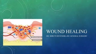
7. INFLAMMATORY PHASE
• This phase lasts for 2-3 days
• When bleeding stops, the platelets then release several cytokines from their
alpha granules. These are platelet-derived growth factor (PDGF), platelet
factor IV and transforming growth factor beta (TGFβ).These attract
inflammatory cells such as polymorphonuclear leukocytes (PMN) and
macrophages Platelets and the local injured tissue release vasoactive amines,
such as histamine, serotonin and prostaglandins, which increase vascular
permeability Macrophages remove devitalised tissue and microorganisms
while regulating fibroblast activity in the proliferative phase of healing. The
initial framework for structural support of cells is provided by fibrin produced
by fibrinogen.
8. PROLIFERATIVE PHASE
• Lasts from 3 days to 3 weeks
• consisting mainly of fibroblast activity with the production of collagen and
ground substance (glycosaminoglycans and proteoglycans), the growth of new
blood vessels as capillary loops (angioneogenesis) and the re-epithelialisation
of the wound surface.
• The wound tissue formed in the early part of this phase is called granulation
tissue. In the latter part of this phase, there is an increase in the tensile strength
of the wound due to increased collagen, which is at first deposited in a random
fashion and consists of type III collagen
9. REMODELLING PHASE
• It is characterised by maturation of collagen (type I replacing type III until a
ratio of 4:1 is achieved). There is a realignment of collagen fibres along the
lines of tension, decreased wound vascularity, and wound contraction due to
fibroblast and myofibroblast activity.
• This maturation of collagen leads to increased tensile strength in the wound
which is maximal at the 12th week post injury and represents approximately
80% of the uninjured skin strength.
11. BONE HEALING
• The phases are similar to the wound healing
• Periosteal and endosteal proliferation leads to the formation of callus, which is
immature bone consisting of osteoid (mineralised by hydroxyapatite and laid down by
osteoblasts).
• In the remodelling phase, cortical structure and the medullary cavity are restored.
• If fracture ends are
• accurately opposed and rigidly fixed, callus formation is minimal and primary healing
occurs. If a gap exists, then secondary healing may lead to delayed union, non-union
or malunion.
12.
13. NERVE
• Distal to the wound, Wallerian degeneration occurs. Proximally, the nerve
suffers traumatic degeneration as far as the last node of Ranvier.
• The regenerating nerve fibres are attracted to their receptors by neurotrophism,
which is mediated by growth factors, hormones and other extracellular matrix
trophins.
• Nerve regeneration is characterised by profuse growth of new nerve fibres
which sprout from the cut proximal end. Overgrowth of these, coupled with
poor approximation, may lead to neuroma formation.
14.
15. TENDON
• Although repair follows the normal pattern of wound healing, there are two
main mechanisms whereby nutrients, cells and new vessels reach the severed
tendon. These are intrinsic, which consists of vincular blood flow and synovial
diffusion, and extrinsic, which depends on the formation of fibrous adhesions
between the tendon and the tendon sheath.
16. Classification of wound closure and healing
● Primary intention
Wound edges opposed
Normal healing
Minimal scar
● Secondary intention
Wound left open
Heals by granulation, contraction and epithelialisation
Increased inflammation and proliferation
Poor scar
● Tertiary intention (also called delayed primary intention)
Wound initially left open
Edges later opposed when healing conditions favourable
Tidy versus untidy wounds.
Tidy Untidy
Incised Crushed or avulsed
Clean Contaminated
Healthy tissue Devitalised tissue
Seldom tissue loss Often tissue loss
17.
18. MANAGING THE ACUTE WOUND
• Examine the whole patient according to acute trauma life support (ATLS)
principles.
• A stab wound in the back can be missed just as easily in the reality of the
accident and emergency room as in a fictitious detective novel.
• Management of pain and tetanus cover.
• In order to facilitate examination, adequate analgesia and/ or anaesthesia
(local, regional or general) are required.
19. • With limb injuries, particularly those of the hand, a tourniquet should be used
in order to facilitate visualisation of all structures.
• After assessment, a thorough debridement is essential. Abrasions, ‘road rash’
(following a fall from a motorbike) and explosions all cause dirt tattooing and
require the use of a scrubbing brush or even excision under magnification.
• Devitalised tissue must be excised until bleeding occurs, with the obvious
exceptions of nerves, vessels and tendons.
20. • Muscle viability is judged by the colour, bleeding pattern and contractility.
In a tidy wound, repair of all damaged structures may be attempted.
• Repair of nerves under magnification (loupes or microscope) using 8/0 or 10/0
monofilament nylon is usual.
• Vessels such as the radial or ulnar artery may be repaired using similar
techniques.
• Tendon repairs, particularly those in the hand, benefit from early active
mobilisation because this minimises adhesions between the tendon and the
tendon sheath.
21. • Skin cover by flap or graft may be required as skin closure should always be
without tension and should allow for the oedema typically associated with
injury and the inflammatory phase of healing.
23. BITES
• Most bites involve either puncture wounds or avulsions.
• Bites from small animals are common in children and
require cleansing and treatment according to the
principles just mentioned.
• Injuries to the ear, tip of nose and lower lip are most
usually seen in victims of human bites.
• A boxing-type injury of
• the metacarpophalangeal joint may result from a
perforating contact with the teeth of a victim.
• Antibiotic prophylaxis is required in bite wounds.
24. PUNCTURE WOUNDS
• Wounds caused by sharp objects should be explored to the limit of tissue blood
staining. Needle-stick injuries should be treated according to the well-
published protocols because of hepatitis and HIV risks. X-ray examination
should be carried out in order to rule out retained foreign bodies in the depth of
the wound.
25. HAEMATOMA
• If large, painful or causing neural deficit, a haematoma may require release by
incision or aspiration. In the gluteal or thigh region, there may be an associated
disruption of fat in the form of a fat fracture, which results in an unsightly
groove but intact skin. An untreated haematoma may also calcify and therefore
require surgical exploration if symptomatic.
26. DEGLOVING
• Degloving occurs when the skin and subcutaneous
fat are stripped by avulsion from the underlying
fascia, leaving neurovascular structures, tendon or
bone exposed.
• It can be open or closed.
• Serial excision until punctate dermal bleeding is
obvious. Split-skin grafts can be harvested from the
degloved non-viable skin and meshed to cover the
raw areas resulting from debridement
27. COMPARTMENT SYNDROMES
• It is characterized by an increase in tissue pressure within a closed
osteofacial space sufficient to compromise microcirculation, leading to
irreversible damage to tissue within that compartment, including death of
muscles and nerves . This occur in association with prolonged limb
ischemia/reperfusion, external pressure, fracture, and burns.
• Patient presents with pain (especially on passive motion), pressure, paralysis,
paraesthesia, pulselessness and pallor (6 P’s)
• Nerve dysfunction and pulselessness presents late and are called “Hard
signs”.
28. • Compartment pressures can be measured using a pressure monitor and a
catheter placed in the muscle compartment. If pressures are constantly greater
than 30 mmHg or if the above clinical signs are present, then fasciotomy
should be performed.
• Fasciotomy involves incising the deep muscle fascia and is best carried out via
longitudinal incisions of skin, fat and fascia. The muscle will then be seen
bulging out through the fasciotomy opening.
• The lower limb can be decompressed via two incisions, each being lateral to
the subcutaneous border of the tibia. This gives access to the two posterior
compartments and to the peroneal and anterior compartments of the leg.
32. LEG ULCERS
• An ulcer can be defined as a break in the epithelial continuity. A prolonged
inflammatory phase leads to overgrowth of granulation tissue, and attempts to
heal by scarring leave a fibrotic margin.
• A chronic ulcer, unresponsive to dressings and simple treatments, should be
biopsied to rule out neoplastic change, a squamous cell carcinoma known as
a Marjolin’s ulcer being the most common.
• Effective treatment of any leg ulcer depends on treating the underlying cause,
and diagnosis is therefore vital. Arterial and venous circulation should be
assessed, as should sensation throughout the lower limb.
33. Aetiology of leg ulcers
● Venous disease leading to local venous hypertension (e.g.varicose veins)
● Arterial disease, either large vessel (atherosclerosis) or small vessel (diabetes)
● Arteritis associated with autoimmune disease (rheumatoid arthritis, lupus, etc.)
● Trauma – could be self-inflicted
● Chronic infection – tuberculosis/syphilis
● Neoplastic – squamous or basal cell carcinoma, sarcoma
34. • Surgical treatment is only indicated if non-operative
treatment has failed or if the patient suffers from
intractable pain.
• Meshed skin grafts are more successful than sheet
grafts and have the advantage of allowing
mobilisation, as any tissue exudate can escape through
the mesh. It should be stressed that the recurrence rate
is high in venous ulceration, and patient compliance
with a regime of hygiene, elevation and elastic
compression is essential.
Meshed split-skin graft.
35. PRESSURE SORES
• These can be defined as tissue necrosis with ulceration due to prolonged
pressure. Less preferable terms are bed sores, pressure ulcers and decubitus
ulcers.
• They should be regarded as preventable but occur in approximately 5% of all
hospitalised patients (range 3–12% in published literature).
• There is a higher incidence in paraplegic patients, in the elderly and in the
severely ill patient.
36. Pressure sore frequency in descending order
● Ischium
● Greater trochanter
● Sacrum
● Heel
● Malleolus (lateral then medial)
● Occiput
37. STAGING OF PRESSURE SORES
Stage Description
1 Non-blanchable erythema without a breach in the
epidermis
2 Partial-thickness skin loss involving the epidermis and dermis
3 Full-thickness skin loss extending into the subcutaneous tissue but not through
underlying fascia
4 Full-thickness skin loss through fascia with extensive
tissue destruction, maybe involving muscle, bone, tendon or joint
38. • If external pressure exceeds the capillary
occlusive pressure (over 30 mmHg), blood
flow to the skin ceases, leading to tissue
anoxia, necrosis and ulceration.
• The best treatment, with good skin care,
special pressure dispersion cushions or foams,
the use of low air loss and air-fluidised beds
and urinary or faecal diversion in selected
cases. Pressure sore awareness is vital, and the
bed-bound patient should be turned at least
every 2 hours, with the wheelchair- bound
patient being taught to lift themselves off their
seat for 10 seconds every 10 minutes.
Pressure ulcer.
39. NECROTISING SOFT-TISSUE
INFECTIONS
• These are rare but often fatal. They are most commonly polymicrobial
infections with Gram-positive aerobes (Staphylococcus aureus, S. pyogenes),
Gram-negative anaerobes (Escherichia coli, Pseudomonas, Clostridium,
Bacteroides) and beta-haemolytic Streptococcus.
• There is usually a history of trauma or surgery with wound contamination.
Sometimes, the patient’s own defence mechanisms may be deficient. These
infections are characterised by sudden presentation and rapid progression.
40. Signs and symptoms of necrotising infections
● Unusual pain
● Oedema beyond area of erythema
● Crepitus
● Skin blistering
● Fever (often absent)
● Greyish drainage (‘dishwater pus’)
● Pink/orange skin staining
● Focal skin gangrene (late sign)
● Shock, coagulopathy and multiorgan failure Necrotising fasciitis of the anterior
abdominal wall.
41. • Treatment consists of appropriate antibiotics with wide surgical excision.
• The raw areas resulting from excision often require skin grafting. Treatment is
surgical excision, with tissue biopsies being sent for culture and diagnosis.
Wide raw areas requiring skin grafting often result.
44. Treatment of hypertrophic and keloid scars
● Pressure – local moulds or elasticated garments
● Silicone gel sheeting (mechanism unknown)
● Intralesional steroid injection (triamcinolone)
● Excision and steroid injections
● Excision and postoperative radiation (external beam or brachytherapy)
● Intralesional excision (keloids only)
● Laser – to reduce redness (which may resolve in any event)
● Vitamin E or palm oil massage (unproven)
*All excisions are associated with high rates of recurrence.
45. CONTRACTURES
• Where scars cross joints or flexion creases, a tight web may form restricting the
range of movement at the joint. This may be referred to as a contracture and can
cause hyperextension or hyperflexion deformity. In the neck, it may interfere with
head extension. Treatment may be simple involving, multiple Z-plasties, or more
complex, requiring the inset of grafts or flaps. Splintage and intensive
physiotherapy are often required postoperatively.
46. Burn contractures showing
hyperextended fingers and
hyperflexed elbow.
Multiple Z-plasty release
of finger contracture.
Post-traumatic (chainsaw)
midline neck contracture.