
Recommandé
Contenu connexe
Tendances
Tendances (20)
Similaire à Chromosome
Similaire à Chromosome (20)
Plus de SoniaBajaj10
Plus de SoniaBajaj10 (20)
Dernier
Dernier (20)
Chromosome
- 1. Shri Shankaracharya Mahavidyalaya, Junwani , Bhilai Structure & Function of Chromosome Dr. Sonia Bajaj, (Head of Department)
- 2. Introduction Chromosomes are thread-like structures present in the nucleus, which carries genetic information from one generation to another. They play a vital role in cell division, heredity, variation, mutation, repair and regeneration. In Eukaryotic cells, genetic material is present in the nucleus in chromosomes, which is made up of highly organized DNA molecules with histone proteins supporting its structure. The term chromosome comes from the Greek words for (chroma) color and body (soma) Chromosome means ‘colored body’ that refers to its staining ability by certain dyes. Karl Nagli in 1842, first observed the rod-like structure present in the nucleus of the plant cell. W. Waldeyer in 1888 coined the term ‘chromosome’. Walter Sutton and Theodor Boveri in 1902 suggested that chromosomes are the physical carrier of genes in the eukaryotic cells. Chromosomes are not visible during interphase under light microscope. During other stages of cell division, they are visible, but are more clearly visible during mitotic metaphase Chromosomes have property of self-duplication, segregation and mutation. The number of chromosome in a gamete is called Genomes.(in human23)
- 3. Chemical Composition Chromosomes vary in shape, size and number in different species of plants and animals. Shapes -Chromosomes have generally three different shapes, viz., rod shape, J shape and V shape. These shapes are observed when the centromere occupies terminal, sub-terminal and median (middle) position on the chromosomes. Size-Chromosome size is measured with the help of micrometer at mitotic metaphase. It is measured in two ways, viz., in length and in diameter. Plants usually have longer chromosomes than animals. The maximum length of chromosome is observed during interphase and minimum during anaphase. Thus chromosome size varies from species to species from 0.5-30 µ, Giant chromosomes have length up to 300 µ and diameter from 0.2-3µ. Number- Nematode -species contains only 2 chromosomes in a cell. Protozoan -species contains as much as 1600 chromosomes in the cell. Plant and animal species -contain 8 to 50 number of chromosomes in its somatic cell. A human cell contains total 23 pair of chromosomes (2n, total 23×2=46), of which 22 are autosomes and 1 sex chromosome.
- 4. Types of Chromosomes 1. Chromosomes are divided into two parts (p and q arms) with a constriction point called a centromere in the middle. 2. Centromere divides the chromosome into two parts, the shorter arm is known as ‘p’ arm and the longer arm is known as ‘q’ arm. (A)Based on the positions of centromeres - Metacentric – centromere is in middle, meaning p and q arms are of comparable length (e.g. chromosomes 1, 3, 16, 19, 20) Sub metacentric – centromere off-center, leading to shorter p arm relative to q arm (e.g. chromosomes 2, 4 – 12, 17, 18, X) Acrocentric – centromere severely off-set from center, leading to much shorter p arm (e.g. chromosomes 13 – 15, 21, 22, Y) Telocentric – centromere found at end of chromosome, meaning no p arm exists (chromosome not found in humans)
- 5. (B) Based on the number of centromeres - 1. Monocentric - only one centromere present. 2. Dicentric- only two centromere present. 3. Polycentric -. More than two centromere present. 4. Acentric- Without centromere.
- 6. Types of Special Chromosome 1. Polytene chromosome/ Giant Chromosomes 1. Balbiani first discovered in 1881. 2. Painter, Heitz and Bauer, rediscovered them in the salivary gland of Drosophila and recognized them as a chromosome. 3. Also known as Salivary gland chromosome. 4. These are called polytene by Kollar due to the presence of many chromonemata in them. 5. These are present in some cells of the larvae of Dipteran insects. 6. These are very large due to the presence of high DNA content. 7. The polytene chromosome of Drosophila’s salivary gland has 1000 DNA molecules Chironomus has 1600 DNA molecules in its each polytene chromosome. 8. There is a series of alternating dark and clear bands called interband. 9. Chromosome puffs or Balbiani rings are present, which are the swelling of bands due to DNA unfolding into open loops. These are the region of the intense transcription or mRNA formation.
- 7. 2. Lampbrush chromosome 1. First discovered in the oocytes of amphibian, fishes and insects. 2. The name is given due to its resemblance with a brush that is used for cleaning lamp, glass chimneys, etc. 3. They occur at the diplotene stage of oocytes in vertebrates and invertebrates. 4. Lampbrush chromosomes are also found in the spermatocytes of many animals and also found in the giant nucleus of an algae Acetabularia. 5. They are present as a bivalent with 4 chromatids 6. Chromosomal axis is formed from highly condensed chromatin and lateral loops extend from the row of chromomeres. 7. Lateral loops of DNA are always symmetrical and formed due to intense RNA synthesis 8. In the oocytes of salamander, there are 10,000 loops present per haploid set of chromosomes. 9. The centromere doesn’t bear any loops.
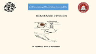
- 8. Structure of Chromosome Each cell has a pair of each kind of chromosome known as a homologous chromosome. Chromosomes are made up of chromatin, which contains a single molecule of DNA and associated proteins. Each chromosome contains hundreds and thousands of genes that can precisely code for several proteins in the cell. Structure of a chromosome can be best seen during cell division. 1. Chromonema- Under light microscope, thread like coiled structures are found in the chromosomes and chromatids which are called chromonema. Two chromatids which are joint at a place called centromere. Chromonema are sub chromatid in nature. 2. Pellicle /Matrix- A mass of acromatic material in which chromonemata are embedded is called matrix. Matrix is enclosed in a sheath which is known as pellicle. Both matrix and pellicle are non-genetic materials. 3. Chromatid- 1. Each chromosome has two symmetrical structures called chromatids or sister chromatids which is visible in mitotic metaphase. 2. Each chromatid contains a single DNA molecule. 3. At the anaphase of mitotic cell division, sister chromatids separate and migrate to opposite poles.
- 9. 4. Centromere and kinetochore- 1. Sister chromatids are joined by the centromere. 2. Spindle fibres during cell division are attached at the centromere. 3. The number and position of the centromere differs in different chromosomes. 4. The centromere is called primary constriction. 5. Centromere divides the chromosome into two parts, the shorter arm is known as ‘p’ arm and the longer arm is known as ‘q’ arm. 6. The centromere contains a disc-shaped kinetochore, which has specific DNA sequence with special proteins bound to them. 7. The kinetochore provides the center for polymerization of tubulin proteins and assembly of microtubules. 8. This is also called kinetochore. .
- 10. 5. Secondary constriction and nucleolar organizers-Other than centromere, chromosomes possess secondary constrictions. Secondary constrictions can be identified from centromere at anaphase because there is bending only at the centromere (primary constriction).Secondary constrictions, which contain genes to form nucleoli are known as the nucleolar organizers. 6. Telomere- 1. Terminal part of a chromosome is known as a telomere. 2. These are not visible in the light or electron microscope, they are rather conceptual structures. 3. Each chromosome has two telomeres. The telomere of one chromosome cannot unite with the telomere of another chromosome due to polarity effect. 7. Satellite: It is an elongated segment that is sometimes present on a chromosome at the secondary constriction. The chromosomes with satellite are known as sat-chromosome.
- 11. Functions of Chromosomes • The main function of chromosomes is to carry the genetic material from one generation to another. • Chromosomes play an important role and act as a guiding force in the growth, reproduction& repair and regeneration process that is important for their survival. • Chromosomes protect the DNA from getting tangled and damaged. • Histone and non-histone proteins help in the regulation of gene expression.