SlideShare une entreprise Scribd logo
1  sur  7
O Arthroscopic Side-to-Side  Rotator Cuff Repair Eugene M. Wolf, M.D., William T. Pennington, M.D., and Vivek Agrawal, M.D. Purpose:  To  study  the  results  of  arthroscopic  repair  of  full-thickness  rotator  cuff  tears  using  a side-to-side  suture  technique  without  fixation  to  bone.  Type of Study:  Case  series  study  of  the long-term results of patients who underwent purely arthroscopic rotator cuff repair with a side-to-side suturing  technique.  Methods:  A  retrospective  review  was  performed  of  patients  who  underwent arthroscopic repair of full-thickness rotator cuff defects. Patients with full-thickness rotator cuff tears repaired in a side-to-side fashion without anchoring the repair to bone were selected. Patients were evaluated using a modified UCLA shoulder scoring system. The data collected were analyzed to determine the outcome in patients with a 4- to 10-year follow-up.  Results:  A total of 105 arthroscopic rotator  cuff  repairs  were  performed  in  104  patients  between  February  1990  and  February  1996. Forty-two  patients  had  a  full-thickness  tear  of  the  rotator  cuff  that  was  repaired  using  a  purely side-to-side  suturing  technique.  The  mean  UCLA  score  of  all  patients  in  this  group  was  33;  23 patients reported excellent results, 18 good results, and 1 poor result according to the UCLA scoring system.  Conclusions:  In this series, 98% of patients qualified as a good to excellent result according to the UCLA shoulder score. This study shows that patients with a full-thickness defect of the rotator cuff tendon with anatomy amenable to side-to-side closure may be effectively treated with a purely arthroscopic  repair  using  only  a  side-to-side  suturing  technique  with  excellent  long-term  clinical results.  Level of Evidence:  Level IV.  Key Words:  Side-to-side suture repair—  Rotator cuff tear Surgical treatment of full-thickness rotator cuff de- fects has focused on recreating the anatomy of the intact rotator cuff with reinsertion and fixation of the tendon to the greater tuberosity of the humerus. Pre- vious authors have presented open and arthroscopic repair techniques that involve recognition of the tear configuration and use a side-to-side repair as a com- ponent of the rotator cuff repair. 2,3,8,9  To date, all reports of purely arthroscopic repair of the rotator cuff have involved securing the repair to the proximal humerus with either suture through bone tunnels or with anchor-based fixation. This article is unique in that we report the results of repairs of full-thickness defects of the rotator cuff with a purely arthroscopic side-to-side technique without anchoring the repair to bone. This repair technique was performed in tears that appeared amenable to this type of repair after thorough arthroscopic visualization and evaluation. METHODS The results of 105 consecutive arthroscopic rotator cuff repairs in 104 patients performed by the senior ther surgeons have reported the results of arthro- scopic  repair  of  full-thickness  defects  of  the rotator cuff. 1-7  These studies support the  use  of arthros- copy in the treatment of symptomatic rotator cuff tears recalcitrant to nonsurgical management. As a result of these reports of successful postoperative outcomes, purely arthroscopic methods to repair full-thickness tears of the rotator cuff are being increasingly used. Proponents of the arthroscopic treatment of rotator cuff pathology cite less morbidity to the surrounding soft- tissue envelope, no deltoid detachment, better visualiza- tion of the pathology of the rotator cuff, and an acceler- ated postoperative rehabilitation period with a lower incidence of postoperative stiffness. 6,7 From  the  California  Pacific  Medical  Center  (E.M.W.),  San Francisco, California; St. Luke’s Medical Center (W.T.P.), Mil- waukee, Wisconsin; and Central Indiana Orthopedics (V.A.), Mun- cie, Indiana, U.S.A. Address  correspondence  and  reprint  requests  to  Eugene  M. Wolf, M.D., 3000 California St, San Francisco, CA 94115, U.S.A. E-mail: genewolfmd@aol.com © 2005 by the Arthroscopy Association of North America 0749-8063/05/2107-2767$30.00/0 doi:10.1016/j.arthro.2005.03.014 881 Arthroscopy: The Journal of Arthroscopic and Related Surgery, Vol 21, No 7 (July), 2005: pp 881-887
author (E.M.W.) over a 6-year period between Febru- ary 1990 and February 1996 were evaluated. Initially, a retrospective clinical chart review was performed for each case. Nine patients were lost to follow-up, leav- ing 96 shoulders in 95 patients available for evaluation with an average follow-up of 73 months (range, 48 to 120 months). Forty-two of these patients who under- went repair of a full-thickness rotator cuff tear in a side-to-side fashion without anchoring the repair to bone were selected for this study. There were 24 male and 18 female patients, and the average age at the time of surgery was 59.8 years (range, 42 to 79 years). All patients had been recalcitrant to conservative therapy and continued to experience unacceptable pain and weakness in the affected shoulder. All patients were clinically evaluated by the senior author. Final outcome assessment was performed by an independent surgeon (W.T.P.) by telephone. Each patient was contacted to assess for pain, function, range of motion, strength, return to work date, and perceived success of the procedure. Outcome was evaluated using a modified UCLA shoulder rating scale 10  ( Table 1 ). This scale designates 10 points each for pain and function and 5 points each for active forward flexion, strength of forward flexion, and pa- tient satisfaction, for a total possible score of 35. Good and excellent results (total UCLA score 28-35 points) are considered satisfactory and fair and poor results (less than 28 points) are considered unsatisfactory. Eighty-one percent of patients had repairs of their dominant shoulder, with 33 right and 9 left repairs. All patients also had arthroscopic subacromial decom- pressions. Seven patients had other procedures per- formed concurrently, including 3 arthroscopic Mum- ford  procedures,  1  SLAP  lesion  debridement,  1 debridement of a biceps tendon rupture, and 2 os acromiale excisions. Surgical Technique Routine  shoulder arthroscopy  was performed with the patient in the lateral decubitus position. Initially, the glenohumeral joint was inspected to evaluate for any significant intra-articular pathology. The cuff was inspected from the articular side and the defect in the rotator cuff tendon was debrided of all frayed, devi- talized tissue. The arthroscope was reconfigured into the subacromial space and a decompression was per- formed using a cutting-block technique. It is important to remove all bursal tissue covering the rotator cuff to be able to evaluate the extent of the tear. The bursec- tomy was also necessary to provide enough visualiza- T ABLE  1. Modified UCLA Shoulder Rating Scale Patient satisfaction 0  Patient feels procedure was not successful 5  Patient feels procedure was a success Active forward flexion range of motion 0  Less than 30° 1  30°-45° 2  45°-90° 3  90°-120° 4  120°-150° 5  Greater than 150° Strength of forward flexion 0  No active contraction 1  Evidence of slight muscle contraction, no active elevation 2  Complete active forward flexion with gravity eliminated 3  Complete active forward flexion against gravity 4  Complete active forward flexion against gravity with some resistance 5  Complete active forward flexion against gravity with full resistance Pain 1  Present always and unbearable, strong medication frequently 2  Present always but bearable, strong medication occasionally 4  None or little at rest, present during light activities; salicylates frequently 6  Present during heavy or particular activities only, salicylates occasionally 8  Occasional and slight 10  None Function 1  Unable to use limb 2  Only light activities possible 3  Able to do light housework or most activities of daily living 6  Most housework, shopping, and driving possible; able to do hair and to dress and undress, including fastening brassiere 8  Slight restriction only, able to work above shoulder level 10  Normal activities Total Excellent: 34-35 Good: 28-33 Fair: 21-27 Poor: 0-20 tion of the cuff and of the suture hooks used in the repair. The region of the greater tuberosity of the humerus was abraded with a full-radius shaver and burr to create a bed of bleeding bone to promote healing of the cuff to the tuberosity. The mobility of the rotator cuff was evaluated with a grasper or nerve hook. Each tear was assessed individually and re- paired with “L” or “V-Y” techniques. All repairs in this report were performed arthroscopically using a side-to-side technique without fixation of the repair to bone. All tears in this series were evaluated with a nerve hook with an attempt to evaluate the anatomic relationship between the margins of the torn cuff 882 E. M. WOLF ET AL.
relationship of this configuration. The repair is then performed with the appropriate suture passer to pass sutures that will approximate the tendon edges, clos- ing the entire defect over the bleeding trough of bone that was previously created on the proximal humerus. It is important to note that during the process of the side-to-side repair, a suture is often placed in what is believed to be the anterior corner of the posterior leaf of the tear followed by passage beneath the transverse humeral ligament and through the coracohumeral lig- ament to help advance the posterior leaf anteriorly. This tendon-to-tendon stitch provides secure approx- imation of the retracted posterior leaf of the tear to the rotator  interval  while  holding  the  entire  repaired stump over the prepared bleeding trough in the greater tuberosity. There were no partial repairs performed; all tears in this series were closed completely and were evaluated arthroscopically from the bursal side of the tear as the shoulder was placed through a full range of motion to ensure the maintenance of the relationship of the tendon repair to the greater tuberosity of the humerus. An average of 4 sutures (range, 1 to 7 sutures) were used per cuff repair. A clinical example of a side-to-side repair of a large U-shaped tear is shown in  Figs 1 - 4 . Repairs were performed exclusively with absorb- able PDS suture in 88% of repairs and nonabsorbable suture in 12%. This includes 36 shoulders repaired with No. 1 PDS and 1 shoulder with No. 0 PDS. Four shoulders were repaired with nonabsorbable No. 2 Ethibond (Ethicon, Somerville, NJ). One repair was performed using No. 2 Mersilene suture. Postopera- tively, patients were placed in a simple immobilizer for 6 weeks but allowed to begin pulley and pendulum F IGURE  1. This view from the midlateral subacromial portal is directed caudally and shows the bursal side of a large U-shaped tear of the supraspinatus tendon. A bed of cancellous bone in the humeral head (H) is seen beneath the large defect in the rotator cuff. The anterior (A) and posterior (P) margins of the torn rotator cuff can be appreciated from this viewing portal. This large tear appears to be amenable to a side-to-side technique of closure. exercises after their first visit 5 days postoperatively. Active exercises began 6 weeks postoperatively. No patients were treated with an abduction brace postop- eratively. RESULTS Ninety-eight percent of patients had good and ex- cellent postoperative scores with 23 excellent (55%), 18 good (43%), and 1 poor result (2%). The average UCLA score was 33. Forty-one of the 42 patients (98%) rated their surgery as being successful and were satisfied with the repair. One patient rated his surgery as unsuccessful. The UCLA shoulder scoring system evaluated for strength, pain, and function ( Table 1 ). The mean re- sponse in all patients grading their strength was 4.6 (range 2-5), mean response for pain was 9.0 (range 2-10), and mean perceived function grade was 9.3 (range 1-10). The average grade for forward flexion of the shoulder was 4.9 (range 1-5). There were 2 patients in whom this arthroscopic repair was a revision of a previous open  rotator cuff repair and 3 patients had previously undergone arthro- scopic assisted mini-open repair on the contralateral shoulder. All 3 of these patients stated that they were 883 ARTHROSCOPIC SIDE-TO-SIDE ROTATOR CUFF REPAIR edges. The nerve hook was believed to be better than a grasper because of its smaller size. Its blunt tip was used to penetrate various points on the edge of the cuff, which was then mobilized to determine the ap- propriate configuration of repair. This evaluation of the cuff tear with a nerve hook is of foremost impor- tance to ensure the optimal restoration of the normal anatomy without producing any undue tension on any part of the repair. Burkhart 9  has suggested that the deepest point of a U-shaped tear does not represent tear retraction but is actually  an  L-shaped  tear  under  physiologic  load. Chronic rotator cuff tears have tapered edges and may well have a “U” or “V” configuration, but this can be appreciated by trying to approximate the deep point of the U-shaped tear to the greater tuberosity with a nerve hook and noticing the creation of “dog ears” in the remainder of the cuff, showing the nonanatomic
F IGURE  2. This arthroscopic view through the midlateral subacro- mial viewing portal shows the passage of a No. 1 PDS suture from posterior to anterior through a long crescent-shaped suture passing hook (Linvatec, Largo, FL) through the apex of the U-shaped tear. more satisfied with their side in which the purely arthroscopic repair was performed and had a percep- tion of a quicker period of recovery and return to function than with their open repair. The 1 patient F IGURE  3.  This image again through the midlateral subacromial viewing portal shows the effect of this initial suture placed through the  apex  of  the  previously  large-appearing  U-shaped  tear.  The arthroscopic  knot  pusher  is  shown  securing  the  initial  Duncan sliding knot of the single suture that was seen in  Fig 2 . Note the effect of this single suture on closing the remaining defect. F IGURE  4. This final arthroscopic view from the midlateral view- ing portal shows complete closure of the rotator cuff defect with the placement of 3 side-to-side No. 1 PDS sutures. with a poor result has failed subsequent open repairs as well. DISCUSSION Rotator cuff  tears  are often attritional in nature and the defect present often involves more than just an avulsion  of  the  musculotendinous  cuff  from  the greater tuberosity of the humerus. Burkhart 9  has elo- quently described a broad classification scheme to which rotator cuff tears can be classified: crescent- shaped or U-shaped tears. He describes the crescent- shaped tear as a disruption of the tendinous insertion from the greater tuberosity of the humerus without a large element of retraction. The U-shaped tear usually appears on initial inspection to be a large retracted tear often medial to the level of the glenoid ( Fig 1 ). Plac- ing the nerve hook in the base of such a tear and attempting to approximate the base to the greater tuberosity usually yields 1 of 2 results with this tear configuration:  approximation  of  the  tendon  is  not achievable to the tuberosity secondary to tension, or approximating the base of the U-shaped tear results in a “dog-eared” appearance of the repaired cuff, indi- cating tension mismatch and a nonanatomic repair. It is important for the surgeon to recognize this tear pattern and use margin convergence as the primary approach to repair. Repairing a U-shaped tear by an- choring the apex of the tear to the tuberosity will result 884 E. M. WOLF ET AL.
in tension overload of the repair, which has been shown by Burkhart et al. to be doomed to failure. 11,12 The use of side-to-side suturing as an element of rotator cuff repair has been described previously by McLaughlin 8  in his open approach to treating large retracted tears of the rotator cuff. Although McLaugh- lin advocated the use of this method to help close large defects, he also was a proponent of final fixation of the tendinous disruption of the cuff to bone in the head of the humerus at the point on the tuberosity that it would reach without undue tension with the arm at the pa- tient’s side. 8  This early description is echoed in the repair techniques employed today with the arthro- scopic approach. That is, anatomic restoration of the cuff without the introduction of tension at the site of the repair. Burkhart et al. have coined the term “mar- gin convergence” to describe the observation that dur- ing side-to-side repair the surgeon can visualize the free margin of the tear converging toward the greater tuberosity with each suture being placed. They agree that using margin convergence in the repair of U- shaped tears decreases the amount of strain at the tendon  bone  interface  of  the  repair  and  therefore should be protective to the tendon bone interface of the repair. 9,13 Cadaveric dissection as well as arthroscopic clinical evaluation has led to an appreciation of the relation- ship of the rotator crescent and rotator cable. Burkhart et al. 14  described a consistently identifiable crescent- shaped insertion of the distal supraspinatus and in- fraspinatus tendons into the greater tuberosity of the humerus bordered on its medial margin by a thickened bundle of fibers oriented perpendicular to the axis of the supraspinatus and infraspinatus tendons. 14  The rotator cable was grossly and histologically confirmed by a cadaveric study performed by Clark and Harry- man. 15  It has been theorized that the thick rotator cable, when intact, provides stress shielding of the rotator cuff crescent much like a suspension bridge. 14 We believe that arthroscopic evaluation of the anat- omy of the  rotator cuff tear  is an essential step in restoring the anatomy of the disrupted rotator cuff. Burkhart 9  suggests that visualization of the tear from different arthroscopic portals allows the surgeon to obtain a 3-dimensional understanding of the tear pat- tern superior to that obtained by open means. We echo this sentiment in that arthroscopic repair of the rotator cuff allows a thorough evaluation of the complete anatomy of the cuff disruption. Furthermore, with each suture passed, the effect may be evaluated by direct visualization of the impact of the suture on the entire cuff. The creation of flaps or “dog ears” indi- cates a nonanatomic repair and tension mismatch that will likely fail under cyclic loading. While other researchers have reported their results of purely arthroscopic repair, this is the first report of a series of patients undergoing arthroscopic repair with purely tendon-to-tendon sutures being placed without  secure  anchor  or  transosseous  fixation  to bone. This technique of repair is used only in cases for which, after thorough arthroscopic evaluation, it is believed that a configuration exists that tear margin convergence can be accomplished with side-to-side closure. All patients in this series had a trough of bleeding bone created in the proximal humerus; when repair was complete, the repaired tendon stump lay directly over this bleeding bed of bone through a full range of motion. All of these repairs were believed to be complete repairs because visualization of the bursal side of the tear after completion of the repair failed to show any remaining defect in the rotator cuff. The 98% good to excellent results compares quite favorably with results previously reported in the liter- ature for surgical treatment of full-thickness defects of the rotator cuff. The minimum duration of follow-up of 4 years with an average of more than 6 years demonstrates an excellent long-term clinical outcome in this subset of patients. As alluded to earlier and described by other inves- tigators, there is an inherent balance of forces through- out the musculotendinous insertion of the rotator cuff into the greater tuberosity of the humerus. Our hy- pothesis to explain such a positive long-term outcome in these patients without secure fixation of the repair to bone is that with the recreation of the anatomy, the natural balance of the rotator cuff musculature pro- vides an environment that is relatively stress free at the tendon-to-bone interface. This allows the distal- most end of the repair that is overlying the bleeding trough of bone to heal to the tuberosity during the period of postoperative convalescence. This hypothe- sis is supported by Burkhart’s suspension bridge con- cept of stress shielding of the rotator crescent by an intact rotator cuff cable. If the cable is intact and able to transfer the stress away from the crescent, this stress-free environment should be conducive to heal- ing of the tendon-to-bone interface. The senior author has previously reported the re- sults of the first 54 purely arthroscopic repairs of full-thickness defects of the rotator cuff. As a compo- nent of this previous study, second-look arthroscopy was performed on 23 patients to evaluate the integrity of the repair at a minimum of 6 months postopera- tively. Nine of these patients had purely side-to-side 885 ARTHROSCOPIC SIDE-TO-SIDE  ROTATOR CUFF REPAIR
repair  of  their  rotator  cuff   and  the  second-look arthroscopy was performed at 5 to 16 months postop- eratively. When examined, all 9 of these tears were completely healed to the greater tuberosity without any evidence of residual defect. Overall, including all 23 arthroscopies performed, 70% of the repaired cuffs were intact at the time of second-look arthroscopy. 1 We believe that these findings support our hypothesis that tendon-to-bone healing does occur despite the absence of secure tendon-to-bone fixation with this technique. Reviewing the literature one would also fi nd ample evidence that the concept of “watertight” closure of the rotator cuff is difficult to achieve re- gardless of the method of fixation of the repaired tendon to the greater tuberosity. Previous studies eval- uating the integrity of the rotator cuff have found residual rotator cuff defects in 34% to 90% of patients who had previously undergone open rotator cuff re- pair, despite secure intraoperative fixation of the re- pair to the greater tuberosity. 16-20 As discussed in the surgical technique section ear- lier, a suture that is often used by the senior author when performing repairs with this technique is passed through the anterior corner of the posterior leaf of the tear and then beneath the transverse humeral ligament and through the coracohumeral ligament anteriorly. After this suture is tied, there is usually secure ap- proximation of the anterior corner of the posterior leaf to the remaining intact rotator cuff anterior to the rotator cuff interval. This suture is often useful in small tears involving only the supraspinatus with ex- tension anterior into the rotator cuff interval. It is also helpful to aid in advancing the retracted posterior leaf anteriorly when closing large L-shaped tears. This study has admitted shortcomings. Although the UCLA shoulder scores are available for the time pe- riod of 4 to 10 years postoperatively, this only signi- fi es wellness at this moment in time after the treatment provided. Ideally, scores during the preoperative pe- riod with sequential scores during the perioperative period would provide conclusive evidence of direct effect of treatment on function of the shoulder. Be- cause some lived at a distance, not all patients could present to the office for physical examination. In these instances, the final physical examination scores at the date of their last follow-up were used along with a detailed telephone interview to confirm the current strength and range of motion of the shoulder. We believe that this study supports the use of ar- throscopy in the treatment of  rotator cuff tears . More importantly, this study shows the excellent clinical success that can be achieved with the correct restora- tion of the rotator cuff anatomy by recognizing the configuration of the tear and performing an anatomic repair of the tendon over a prepared bed of bleeding bone. As a multitude of studies continue to appear at meetings and in the literature evaluating the pullout strengths of different types of fixation techniques of the musculotendinous unit to bone, this study enforces the importance of methodically approaching each tear individually to first recognize and then restore the correct anatomic relationship of the rotator cuff. Just as the orthopaedic surgeon is trained to recognize the personality of a fracture and use this personality to an advantage when performing stabilization,  one  may also keep in mind that, similarly, each rotator cuff tear has a personality of its own with its own subtleties that should be appreciated and used to help achieve opti- mal end results. Although this report is focused on the use of the side-to-side repair technique as a sole treat- ment of certain rotator cuff tears, the use of tendon- to-bone fixation is obviously still used in the treatment of the majority of the tears that we encounter. In this series, 98% of patients had a good to excellent result according to the UCLA shoulder score. This study shows that patients with a full-thickness defect of the rotator cuff tendon, with anatomy amenable to side- to-side closure, may be effectively treated with a purely arthroscopic repair using solely a side-to-side suturing technique, and have excellent long-term clin- ical results. REFERENCES 1.  Baylis RV, Wolf EM. Arthroscopic rotator cuff repair: Clinical and second look assessment. Presented at the Annual Meeting of the Arthroscopy Association of North America, San Fran- cisco, CA, May 1995. 2.  Gartsman GM, Khan M, Hammerman SM. Arthroscopic repair of full-thickness tears of the rotator cuff.  J Bone Joint Surg Am 1998;80:832-840. 3.  Tauro JC. Arthroscopic rotator cuff repair: Analysis of tech- nique and results at a 2- and 3-year follow-up.  Arthroscopy 1998;14:45-51. 4.  Gazielly  DF,  Gleyze  P,  Montagnon  C,  Thomas  T.  Arthro- scopic repair of distal supraspinatus tears with Revo screw and permanent mattress sutures—A preliminary report. Presented at the Annual Meeting of the American Shoulder and Elbow Surgeons, Amelia Island, FL, March 1996. 5.  Snyder SJ, Mileski RA, Karzel RP. Results of arthroscopic repair.  Presented  at  the  Annual  Meeting  of  the  American Shoulder  and  Elbow  Surgeons,  Amelia  Island,  FL,  March 1996. 6.  Weber SC. Arthroscopic versus mini-open rotator cuff repair. Presented at the Arthroscopy Association of North America Fall Course, San Diego, CA, October 1999. 7.  Wolf EM, Pennington WT, Agrawal V. Arthroscopic rotator cuff repair: 4- to 10-year results.  Arthroscopy  2004;20:5-12. 886 E. M. WOLF ET AL.
8.  McLaughlin HL. Lesions of the musculotendinous cuff of the shoulder: The exposure and treatment of tears with retraction. J Bone Joint Surg  1944;26:31-51. 9.  Burkhart SS. A stepwise approach to arthroscopic rotator cuff repair based on biomechanical principles.  Arthroscopy 2000;16:82-90. 10.  Ellman H, Hanker G, Bayer M. Repair of the rotator cuff: End-result study of factors influencing reconstruction.  J Bone Joint Surg Am  1986;68:1136-1144. 11.  Burkhart SS, Johnson TC, Wirth MA, Athanasiou KA. Cyclic loading of transosseous rotator cuff repairs: “Tension overload” as a possible cause of failure.  Arthroscopy  1997;13:172-176. 12.  Burkhart  SS,  Diaz  Pagan  JL,  Wirth  MA,  Athanasiou  KA. Cyclic loading of anchor-based rotator cuff repairs: Confirma- tion of the tension overload phenomenon and comparison of suture anchor fixation with transosseous fixation.  Arthroscopy 1997;13:720-724. 13.  Burkhart  SS,  Athanasiou  KA,  Wirth  MA.  Margin  conver- gence: A method of reducing strain in massive rotator cuff tears.  Arthroscopy  1996;13:335-338. 14.  Burkhart SS, Esch JC, Jolson SJ. The rotator crescent and rotator  cable:  An  anatomic  description  of  the  shoulder’s “ suspension bridge.”  Arthroscopy  1993;9:611-616. 15. 16. 17. 18. 19. 20. Clark JM, Harryman DT. Tendons, ligaments and capsule of the rotator cuff. Gross and microscopic anatomy.  J Bone Joint Surg Am  1992;74:713-725. Liu SH, Baker CL. Arthroscopically assisted rotator cuff re- pair: Correlation of functional results with integrity of the cuff. Arthroscopy  1994;10:54-60. Lundberg BJ. The correlation of clinical evaluation with op- erative  findings  and  prognosis  in  rotator  cuff  rupture,  In: Bayley JI, Kessel L, eds.  Shoulder surgery . Berlin: Springer, 1982;35-38. Calvert PT, Packer NP, Stoker DJ, Bayley JI, Kessel L. Ar- thrography of the shoulder after operative repair of the torn rotator cuff.  J Bone Joint Surg Br  1986;68:147-150. Harryman DT, Mack LA, Wang KY, Jackins SE, Richardson ML, Matsen FA. Repairs of the rotator cuff. Correlation of functional results with integrity of the cuff.  J Bone Joint Surg Am  1991;73:982-989. Gazielly DF, Gleyze P, Montagnon C. Functional and anatom- ical results after rotator cuff repair.  Clin Orthop  1994;304:43- 53. 887 ARTHROSCOPIC SIDE-TO-SIDE ROTATOR CUFF REPAIR

Contenu connexe

Tendances

13. Lelli's Test
13. Lelli's Test 13. Lelli's Test
13. Lelli's Test
drajun
 
12. Meniscal flounce
12. Meniscal flounce12. Meniscal flounce
12. Meniscal flounce
drajun
 
Arthroscopic cuff repair
Arthroscopic cuff repairArthroscopic cuff repair
Arthroscopic cuff repair
orthoprince
 

Tendances (20)

13. Lelli's Test
13. Lelli's Test 13. Lelli's Test
13. Lelli's Test
 
Current Concepts in Shoulder Replacement
Current Concepts in Shoulder ReplacementCurrent Concepts in Shoulder Replacement
Current Concepts in Shoulder Replacement
 
12. Meniscal flounce
12. Meniscal flounce12. Meniscal flounce
12. Meniscal flounce
 
Rotator cuff Repair - New Techniques and Challenges
Rotator cuff Repair - New Techniques and ChallengesRotator cuff Repair - New Techniques and Challenges
Rotator cuff Repair - New Techniques and Challenges
 
U01 clavicle ac_sc_joints1
U01 clavicle ac_sc_joints1U01 clavicle ac_sc_joints1
U01 clavicle ac_sc_joints1
 
eidelman2016.pdf
eidelman2016.pdfeidelman2016.pdf
eidelman2016.pdf
 
Arthroscopic cuff repair
Arthroscopic cuff repairArthroscopic cuff repair
Arthroscopic cuff repair
 
ΑΝΑΘΕΩΡΗΣΗ ΣΥΝΔΕΣΜΟΠΛΑΣΤΙΚΗΣ ΠΡΟΣΘΙΟΥ ΧΙΑΣΤΟΥ ΣΥΝΔΕΣΜΟΥ ΓΟΝΑΤΟΣ ΜΕ ΑΝΑΤΟΜΙΚΗ ...
ΑΝΑΘΕΩΡΗΣΗ ΣΥΝΔΕΣΜΟΠΛΑΣΤΙΚΗΣ ΠΡΟΣΘΙΟΥ ΧΙΑΣΤΟΥ ΣΥΝΔΕΣΜΟΥ ΓΟΝΑΤΟΣ ΜΕ ΑΝΑΤΟΜΙΚΗ ...ΑΝΑΘΕΩΡΗΣΗ ΣΥΝΔΕΣΜΟΠΛΑΣΤΙΚΗΣ ΠΡΟΣΘΙΟΥ ΧΙΑΣΤΟΥ ΣΥΝΔΕΣΜΟΥ ΓΟΝΑΤΟΣ ΜΕ ΑΝΑΤΟΜΙΚΗ ...
ΑΝΑΘΕΩΡΗΣΗ ΣΥΝΔΕΣΜΟΠΛΑΣΤΙΚΗΣ ΠΡΟΣΘΙΟΥ ΧΙΑΣΤΟΥ ΣΥΝΔΕΣΜΟΥ ΓΟΝΑΤΟΣ ΜΕ ΑΝΑΤΟΜΙΚΗ ...
 
Arthrolatarjet (Arthroscopic Latarjet Proc) Dr Sujit Jos kerala
Arthrolatarjet (Arthroscopic Latarjet Proc) Dr Sujit Jos keralaArthrolatarjet (Arthroscopic Latarjet Proc) Dr Sujit Jos kerala
Arthrolatarjet (Arthroscopic Latarjet Proc) Dr Sujit Jos kerala
 
Arthroscopic Transosseous(No Implant) Rotator Cuff Repair-Dr. Raghuveer Reddy .K
Arthroscopic Transosseous(No Implant) Rotator Cuff Repair-Dr. Raghuveer Reddy .KArthroscopic Transosseous(No Implant) Rotator Cuff Repair-Dr. Raghuveer Reddy .K
Arthroscopic Transosseous(No Implant) Rotator Cuff Repair-Dr. Raghuveer Reddy .K
 
Complications in ACL reconstruction 2014
Complications in ACL reconstruction 2014Complications in ACL reconstruction 2014
Complications in ACL reconstruction 2014
 
Ortho Journal Club 11 by Dr Saumya Agarwal
Ortho Journal Club 11 by Dr Saumya AgarwalOrtho Journal Club 11 by Dr Saumya Agarwal
Ortho Journal Club 11 by Dr Saumya Agarwal
 
Retrospective analysis on mini-open technique for Achilles tendon repair
Retrospective analysis on mini-open technique for Achilles tendon repairRetrospective analysis on mini-open technique for Achilles tendon repair
Retrospective analysis on mini-open technique for Achilles tendon repair
 
Latarjet – the panacea for traumatic anterior shoulder
Latarjet – the panacea for traumatic anterior shoulderLatarjet – the panacea for traumatic anterior shoulder
Latarjet – the panacea for traumatic anterior shoulder
 
Osteotomia de Akin asociada a otras técnicas para el tratamiento del hallux v...
Osteotomia de Akin asociada a otras técnicas para el tratamiento del hallux v...Osteotomia de Akin asociada a otras técnicas para el tratamiento del hallux v...
Osteotomia de Akin asociada a otras técnicas para el tratamiento del hallux v...
 
Evolution of tunnel placement in ACL reconstruction
Evolution of tunnel placement in ACL reconstructionEvolution of tunnel placement in ACL reconstruction
Evolution of tunnel placement in ACL reconstruction
 
Clavicle fractures
Clavicle fracturesClavicle fractures
Clavicle fractures
 
Patella in total knee arthroplasty to resurface or not is the question
Patella in total knee arthroplasty  to resurface or not is the  questionPatella in total knee arthroplasty  to resurface or not is the  question
Patella in total knee arthroplasty to resurface or not is the question
 
Per op. in tha
Per op. in thaPer op. in tha
Per op. in tha
 
Full thickness rotator cuff: Diagnosis and Management
Full thickness rotator cuff: Diagnosis and ManagementFull thickness rotator cuff: Diagnosis and Management
Full thickness rotator cuff: Diagnosis and Management
 

Similaire à Rotator cuff-repair-study

Rotator cuff-study
Rotator cuff-studyRotator cuff-study
Rotator cuff-study
SoulderPain
 
Open debridement and radiocapitellar replacement in primary and post-traumati...
Open debridement and radiocapitellar replacement in primary and post-traumati...Open debridement and radiocapitellar replacement in primary and post-traumati...
Open debridement and radiocapitellar replacement in primary and post-traumati...
Alberto Mantovani
 
Surgical Treatment of Primary Malignant Tumours of the Distal Tibia.pptx
Surgical Treatment of Primary Malignant Tumours of the Distal Tibia.pptxSurgical Treatment of Primary Malignant Tumours of the Distal Tibia.pptx
Surgical Treatment of Primary Malignant Tumours of the Distal Tibia.pptx
PunyaChopra1
 
5. PCL repair
5. PCL repair5. PCL repair
5. PCL repair
drajun
 
(Knee plc surg tech) la prade rf
(Knee plc surg tech) la prade rf(Knee plc surg tech) la prade rf
(Knee plc surg tech) la prade rf
Ricardo Vieta
 
6 Calcaneum fracture
6 Calcaneum fracture6 Calcaneum fracture
6 Calcaneum fracture
drajun
 

Similaire à Rotator cuff-repair-study (20)

Rotator cuff-study
Rotator cuff-studyRotator cuff-study
Rotator cuff-study
 
Rotator cuff-study
Rotator cuff-studyRotator cuff-study
Rotator cuff-study
 
Arthroscopic Anterior Cruciate Ligament Reconstruction Using Four-Strand Hams...
Arthroscopic Anterior Cruciate Ligament Reconstruction Using Four-Strand Hams...Arthroscopic Anterior Cruciate Ligament Reconstruction Using Four-Strand Hams...
Arthroscopic Anterior Cruciate Ligament Reconstruction Using Four-Strand Hams...
 
GOODAY.ARTICLE.Final
GOODAY.ARTICLE.FinalGOODAY.ARTICLE.Final
GOODAY.ARTICLE.Final
 
Open debridement and radiocapitellar replacement in primary and post-traumati...
Open debridement and radiocapitellar replacement in primary and post-traumati...Open debridement and radiocapitellar replacement in primary and post-traumati...
Open debridement and radiocapitellar replacement in primary and post-traumati...
 
Surgical Treatment of Primary Malignant Tumours of the Distal Tibia.pptx
Surgical Treatment of Primary Malignant Tumours of the Distal Tibia.pptxSurgical Treatment of Primary Malignant Tumours of the Distal Tibia.pptx
Surgical Treatment of Primary Malignant Tumours of the Distal Tibia.pptx
 
Study of functional outcome following arthroscopic anatomical ACL reconstruct...
Study of functional outcome following arthroscopic anatomical ACL reconstruct...Study of functional outcome following arthroscopic anatomical ACL reconstruct...
Study of functional outcome following arthroscopic anatomical ACL reconstruct...
 
TKA for severe valgus
TKA for severe valgusTKA for severe valgus
TKA for severe valgus
 
5. PCL repair
5. PCL repair5. PCL repair
5. PCL repair
 
(Knee plc surg tech) la prade rf
(Knee plc surg tech) la prade rf(Knee plc surg tech) la prade rf
(Knee plc surg tech) la prade rf
 
Arthroscopic Single Row Versus Double-Row Suture Anchor Rotator Cuff Repair |...
Arthroscopic Single Row Versus Double-Row Suture Anchor Rotator Cuff Repair |...Arthroscopic Single Row Versus Double-Row Suture Anchor Rotator Cuff Repair |...
Arthroscopic Single Row Versus Double-Row Suture Anchor Rotator Cuff Repair |...
 
Ortho Journal Club 3 by Dr Saumya Agarwal
Ortho Journal Club 3 by Dr Saumya AgarwalOrtho Journal Club 3 by Dr Saumya Agarwal
Ortho Journal Club 3 by Dr Saumya Agarwal
 
Birmingham mid-head resection arthroplasty of hip for avascular necrosis of f...
Birmingham mid-head resection arthroplasty of hip for avascular necrosis of f...Birmingham mid-head resection arthroplasty of hip for avascular necrosis of f...
Birmingham mid-head resection arthroplasty of hip for avascular necrosis of f...
 
V60 n3 4-3
V60 n3 4-3V60 n3 4-3
V60 n3 4-3
 
Treatment For Sarcoma Cancer
Treatment For Sarcoma Cancer Treatment For Sarcoma Cancer
Treatment For Sarcoma Cancer
 
CLAVICLE FRACTURE BIOMECHANICS AND SURGERY
CLAVICLE FRACTURE BIOMECHANICS AND SURGERYCLAVICLE FRACTURE BIOMECHANICS AND SURGERY
CLAVICLE FRACTURE BIOMECHANICS AND SURGERY
 
6 Calcaneum fracture
6 Calcaneum fracture6 Calcaneum fracture
6 Calcaneum fracture
 
Anotomic-Biological Reconstruction of Acromio-Clavicular Joint Injuries-Dr. U...
Anotomic-Biological Reconstruction of Acromio-Clavicular Joint Injuries-Dr. U...Anotomic-Biological Reconstruction of Acromio-Clavicular Joint Injuries-Dr. U...
Anotomic-Biological Reconstruction of Acromio-Clavicular Joint Injuries-Dr. U...
 
Two treatment methods for spiral fracture of the lower third of the tibia sha...
Two treatment methods for spiral fracture of the lower third of the tibia sha...Two treatment methods for spiral fracture of the lower third of the tibia sha...
Two treatment methods for spiral fracture of the lower third of the tibia sha...
 
JC Acromioclavicular joint injuries.pptx
JC Acromioclavicular joint injuries.pptxJC Acromioclavicular joint injuries.pptx
JC Acromioclavicular joint injuries.pptx
 

Plus de SoulderPain (10)

Rotator cuff-repair
Rotator cuff-repairRotator cuff-repair
Rotator cuff-repair
 
Pinched nerve-shoulder
Pinched nerve-shoulderPinched nerve-shoulder
Pinched nerve-shoulder
 
Ac shoulder-dislocation
Ac shoulder-dislocationAc shoulder-dislocation
Ac shoulder-dislocation
 
Rotator cuff-repair
Rotator cuff-repairRotator cuff-repair
Rotator cuff-repair
 
Pinched nerve-shoulder
Pinched nerve-shoulderPinched nerve-shoulder
Pinched nerve-shoulder
 
Ac shoulder-dislocation
Ac shoulder-dislocationAc shoulder-dislocation
Ac shoulder-dislocation
 
Shoulder replacement-surgery
Shoulder replacement-surgeryShoulder replacement-surgery
Shoulder replacement-surgery
 
Rotator cuff-repair
Rotator cuff-repairRotator cuff-repair
Rotator cuff-repair
 
Pinched nerve-shoulder
Pinched nerve-shoulderPinched nerve-shoulder
Pinched nerve-shoulder
 
Ac shoulder-dislocation
Ac shoulder-dislocationAc shoulder-dislocation
Ac shoulder-dislocation
 

Dernier

Difference Between Skeletal Smooth and Cardiac Muscles
Difference Between Skeletal Smooth and Cardiac MusclesDifference Between Skeletal Smooth and Cardiac Muscles
Difference Between Skeletal Smooth and Cardiac Muscles
MedicoseAcademics
 
👉 Chennai Sexy Aunty’s WhatsApp Number 👉📞 7427069034 👉📞 Just📲 Call Ruhi Colle...
👉 Chennai Sexy Aunty’s WhatsApp Number 👉📞 7427069034 👉📞 Just📲 Call Ruhi Colle...👉 Chennai Sexy Aunty’s WhatsApp Number 👉📞 7427069034 👉📞 Just📲 Call Ruhi Colle...
👉 Chennai Sexy Aunty’s WhatsApp Number 👉📞 7427069034 👉📞 Just📲 Call Ruhi Colle...
rajnisinghkjn
 
Call Girl in Chennai | Whatsapp No 📞 7427069034 📞 VIP Escorts Service Availab...
Call Girl in Chennai | Whatsapp No 📞 7427069034 📞 VIP Escorts Service Availab...Call Girl in Chennai | Whatsapp No 📞 7427069034 📞 VIP Escorts Service Availab...
Call Girl in Chennai | Whatsapp No 📞 7427069034 📞 VIP Escorts Service Availab...
amritaverma53
 
Gorgeous Call Girls Dehradun {8854095900} ❤️VVIP ROCKY Call Girls in Dehradun...
Gorgeous Call Girls Dehradun {8854095900} ❤️VVIP ROCKY Call Girls in Dehradun...Gorgeous Call Girls Dehradun {8854095900} ❤️VVIP ROCKY Call Girls in Dehradun...
Gorgeous Call Girls Dehradun {8854095900} ❤️VVIP ROCKY Call Girls in Dehradun...
Sheetaleventcompany
 
Kolkata Call Girls Service ❤️🍑 9xx000xx09 👄🫦 Independent Escort Service Kolka...
Kolkata Call Girls Service ❤️🍑 9xx000xx09 👄🫦 Independent Escort Service Kolka...Kolkata Call Girls Service ❤️🍑 9xx000xx09 👄🫦 Independent Escort Service Kolka...
Kolkata Call Girls Service ❤️🍑 9xx000xx09 👄🫦 Independent Escort Service Kolka...
Sheetaleventcompany
 
Call Girl In Indore 📞9235973566📞 Just📲 Call Inaaya Indore Call Girls Service ...
Call Girl In Indore 📞9235973566📞 Just📲 Call Inaaya Indore Call Girls Service ...Call Girl In Indore 📞9235973566📞 Just📲 Call Inaaya Indore Call Girls Service ...
Call Girl In Indore 📞9235973566📞 Just📲 Call Inaaya Indore Call Girls Service ...
Sheetaleventcompany
 
💚Chandigarh Call Girls Service 💯Piya 📲🔝8868886958🔝Call Girls In Chandigarh No...
💚Chandigarh Call Girls Service 💯Piya 📲🔝8868886958🔝Call Girls In Chandigarh No...💚Chandigarh Call Girls Service 💯Piya 📲🔝8868886958🔝Call Girls In Chandigarh No...
💚Chandigarh Call Girls Service 💯Piya 📲🔝8868886958🔝Call Girls In Chandigarh No...
Sheetaleventcompany
 
Goa Call Girl Service 📞9xx000xx09📞Just Call Divya📲 Call Girl In Goa No💰Advanc...
Goa Call Girl Service 📞9xx000xx09📞Just Call Divya📲 Call Girl In Goa No💰Advanc...Goa Call Girl Service 📞9xx000xx09📞Just Call Divya📲 Call Girl In Goa No💰Advanc...
Goa Call Girl Service 📞9xx000xx09📞Just Call Divya📲 Call Girl In Goa No💰Advanc...
Sheetaleventcompany
 

Dernier (20)

Cardiac Output, Venous Return, and Their Regulation
Cardiac Output, Venous Return, and Their RegulationCardiac Output, Venous Return, and Their Regulation
Cardiac Output, Venous Return, and Their Regulation
 
Chandigarh Call Girls Service ❤️🍑 9809698092 👄🫦Independent Escort Service Cha...
Chandigarh Call Girls Service ❤️🍑 9809698092 👄🫦Independent Escort Service Cha...Chandigarh Call Girls Service ❤️🍑 9809698092 👄🫦Independent Escort Service Cha...
Chandigarh Call Girls Service ❤️🍑 9809698092 👄🫦Independent Escort Service Cha...
 
Call Girls Kathua Just Call 8250077686 Top Class Call Girl Service Available
Call Girls Kathua Just Call 8250077686 Top Class Call Girl Service AvailableCall Girls Kathua Just Call 8250077686 Top Class Call Girl Service Available
Call Girls Kathua Just Call 8250077686 Top Class Call Girl Service Available
 
Difference Between Skeletal Smooth and Cardiac Muscles
Difference Between Skeletal Smooth and Cardiac MusclesDifference Between Skeletal Smooth and Cardiac Muscles
Difference Between Skeletal Smooth and Cardiac Muscles
 
👉 Chennai Sexy Aunty’s WhatsApp Number 👉📞 7427069034 👉📞 Just📲 Call Ruhi Colle...
👉 Chennai Sexy Aunty’s WhatsApp Number 👉📞 7427069034 👉📞 Just📲 Call Ruhi Colle...👉 Chennai Sexy Aunty’s WhatsApp Number 👉📞 7427069034 👉📞 Just📲 Call Ruhi Colle...
👉 Chennai Sexy Aunty’s WhatsApp Number 👉📞 7427069034 👉📞 Just📲 Call Ruhi Colle...
 
Call Girls Bangalore - 450+ Call Girl Cash Payment 💯Call Us 🔝 6378878445 🔝 💃 ...
Call Girls Bangalore - 450+ Call Girl Cash Payment 💯Call Us 🔝 6378878445 🔝 💃 ...Call Girls Bangalore - 450+ Call Girl Cash Payment 💯Call Us 🔝 6378878445 🔝 💃 ...
Call Girls Bangalore - 450+ Call Girl Cash Payment 💯Call Us 🔝 6378878445 🔝 💃 ...
 
Call Girl in Chennai | Whatsapp No 📞 7427069034 📞 VIP Escorts Service Availab...
Call Girl in Chennai | Whatsapp No 📞 7427069034 📞 VIP Escorts Service Availab...Call Girl in Chennai | Whatsapp No 📞 7427069034 📞 VIP Escorts Service Availab...
Call Girl in Chennai | Whatsapp No 📞 7427069034 📞 VIP Escorts Service Availab...
 
Call 8250092165 Patna Call Girls ₹4.5k Cash Payment With Room Delivery
Call 8250092165 Patna Call Girls ₹4.5k Cash Payment With Room DeliveryCall 8250092165 Patna Call Girls ₹4.5k Cash Payment With Room Delivery
Call 8250092165 Patna Call Girls ₹4.5k Cash Payment With Room Delivery
 
Call Girl In Chandigarh 📞9809698092📞 Just📲 Call Inaaya Chandigarh Call Girls ...
Call Girl In Chandigarh 📞9809698092📞 Just📲 Call Inaaya Chandigarh Call Girls ...Call Girl In Chandigarh 📞9809698092📞 Just📲 Call Inaaya Chandigarh Call Girls ...
Call Girl In Chandigarh 📞9809698092📞 Just📲 Call Inaaya Chandigarh Call Girls ...
 
Gorgeous Call Girls Dehradun {8854095900} ❤️VVIP ROCKY Call Girls in Dehradun...
Gorgeous Call Girls Dehradun {8854095900} ❤️VVIP ROCKY Call Girls in Dehradun...Gorgeous Call Girls Dehradun {8854095900} ❤️VVIP ROCKY Call Girls in Dehradun...
Gorgeous Call Girls Dehradun {8854095900} ❤️VVIP ROCKY Call Girls in Dehradun...
 
Low Cost Call Girls Bangalore {9179660964} ❤️VVIP NISHA Call Girls in Bangalo...
Low Cost Call Girls Bangalore {9179660964} ❤️VVIP NISHA Call Girls in Bangalo...Low Cost Call Girls Bangalore {9179660964} ❤️VVIP NISHA Call Girls in Bangalo...
Low Cost Call Girls Bangalore {9179660964} ❤️VVIP NISHA Call Girls in Bangalo...
 
Kolkata Call Girls Service ❤️🍑 9xx000xx09 👄🫦 Independent Escort Service Kolka...
Kolkata Call Girls Service ❤️🍑 9xx000xx09 👄🫦 Independent Escort Service Kolka...Kolkata Call Girls Service ❤️🍑 9xx000xx09 👄🫦 Independent Escort Service Kolka...
Kolkata Call Girls Service ❤️🍑 9xx000xx09 👄🫦 Independent Escort Service Kolka...
 
Call Girl In Indore 📞9235973566📞 Just📲 Call Inaaya Indore Call Girls Service ...
Call Girl In Indore 📞9235973566📞 Just📲 Call Inaaya Indore Call Girls Service ...Call Girl In Indore 📞9235973566📞 Just📲 Call Inaaya Indore Call Girls Service ...
Call Girl In Indore 📞9235973566📞 Just📲 Call Inaaya Indore Call Girls Service ...
 
Gastric Cancer: Сlinical Implementation of Artificial Intelligence, Synergeti...
Gastric Cancer: Сlinical Implementation of Artificial Intelligence, Synergeti...Gastric Cancer: Сlinical Implementation of Artificial Intelligence, Synergeti...
Gastric Cancer: Сlinical Implementation of Artificial Intelligence, Synergeti...
 
Call Girls Mussoorie Just Call 8854095900 Top Class Call Girl Service Available
Call Girls Mussoorie Just Call 8854095900 Top Class Call Girl Service AvailableCall Girls Mussoorie Just Call 8854095900 Top Class Call Girl Service Available
Call Girls Mussoorie Just Call 8854095900 Top Class Call Girl Service Available
 
💚Chandigarh Call Girls Service 💯Piya 📲🔝8868886958🔝Call Girls In Chandigarh No...
💚Chandigarh Call Girls Service 💯Piya 📲🔝8868886958🔝Call Girls In Chandigarh No...💚Chandigarh Call Girls Service 💯Piya 📲🔝8868886958🔝Call Girls In Chandigarh No...
💚Chandigarh Call Girls Service 💯Piya 📲🔝8868886958🔝Call Girls In Chandigarh No...
 
💰Call Girl In Bangalore☎️7304373326💰 Call Girl service in Bangalore☎️Bangalor...
💰Call Girl In Bangalore☎️7304373326💰 Call Girl service in Bangalore☎️Bangalor...💰Call Girl In Bangalore☎️7304373326💰 Call Girl service in Bangalore☎️Bangalor...
💰Call Girl In Bangalore☎️7304373326💰 Call Girl service in Bangalore☎️Bangalor...
 
Goa Call Girl Service 📞9xx000xx09📞Just Call Divya📲 Call Girl In Goa No💰Advanc...
Goa Call Girl Service 📞9xx000xx09📞Just Call Divya📲 Call Girl In Goa No💰Advanc...Goa Call Girl Service 📞9xx000xx09📞Just Call Divya📲 Call Girl In Goa No💰Advanc...
Goa Call Girl Service 📞9xx000xx09📞Just Call Divya📲 Call Girl In Goa No💰Advanc...
 
Exclusive Call Girls Bangalore {7304373326} ❤️VVIP POOJA Call Girls in Bangal...
Exclusive Call Girls Bangalore {7304373326} ❤️VVIP POOJA Call Girls in Bangal...Exclusive Call Girls Bangalore {7304373326} ❤️VVIP POOJA Call Girls in Bangal...
Exclusive Call Girls Bangalore {7304373326} ❤️VVIP POOJA Call Girls in Bangal...
 
Race Course Road } Book Call Girls in Bangalore | Whatsapp No 6378878445 VIP ...
Race Course Road } Book Call Girls in Bangalore | Whatsapp No 6378878445 VIP ...Race Course Road } Book Call Girls in Bangalore | Whatsapp No 6378878445 VIP ...
Race Course Road } Book Call Girls in Bangalore | Whatsapp No 6378878445 VIP ...
 

Rotator cuff-repair-study

  • 1. O Arthroscopic Side-to-Side Rotator Cuff Repair Eugene M. Wolf, M.D., William T. Pennington, M.D., and Vivek Agrawal, M.D. Purpose: To study the results of arthroscopic repair of full-thickness rotator cuff tears using a side-to-side suture technique without fixation to bone. Type of Study: Case series study of the long-term results of patients who underwent purely arthroscopic rotator cuff repair with a side-to-side suturing technique. Methods: A retrospective review was performed of patients who underwent arthroscopic repair of full-thickness rotator cuff defects. Patients with full-thickness rotator cuff tears repaired in a side-to-side fashion without anchoring the repair to bone were selected. Patients were evaluated using a modified UCLA shoulder scoring system. The data collected were analyzed to determine the outcome in patients with a 4- to 10-year follow-up. Results: A total of 105 arthroscopic rotator cuff repairs were performed in 104 patients between February 1990 and February 1996. Forty-two patients had a full-thickness tear of the rotator cuff that was repaired using a purely side-to-side suturing technique. The mean UCLA score of all patients in this group was 33; 23 patients reported excellent results, 18 good results, and 1 poor result according to the UCLA scoring system. Conclusions: In this series, 98% of patients qualified as a good to excellent result according to the UCLA shoulder score. This study shows that patients with a full-thickness defect of the rotator cuff tendon with anatomy amenable to side-to-side closure may be effectively treated with a purely arthroscopic repair using only a side-to-side suturing technique with excellent long-term clinical results. Level of Evidence: Level IV. Key Words: Side-to-side suture repair— Rotator cuff tear Surgical treatment of full-thickness rotator cuff de- fects has focused on recreating the anatomy of the intact rotator cuff with reinsertion and fixation of the tendon to the greater tuberosity of the humerus. Pre- vious authors have presented open and arthroscopic repair techniques that involve recognition of the tear configuration and use a side-to-side repair as a com- ponent of the rotator cuff repair. 2,3,8,9 To date, all reports of purely arthroscopic repair of the rotator cuff have involved securing the repair to the proximal humerus with either suture through bone tunnels or with anchor-based fixation. This article is unique in that we report the results of repairs of full-thickness defects of the rotator cuff with a purely arthroscopic side-to-side technique without anchoring the repair to bone. This repair technique was performed in tears that appeared amenable to this type of repair after thorough arthroscopic visualization and evaluation. METHODS The results of 105 consecutive arthroscopic rotator cuff repairs in 104 patients performed by the senior ther surgeons have reported the results of arthro- scopic repair of full-thickness defects of the rotator cuff. 1-7 These studies support the use of arthros- copy in the treatment of symptomatic rotator cuff tears recalcitrant to nonsurgical management. As a result of these reports of successful postoperative outcomes, purely arthroscopic methods to repair full-thickness tears of the rotator cuff are being increasingly used. Proponents of the arthroscopic treatment of rotator cuff pathology cite less morbidity to the surrounding soft- tissue envelope, no deltoid detachment, better visualiza- tion of the pathology of the rotator cuff, and an acceler- ated postoperative rehabilitation period with a lower incidence of postoperative stiffness. 6,7 From the California Pacific Medical Center (E.M.W.), San Francisco, California; St. Luke’s Medical Center (W.T.P.), Mil- waukee, Wisconsin; and Central Indiana Orthopedics (V.A.), Mun- cie, Indiana, U.S.A. Address correspondence and reprint requests to Eugene M. Wolf, M.D., 3000 California St, San Francisco, CA 94115, U.S.A. E-mail: genewolfmd@aol.com © 2005 by the Arthroscopy Association of North America 0749-8063/05/2107-2767$30.00/0 doi:10.1016/j.arthro.2005.03.014 881 Arthroscopy: The Journal of Arthroscopic and Related Surgery, Vol 21, No 7 (July), 2005: pp 881-887
  • 2. author (E.M.W.) over a 6-year period between Febru- ary 1990 and February 1996 were evaluated. Initially, a retrospective clinical chart review was performed for each case. Nine patients were lost to follow-up, leav- ing 96 shoulders in 95 patients available for evaluation with an average follow-up of 73 months (range, 48 to 120 months). Forty-two of these patients who under- went repair of a full-thickness rotator cuff tear in a side-to-side fashion without anchoring the repair to bone were selected for this study. There were 24 male and 18 female patients, and the average age at the time of surgery was 59.8 years (range, 42 to 79 years). All patients had been recalcitrant to conservative therapy and continued to experience unacceptable pain and weakness in the affected shoulder. All patients were clinically evaluated by the senior author. Final outcome assessment was performed by an independent surgeon (W.T.P.) by telephone. Each patient was contacted to assess for pain, function, range of motion, strength, return to work date, and perceived success of the procedure. Outcome was evaluated using a modified UCLA shoulder rating scale 10 ( Table 1 ). This scale designates 10 points each for pain and function and 5 points each for active forward flexion, strength of forward flexion, and pa- tient satisfaction, for a total possible score of 35. Good and excellent results (total UCLA score 28-35 points) are considered satisfactory and fair and poor results (less than 28 points) are considered unsatisfactory. Eighty-one percent of patients had repairs of their dominant shoulder, with 33 right and 9 left repairs. All patients also had arthroscopic subacromial decom- pressions. Seven patients had other procedures per- formed concurrently, including 3 arthroscopic Mum- ford procedures, 1 SLAP lesion debridement, 1 debridement of a biceps tendon rupture, and 2 os acromiale excisions. Surgical Technique Routine shoulder arthroscopy was performed with the patient in the lateral decubitus position. Initially, the glenohumeral joint was inspected to evaluate for any significant intra-articular pathology. The cuff was inspected from the articular side and the defect in the rotator cuff tendon was debrided of all frayed, devi- talized tissue. The arthroscope was reconfigured into the subacromial space and a decompression was per- formed using a cutting-block technique. It is important to remove all bursal tissue covering the rotator cuff to be able to evaluate the extent of the tear. The bursec- tomy was also necessary to provide enough visualiza- T ABLE 1. Modified UCLA Shoulder Rating Scale Patient satisfaction 0 Patient feels procedure was not successful 5 Patient feels procedure was a success Active forward flexion range of motion 0 Less than 30° 1 30°-45° 2 45°-90° 3 90°-120° 4 120°-150° 5 Greater than 150° Strength of forward flexion 0 No active contraction 1 Evidence of slight muscle contraction, no active elevation 2 Complete active forward flexion with gravity eliminated 3 Complete active forward flexion against gravity 4 Complete active forward flexion against gravity with some resistance 5 Complete active forward flexion against gravity with full resistance Pain 1 Present always and unbearable, strong medication frequently 2 Present always but bearable, strong medication occasionally 4 None or little at rest, present during light activities; salicylates frequently 6 Present during heavy or particular activities only, salicylates occasionally 8 Occasional and slight 10 None Function 1 Unable to use limb 2 Only light activities possible 3 Able to do light housework or most activities of daily living 6 Most housework, shopping, and driving possible; able to do hair and to dress and undress, including fastening brassiere 8 Slight restriction only, able to work above shoulder level 10 Normal activities Total Excellent: 34-35 Good: 28-33 Fair: 21-27 Poor: 0-20 tion of the cuff and of the suture hooks used in the repair. The region of the greater tuberosity of the humerus was abraded with a full-radius shaver and burr to create a bed of bleeding bone to promote healing of the cuff to the tuberosity. The mobility of the rotator cuff was evaluated with a grasper or nerve hook. Each tear was assessed individually and re- paired with “L” or “V-Y” techniques. All repairs in this report were performed arthroscopically using a side-to-side technique without fixation of the repair to bone. All tears in this series were evaluated with a nerve hook with an attempt to evaluate the anatomic relationship between the margins of the torn cuff 882 E. M. WOLF ET AL.
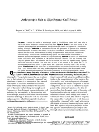
  • 3. relationship of this configuration. The repair is then performed with the appropriate suture passer to pass sutures that will approximate the tendon edges, clos- ing the entire defect over the bleeding trough of bone that was previously created on the proximal humerus. It is important to note that during the process of the side-to-side repair, a suture is often placed in what is believed to be the anterior corner of the posterior leaf of the tear followed by passage beneath the transverse humeral ligament and through the coracohumeral lig- ament to help advance the posterior leaf anteriorly. This tendon-to-tendon stitch provides secure approx- imation of the retracted posterior leaf of the tear to the rotator interval while holding the entire repaired stump over the prepared bleeding trough in the greater tuberosity. There were no partial repairs performed; all tears in this series were closed completely and were evaluated arthroscopically from the bursal side of the tear as the shoulder was placed through a full range of motion to ensure the maintenance of the relationship of the tendon repair to the greater tuberosity of the humerus. An average of 4 sutures (range, 1 to 7 sutures) were used per cuff repair. A clinical example of a side-to-side repair of a large U-shaped tear is shown in Figs 1 - 4 . Repairs were performed exclusively with absorb- able PDS suture in 88% of repairs and nonabsorbable suture in 12%. This includes 36 shoulders repaired with No. 1 PDS and 1 shoulder with No. 0 PDS. Four shoulders were repaired with nonabsorbable No. 2 Ethibond (Ethicon, Somerville, NJ). One repair was performed using No. 2 Mersilene suture. Postopera- tively, patients were placed in a simple immobilizer for 6 weeks but allowed to begin pulley and pendulum F IGURE 1. This view from the midlateral subacromial portal is directed caudally and shows the bursal side of a large U-shaped tear of the supraspinatus tendon. A bed of cancellous bone in the humeral head (H) is seen beneath the large defect in the rotator cuff. The anterior (A) and posterior (P) margins of the torn rotator cuff can be appreciated from this viewing portal. This large tear appears to be amenable to a side-to-side technique of closure. exercises after their first visit 5 days postoperatively. Active exercises began 6 weeks postoperatively. No patients were treated with an abduction brace postop- eratively. RESULTS Ninety-eight percent of patients had good and ex- cellent postoperative scores with 23 excellent (55%), 18 good (43%), and 1 poor result (2%). The average UCLA score was 33. Forty-one of the 42 patients (98%) rated their surgery as being successful and were satisfied with the repair. One patient rated his surgery as unsuccessful. The UCLA shoulder scoring system evaluated for strength, pain, and function ( Table 1 ). The mean re- sponse in all patients grading their strength was 4.6 (range 2-5), mean response for pain was 9.0 (range 2-10), and mean perceived function grade was 9.3 (range 1-10). The average grade for forward flexion of the shoulder was 4.9 (range 1-5). There were 2 patients in whom this arthroscopic repair was a revision of a previous open rotator cuff repair and 3 patients had previously undergone arthro- scopic assisted mini-open repair on the contralateral shoulder. All 3 of these patients stated that they were 883 ARTHROSCOPIC SIDE-TO-SIDE ROTATOR CUFF REPAIR edges. The nerve hook was believed to be better than a grasper because of its smaller size. Its blunt tip was used to penetrate various points on the edge of the cuff, which was then mobilized to determine the ap- propriate configuration of repair. This evaluation of the cuff tear with a nerve hook is of foremost impor- tance to ensure the optimal restoration of the normal anatomy without producing any undue tension on any part of the repair. Burkhart 9 has suggested that the deepest point of a U-shaped tear does not represent tear retraction but is actually an L-shaped tear under physiologic load. Chronic rotator cuff tears have tapered edges and may well have a “U” or “V” configuration, but this can be appreciated by trying to approximate the deep point of the U-shaped tear to the greater tuberosity with a nerve hook and noticing the creation of “dog ears” in the remainder of the cuff, showing the nonanatomic
  • 4. F IGURE 2. This arthroscopic view through the midlateral subacro- mial viewing portal shows the passage of a No. 1 PDS suture from posterior to anterior through a long crescent-shaped suture passing hook (Linvatec, Largo, FL) through the apex of the U-shaped tear. more satisfied with their side in which the purely arthroscopic repair was performed and had a percep- tion of a quicker period of recovery and return to function than with their open repair. The 1 patient F IGURE 3. This image again through the midlateral subacromial viewing portal shows the effect of this initial suture placed through the apex of the previously large-appearing U-shaped tear. The arthroscopic knot pusher is shown securing the initial Duncan sliding knot of the single suture that was seen in Fig 2 . Note the effect of this single suture on closing the remaining defect. F IGURE 4. This final arthroscopic view from the midlateral view- ing portal shows complete closure of the rotator cuff defect with the placement of 3 side-to-side No. 1 PDS sutures. with a poor result has failed subsequent open repairs as well. DISCUSSION Rotator cuff tears are often attritional in nature and the defect present often involves more than just an avulsion of the musculotendinous cuff from the greater tuberosity of the humerus. Burkhart 9 has elo- quently described a broad classification scheme to which rotator cuff tears can be classified: crescent- shaped or U-shaped tears. He describes the crescent- shaped tear as a disruption of the tendinous insertion from the greater tuberosity of the humerus without a large element of retraction. The U-shaped tear usually appears on initial inspection to be a large retracted tear often medial to the level of the glenoid ( Fig 1 ). Plac- ing the nerve hook in the base of such a tear and attempting to approximate the base to the greater tuberosity usually yields 1 of 2 results with this tear configuration: approximation of the tendon is not achievable to the tuberosity secondary to tension, or approximating the base of the U-shaped tear results in a “dog-eared” appearance of the repaired cuff, indi- cating tension mismatch and a nonanatomic repair. It is important for the surgeon to recognize this tear pattern and use margin convergence as the primary approach to repair. Repairing a U-shaped tear by an- choring the apex of the tear to the tuberosity will result 884 E. M. WOLF ET AL.
  • 5. in tension overload of the repair, which has been shown by Burkhart et al. to be doomed to failure. 11,12 The use of side-to-side suturing as an element of rotator cuff repair has been described previously by McLaughlin 8 in his open approach to treating large retracted tears of the rotator cuff. Although McLaugh- lin advocated the use of this method to help close large defects, he also was a proponent of final fixation of the tendinous disruption of the cuff to bone in the head of the humerus at the point on the tuberosity that it would reach without undue tension with the arm at the pa- tient’s side. 8 This early description is echoed in the repair techniques employed today with the arthro- scopic approach. That is, anatomic restoration of the cuff without the introduction of tension at the site of the repair. Burkhart et al. have coined the term “mar- gin convergence” to describe the observation that dur- ing side-to-side repair the surgeon can visualize the free margin of the tear converging toward the greater tuberosity with each suture being placed. They agree that using margin convergence in the repair of U- shaped tears decreases the amount of strain at the tendon bone interface of the repair and therefore should be protective to the tendon bone interface of the repair. 9,13 Cadaveric dissection as well as arthroscopic clinical evaluation has led to an appreciation of the relation- ship of the rotator crescent and rotator cable. Burkhart et al. 14 described a consistently identifiable crescent- shaped insertion of the distal supraspinatus and in- fraspinatus tendons into the greater tuberosity of the humerus bordered on its medial margin by a thickened bundle of fibers oriented perpendicular to the axis of the supraspinatus and infraspinatus tendons. 14 The rotator cable was grossly and histologically confirmed by a cadaveric study performed by Clark and Harry- man. 15 It has been theorized that the thick rotator cable, when intact, provides stress shielding of the rotator cuff crescent much like a suspension bridge. 14 We believe that arthroscopic evaluation of the anat- omy of the rotator cuff tear is an essential step in restoring the anatomy of the disrupted rotator cuff. Burkhart 9 suggests that visualization of the tear from different arthroscopic portals allows the surgeon to obtain a 3-dimensional understanding of the tear pat- tern superior to that obtained by open means. We echo this sentiment in that arthroscopic repair of the rotator cuff allows a thorough evaluation of the complete anatomy of the cuff disruption. Furthermore, with each suture passed, the effect may be evaluated by direct visualization of the impact of the suture on the entire cuff. The creation of flaps or “dog ears” indi- cates a nonanatomic repair and tension mismatch that will likely fail under cyclic loading. While other researchers have reported their results of purely arthroscopic repair, this is the first report of a series of patients undergoing arthroscopic repair with purely tendon-to-tendon sutures being placed without secure anchor or transosseous fixation to bone. This technique of repair is used only in cases for which, after thorough arthroscopic evaluation, it is believed that a configuration exists that tear margin convergence can be accomplished with side-to-side closure. All patients in this series had a trough of bleeding bone created in the proximal humerus; when repair was complete, the repaired tendon stump lay directly over this bleeding bed of bone through a full range of motion. All of these repairs were believed to be complete repairs because visualization of the bursal side of the tear after completion of the repair failed to show any remaining defect in the rotator cuff. The 98% good to excellent results compares quite favorably with results previously reported in the liter- ature for surgical treatment of full-thickness defects of the rotator cuff. The minimum duration of follow-up of 4 years with an average of more than 6 years demonstrates an excellent long-term clinical outcome in this subset of patients. As alluded to earlier and described by other inves- tigators, there is an inherent balance of forces through- out the musculotendinous insertion of the rotator cuff into the greater tuberosity of the humerus. Our hy- pothesis to explain such a positive long-term outcome in these patients without secure fixation of the repair to bone is that with the recreation of the anatomy, the natural balance of the rotator cuff musculature pro- vides an environment that is relatively stress free at the tendon-to-bone interface. This allows the distal- most end of the repair that is overlying the bleeding trough of bone to heal to the tuberosity during the period of postoperative convalescence. This hypothe- sis is supported by Burkhart’s suspension bridge con- cept of stress shielding of the rotator crescent by an intact rotator cuff cable. If the cable is intact and able to transfer the stress away from the crescent, this stress-free environment should be conducive to heal- ing of the tendon-to-bone interface. The senior author has previously reported the re- sults of the first 54 purely arthroscopic repairs of full-thickness defects of the rotator cuff. As a compo- nent of this previous study, second-look arthroscopy was performed on 23 patients to evaluate the integrity of the repair at a minimum of 6 months postopera- tively. Nine of these patients had purely side-to-side 885 ARTHROSCOPIC SIDE-TO-SIDE ROTATOR CUFF REPAIR
  • 6. repair of their rotator cuff and the second-look arthroscopy was performed at 5 to 16 months postop- eratively. When examined, all 9 of these tears were completely healed to the greater tuberosity without any evidence of residual defect. Overall, including all 23 arthroscopies performed, 70% of the repaired cuffs were intact at the time of second-look arthroscopy. 1 We believe that these findings support our hypothesis that tendon-to-bone healing does occur despite the absence of secure tendon-to-bone fixation with this technique. Reviewing the literature one would also fi nd ample evidence that the concept of “watertight” closure of the rotator cuff is difficult to achieve re- gardless of the method of fixation of the repaired tendon to the greater tuberosity. Previous studies eval- uating the integrity of the rotator cuff have found residual rotator cuff defects in 34% to 90% of patients who had previously undergone open rotator cuff re- pair, despite secure intraoperative fixation of the re- pair to the greater tuberosity. 16-20 As discussed in the surgical technique section ear- lier, a suture that is often used by the senior author when performing repairs with this technique is passed through the anterior corner of the posterior leaf of the tear and then beneath the transverse humeral ligament and through the coracohumeral ligament anteriorly. After this suture is tied, there is usually secure ap- proximation of the anterior corner of the posterior leaf to the remaining intact rotator cuff anterior to the rotator cuff interval. This suture is often useful in small tears involving only the supraspinatus with ex- tension anterior into the rotator cuff interval. It is also helpful to aid in advancing the retracted posterior leaf anteriorly when closing large L-shaped tears. This study has admitted shortcomings. Although the UCLA shoulder scores are available for the time pe- riod of 4 to 10 years postoperatively, this only signi- fi es wellness at this moment in time after the treatment provided. Ideally, scores during the preoperative pe- riod with sequential scores during the perioperative period would provide conclusive evidence of direct effect of treatment on function of the shoulder. Be- cause some lived at a distance, not all patients could present to the office for physical examination. In these instances, the final physical examination scores at the date of their last follow-up were used along with a detailed telephone interview to confirm the current strength and range of motion of the shoulder. We believe that this study supports the use of ar- throscopy in the treatment of rotator cuff tears . More importantly, this study shows the excellent clinical success that can be achieved with the correct restora- tion of the rotator cuff anatomy by recognizing the configuration of the tear and performing an anatomic repair of the tendon over a prepared bed of bleeding bone. As a multitude of studies continue to appear at meetings and in the literature evaluating the pullout strengths of different types of fixation techniques of the musculotendinous unit to bone, this study enforces the importance of methodically approaching each tear individually to first recognize and then restore the correct anatomic relationship of the rotator cuff. Just as the orthopaedic surgeon is trained to recognize the personality of a fracture and use this personality to an advantage when performing stabilization, one may also keep in mind that, similarly, each rotator cuff tear has a personality of its own with its own subtleties that should be appreciated and used to help achieve opti- mal end results. Although this report is focused on the use of the side-to-side repair technique as a sole treat- ment of certain rotator cuff tears, the use of tendon- to-bone fixation is obviously still used in the treatment of the majority of the tears that we encounter. In this series, 98% of patients had a good to excellent result according to the UCLA shoulder score. This study shows that patients with a full-thickness defect of the rotator cuff tendon, with anatomy amenable to side- to-side closure, may be effectively treated with a purely arthroscopic repair using solely a side-to-side suturing technique, and have excellent long-term clin- ical results. REFERENCES 1. Baylis RV, Wolf EM. Arthroscopic rotator cuff repair: Clinical and second look assessment. Presented at the Annual Meeting of the Arthroscopy Association of North America, San Fran- cisco, CA, May 1995. 2. Gartsman GM, Khan M, Hammerman SM. Arthroscopic repair of full-thickness tears of the rotator cuff. J Bone Joint Surg Am 1998;80:832-840. 3. Tauro JC. Arthroscopic rotator cuff repair: Analysis of tech- nique and results at a 2- and 3-year follow-up. Arthroscopy 1998;14:45-51. 4. Gazielly DF, Gleyze P, Montagnon C, Thomas T. Arthro- scopic repair of distal supraspinatus tears with Revo screw and permanent mattress sutures—A preliminary report. Presented at the Annual Meeting of the American Shoulder and Elbow Surgeons, Amelia Island, FL, March 1996. 5. Snyder SJ, Mileski RA, Karzel RP. Results of arthroscopic repair. Presented at the Annual Meeting of the American Shoulder and Elbow Surgeons, Amelia Island, FL, March 1996. 6. Weber SC. Arthroscopic versus mini-open rotator cuff repair. Presented at the Arthroscopy Association of North America Fall Course, San Diego, CA, October 1999. 7. Wolf EM, Pennington WT, Agrawal V. Arthroscopic rotator cuff repair: 4- to 10-year results. Arthroscopy 2004;20:5-12. 886 E. M. WOLF ET AL.
  • 7. 8. McLaughlin HL. Lesions of the musculotendinous cuff of the shoulder: The exposure and treatment of tears with retraction. J Bone Joint Surg 1944;26:31-51. 9. Burkhart SS. A stepwise approach to arthroscopic rotator cuff repair based on biomechanical principles. Arthroscopy 2000;16:82-90. 10. Ellman H, Hanker G, Bayer M. Repair of the rotator cuff: End-result study of factors influencing reconstruction. J Bone Joint Surg Am 1986;68:1136-1144. 11. Burkhart SS, Johnson TC, Wirth MA, Athanasiou KA. Cyclic loading of transosseous rotator cuff repairs: “Tension overload” as a possible cause of failure. Arthroscopy 1997;13:172-176. 12. Burkhart SS, Diaz Pagan JL, Wirth MA, Athanasiou KA. Cyclic loading of anchor-based rotator cuff repairs: Confirma- tion of the tension overload phenomenon and comparison of suture anchor fixation with transosseous fixation. Arthroscopy 1997;13:720-724. 13. Burkhart SS, Athanasiou KA, Wirth MA. Margin conver- gence: A method of reducing strain in massive rotator cuff tears. Arthroscopy 1996;13:335-338. 14. Burkhart SS, Esch JC, Jolson SJ. The rotator crescent and rotator cable: An anatomic description of the shoulder’s “ suspension bridge.” Arthroscopy 1993;9:611-616. 15. 16. 17. 18. 19. 20. Clark JM, Harryman DT. Tendons, ligaments and capsule of the rotator cuff. Gross and microscopic anatomy. J Bone Joint Surg Am 1992;74:713-725. Liu SH, Baker CL. Arthroscopically assisted rotator cuff re- pair: Correlation of functional results with integrity of the cuff. Arthroscopy 1994;10:54-60. Lundberg BJ. The correlation of clinical evaluation with op- erative findings and prognosis in rotator cuff rupture, In: Bayley JI, Kessel L, eds. Shoulder surgery . Berlin: Springer, 1982;35-38. Calvert PT, Packer NP, Stoker DJ, Bayley JI, Kessel L. Ar- thrography of the shoulder after operative repair of the torn rotator cuff. J Bone Joint Surg Br 1986;68:147-150. Harryman DT, Mack LA, Wang KY, Jackins SE, Richardson ML, Matsen FA. Repairs of the rotator cuff. Correlation of functional results with integrity of the cuff. J Bone Joint Surg Am 1991;73:982-989. Gazielly DF, Gleyze P, Montagnon C. Functional and anatom- ical results after rotator cuff repair. Clin Orthop 1994;304:43- 53. 887 ARTHROSCOPIC SIDE-TO-SIDE ROTATOR CUFF REPAIR