
Recommandé
Contenu connexe
Similaire à Cardiovascular System.pptx
Similaire à Cardiovascular System.pptx (20)
Dernier
Dernier (20)
Cardiovascular System.pptx
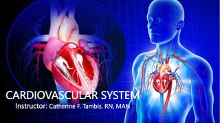
- 1. CARDIOVASCULAR SYSTEM Instructor: Catherine F. Tambis, RN, MAN
- 2. CARDIOVASCULAR SYSTEM The almost continuous traffic into and out of a busy factory at rush hour occurs at a snails’ pace compared to the endless activity going on within our bodies. Like the bustling factory, the body must have a transportation system to carry its various cargos back and forth, and this is where the cardiovascular system steps in
- 3. CARDIOVASCULAR SYSTEM Functions of the Heart 1. Managing blood supply. Variations in the rate and force of heart contraction match blood flow to the changing metabolic needs of the tissues during rest, exercise, and changes in body position. 2. Producing blood pressure. Contractions of the heart produce blood pressure, which is needed for blood flow through the blood vessels. 3. Securing one-way blood flow. The valves of the heart secure a one-way blood flow through the heart and blood vessels. 4. Transmitting blood. The heart separates the pulmonary and systemic circulations, which ensures the flow of oxygenated blood to tissues.
- 4. CARDIOVASCULAR SYSTEM Heart Structure and Functions The modest size and weight of the heart give few hints of its incredible strength. Weight: Approximately the size of a person’s fist, the hollow, cone-shaped heart weighs less than a pound. Mediastinum: Snugly enclosed within the inferior mediastinum, the medial cavity of the thorax, the heart is flanked on each side by the lungs. Apex: It’s more pointed apex is directed toward the left hip and rests on the diaphragm, approximately at the level of the fifth intercostal space. Base: Its broad posterosuperior aspect, or base, from which the great vessels of the body emerge, points toward the right shoulder and lies beneath the second rib.
- 6. CARDIOVASCULAR SYSTEM Heart Structure and Functions Pericardium: The heart is enclosed in a double-walled sac called the pericardium and is the outermost layer of the heart. a. Fibrous pericardium: The superficial part which helps protect the heart and anchors it to surrounding structures such as the diaphragm and sternum. b. Serous pericardium: Deep to the fibrous pericardium is the slippery, two-layer serous pericardium, where its parietal layer lines the interior of the fibrous pericardium.
- 7. CARDIOVASCULAR SYSTEM Layers of the Heart The modest size and weight of the heart give few hints of its incredible strength. The heart muscle has three layers and they are as follows: I. Epicardium: The visceral and outermost layer is actually a part of the heart wall. II. Myocardium: It consists of thick bundles of cardiac muscle twisted and whirled into ringlike arrangements and it is the layer that actually contracts. III. Endocardium: It is the innermost layer of the heart and is a thin, glistening sheet of endothelium that lines the heart chambers.
- 9. CARDIOVASCULAR SYSTEM Chambers of the Heart The heart has four hollow chambers, or cavities: two atria and two ventricles. Receiving chambers The two superior atria are primarily the receiving chambers, they play a lighter role in the pumping activity of the heart. (Right atrium and left atrium) Discharging chambers The two inferior, thick-walled ventricles are the discharging chambers, or actual pumps of the heart wherein when they contract, blood is propelled out of the heart and into the circulation (Right ventricle and left ventricle) Septum: The septum divides the heart longitudinally (the interventricular septum or the interatrial septum) depending on which chamber it separates.
- 11. CARDIOVASCULAR SYSTEM Associated Great Vessels The great blood vessels provide a pathway for the entire cardiac circulation to proceed. Superior and inferior vena cava: The heart receives relatively oxygen-poor blood from the veins of the body through the large superior and inferior vena cava and pumps it through the pulmonary trunk. Pulmonary arteries: The pulmonary trunk splits into the right and left pulmonary arteries, which carry blood to the lungs, where oxygen is picked up and carbon dioxide is unloaded. Pulmonary veins: Oxygen-rich blood drains from the lungs and is returned to the left side of the heart through the four pulmonary veins. Aorta: Blood returned to the left side of the heart is pumped out of the heart into the aorta from which the systemic arteries branch to supply essentially all body tissues.
- 13. CARDIOVASCULAR SYSTEM Heart Valves The heart is equipped with four valves, which allow blood to flow in only one direction through the heart chambers. 1) Atrioventricular valves: Atrioventricular or AV valves are located between the atrial and ventricular chambers on each side, and they prevent backflow into the atria when the ventricles contract. 2) Bicuspid valves: The left AV valve- the bicuspid or mitral valve, consists of two flaps, or cusps, of endocardium. 3) Tricuspid valve: The right AV valve, the tricuspid valve, has three flaps. 4) Semilunar valve: The second set of valves, the semilunar valves, guards the bases of the two large arteries leaving the ventricular chambers, thus they are known as the pulmonary and aortic semilunar valves.
- 15. CARDIOVASCULAR SYSTEM Cardiac Circulation Vessels Although the heart chambers are bathed with blood almost continuously, the blood contained in the heart does not nourish the myocardium. • Coronary arteries: The coronary arteries branch from the base of the aorta and encircle the heart in the coronary sulcus (atrioventricular groove) at the junction of the atria and ventricles, and these arteries are compressed when the ventricles are contracting and fill when the heart is relaxed. • Cardiac veins. The myocardium is drained by several cardiac veins, which empty into an enlarged vessel on the posterior of the heart called the coronary sinus.
- 19. CARDIOVASCULAR SYSTEM Systemic Circulation SVC&IVC Right Atrium Tricuspid valve Right Ventricle Pulmonary Artery LUNGS Pulmonary Vein Left Atrium Bicuspid valve Left Ventricle Aorta Arteries Arterioles CAPILLARIES Venules Veins
- 20. CARDIOVASCULAR SYSTEM Fetal Circulation During pregnancy, the fetal circulatory system works differently than after birth: The fetus is connected by the umbilical cord to the placenta. This is the organ that develops and implants in the mother's uterus during pregnancy. Through the blood vessels in the umbilical cord, the fetus gets all needed nutrition and oxygen. The fetus gets life support from the mother through the placenta. Waste products and carbon dioxide from the fetus are sent back through the umbilical cord and placenta to the mother's circulation to be removed.
- 21. CARDIOVASCULAR SYSTEM Fetal Circulation ( Right to Left side) The fetal circulatory system uses 3 shunts. These are small passages that direct blood that needs to be oxygenated. The purpose of these shunts is to bypass the lungs and liver. That's because these organs will not work fully until after birth. 1. Ductus venosus: This shunt lets the highly oxygenated blood bypass the liver to the inferior vena cava and then to the right atrium of the heart. (ligamentum venosum) 2. Foramen ovale: This shunt bypasses the lungs and moves blood from the right atrium of the heart to the left atrium. (Fossa ovalis) 3. Ductus arteriosus: This shunt moves blood from the pulmonary artery to the aorta. (Ligamentum arteriosum)
- 22. CARDIOVASCULAR SYSTEM Fetal Circulation Placenta Umbilical vein Ductus Venosus Bypass the LUNGS Pulmonary Vein Left Atrium Bicuspid valve Left Ventricle Aorta Bypass the liver SVC& IVC Right Atrium Tricuspid valve Right Ventricle Pulmonary Artery Fossa Ovalis Ductus arteriosus Systemic Circulation 1 2 3
- 24. CARDIOVASCULAR SYSTEM As the heart beats or contracts, the blood makes continuous round trips- into and out of the heart, through the rest of the body, and then back to the heart- only to be sent out again.
- 25. CARDIOVASCULAR SYSTEM Conduction System of the Heart The spontaneous contractions of the cardiac muscle cells occurs in a regular and continuous way, giving rhythm to the heart. Cardiac muscle cells: They can and do contract spontaneously and independently, even if all nervous connections are severed. Although cardiac muscles can beat independently, the muscle cells in the different areas of the heart have different rhythms. The atrial cells beat about 60x per minute, but the ventricular cells contract much more slowly (20-40/min).
- 26. CARDIOVASCULAR SYSTEM Types of Controlling Systems Act to Regulate Heart Activity 1. Nerves of the autonomic nervous system that act like “brakes and accelerators” to decrease or increase the heart rate 2. Intrinsic Conduction System/Nodal System The intrinsic conduction system is built into the heart tissue and sets the basic rhythm. It is much like a cross between a muscle and nervous tissue. This system causes heart muscle depolarization in only one direction- from the atria to the ventricles; it enforces a contraction rate of approximately 75 beats per minute on the heart, thus the heart beats as a coordinated unit.
- 27. CARDIOVASCULAR SYSTEM Types of Controlling Systems Act to Regulate Heart Activity 2. Intrinsic Conduction System/Nodal System Parts: a. Sinoatrial (SA) node: The SA node is located in the right atrium and has the highest rate of depolarization in the whole system, so it can start the beat and set the pace for the whole heart thus it is often called the “pacemaker“. b. Atrial contraction: From the SA node, the impulse spread through the atria to the AV node, and then the atria contract.
- 28. CARDIOVASCULAR SYSTEM Types of Controlling Systems Act to Regulate Heart Activity 2. Intrinsic Conduction System/Nodal System Parts: c. Ventricular contraction: It then passes through the AV bundle, the bundle branches, and the Purkinje fibers, resulting in a “wringing” contraction of the ventricles that begins at the heart apex and moves toward the atria. d. Ejection: This contraction effectively ejects blood superiorly into the large arteries leaving the heart.
- 29. CARDIOVASCULAR SYSTEM The Pathway of the Conduction System
- 30. CARDIOVASCULAR SYSTEM The Pathway of the Conduction System • SA node. The depolarization wave is initiated by the sinoatrial node. • Atrial myocardium. The wave then successively passes through the atrial myocardium. • Atrioventricular node. The depolarization wave then spreads to the AV node, and then the atria contract. • AV bundle. It then passes rapidly through the AV bundle. • Bundle branches and Purkinje fibers. The wave then continues on through the right and left bundle branches, and then to the Purkinje fibers in the ventricular walls, resulting in a contraction that ejects blood, leaving the heart.
- 31. CARDIOVASCULAR SYSTEM Cardiac Cycle and Heart Sounds In a healthy heart, the atria contract simultaneously, then, as they start to relax, contraction of the ventricles begin. • Systole means heart contraction. • Diastole means heart relaxation. • Cardiac cycle. The term cardiac cycle refers to the events of one complete heart beat, during which both atria and ventricles contract and then relax.
- 32. CARDIOVASCULAR SYSTEM Cardiac Cycle and Heart Sounds • Length. The average heart beats approximately 75 times per minute, so the length of the cardiac cycle is normally about 0.8 second. • First heart sound. The first heart sound, “lub”, is caused by the closing of the AV valves. • Second heart sound. The second heart sound, “dub”, occurs when the semilunar valves close at the end of systole. • Cardiac output is the amount of blood pumped out by each side of the heart in one minute. It is the product of the heart rate and the stroke volume.
- 33. CARDIOVASCULAR SYSTEM Blood Vessels Blood circulates inside the blood vessels, which form a closed transport system, the so-called vascular system. Arteries: As the heart beats, blood is propelled into large arteries leaving the heart; carry oxygenated blood Arterioles: It then moves into successively smaller and smaller arteries and then into arterioles, which feed the capillary beds in the tissues. Veins: Capillary beds are drained by venules, which in turn empty into veins that finally empty into the great veins entering the heart; carry deoxygenated blood
- 34. CARDIOVASCULAR SYSTEM Tunics Except for the microscopic capillaries, the walls of the blood vessels have three coats or tunics. 1. Tunica intima: The tunica intima, lines the lumen or interior of the vessels. It is a thin layer of endothelium resting on a basement membrane and decreases friction as blood flows through the vessel lumen. 2. Tunica media: The tunica media is the bulky middle coat which mostly consists of smooth muscle and elastic fibers that constrict or dilate, making the blood pressure increase or decrease. 3. Tunica externa: The tunica externa is the outermost tunic composed largely of fibrous connective tissue, and its function is basically to support and protect the vessels.
- 36. CARDIOVASCULAR SYSTEM Major Arteries of the Systemic Circulation Arterial Branches of the Ascending Aorta The aorta springs upward from the left ventricle of heart as the ascending aorta. 1. Coronary arteries. The only branches of the ascending aorta are the right and left coronary arteries, which serve the heart.
- 37. CARDIOVASCULAR SYSTEM Major Arteries of the Systemic Circulation Arterial Branches of the Aortic Arch The aorta arches to the left as the aortic arch. • Brachiocephalic trunk. The brachiocephalic trunk, the first branch off the aortic arch, splits into the right common carotid artery and right subclavian artery. • Left common carotid artery. The left common carotid artery is the second branch off the aortic arch and it divides, forming the left internal carotid, which serves the brain, and the left external carotid, which serves the skin and muscles of the head and neck.
- 38. CARDIOVASCULAR SYSTEM Major Arteries of the Systemic Circulation. Arterial Branches of the Aortic Arch • Left subclavian artery. The third branch of the aortic arch, the left subclavian artery, gives off an important branch- the vertebral artery, which serves part of the brain. • Axillary artery. In the axilla, the subclavian artery becomes the axillary artery. • Brachial artery. the subclavian artery continues into the arm as the brachial artery, which supplies the arm. • Radial and ulnar arteries. At the elbow, the brachial artery splits to form the radial and ulnar arteries, which serve the forearm.
- 40. CARDIOVASCULAR SYSTEM Major Veins of the Systemic Circulation Major veins converge on the venae cavae, which enter the right atrium of the heart. Veins Draining into the Superior Vena Cava Veins draining into the superior vena cava are named in a distal-to-proximal direction; that is, in the same direction the blood flows into the superior vena cava. Radial and ulnar veins. The radial and ulnar veins are deep veins draining the forearm; they unite to form the deep brachial vein, which drains the arm and empties into the axillary vein in the axillary region. Cephalic vein. The cephalic vein provides for the superficial drainage of the lateral aspect of the arm and empties into the axillary vein. Basilic vein. The basilic vein is a superficial vein that drains the medial aspect of the arm and empties into the brachial vein proximally.
- 41. CARDIOVASCULAR SYSTEM Major Arteries of the Systemic Circulation Veins Draining into the Superior Vena Cava Median cubital vein. The basilic and cephalic veins are joined at the anterior aspect of the elbow by the median cubital vein, often chosen as the site for blood removal for the purpose of blood testing. Subclavian vein. The subclavian vein receives venous blood from the arm through the axillary vein and from the skin and muscles of the head through the external jugular vein. Vertebral vein. The vertebral vein drains the posterior part of the head. Internal jugular vein. The internal jugular vein drains the dural sinuses of the brain. Brachiocephalic veins. The right and left brachiocephalic veins are large veins that receive venous drainage from the subclavian, vertebral, and internal jugular veins on their respective sides. Azygos vein. The azygos vein is a single vein that drains the thorax and enters the superior vena cava just before it joins the heart.
- 42. CARDIOVASCULAR SYSTEM Major Arteries of the Systemic Circulation Veins Draining into the Inferior Vena Cava The inferior vena cava, which is much longer than the superior vena cava, returns blood to the heart from all body regions below the diaphragm. Tibial veins. The anterior and posterior tibial veins and the fibular vein drain the leg; the posterior tibial veins becomes the popliteal vein at the knee and then the femoral vein in the thigh; the femoral vein becomes the external iliac vein as it enters the pelvis. Great saphenous veins. The great saphenous veins are the longest veins in the body; they begin at the dorsal venous arch in the foot and travel up the medial aspect of the leg to empty into the femoral vein in the thigh.
- 43. CARDIOVASCULAR SYSTEM Major Arteries of the Systemic Circulation Veins Draining into the Inferior Vena Cava Common iliac vein. Each common iliac vein is formed by the union of the external iliac vein and the internal iliac vein which drains the pelvis. Gonadal vein. The right gonadal vein drains the right ovary in females and the right testicles in males; the left gonadal veins empties into the left renal veins superiorly. Renal veins. The right and left renal veins drain the kidneys. Hepatic portal vein. The hepatic portal vein is a single vein that drains the digestive tract organs and carries this blood through the liver before it enters the systemic circulation. Hepatic veins. The hepatic veins drain the liver.
- 47. CARDIOVASCULAR SYSTEM Cardiovascular Vital Signs Arterial pulse. The alternating expansion and recoil of an artery that occurs with each beat of the left ventricle creates a pressure wave-a pulse- that travels through the entire arterial system. Normal pulse rate. Normally, the pulse rate (pressure surges per minute) equals the heart rate, so the pulse averages 70 to 76 beats per minute in a normal resting person. Pressure points. There are several clinically important arterial pulse points, and these are the same points that are compressed to stop blood flow into distal tissues during hemorrhage, referred to as pressure points.
- 49. CARDIOVASCULAR SYSTEM Cardiovascular Vital Signs Blood pressure. Blood pressure is the pressure the blood exerts against the inner walls of the blood vessels, and it is the force that keeps blood circulating continuously even between heartbeats. Systolic pressure (the pressure in the arteries at the peak of ventricular contraction) and diastolic pressure (the pressure when the ventricles are relaxing). Peripheral resistance. Peripheral resistance is the amount of friction the blood encounters as it flows through the blood vessels.
Notes de l'éditeur
- https://www.youtube.com/watch?v=z4Eqyylf2_4
- https://www.youtube.com/watch?v=TnFoJ7Hhi-M