ADULT NEURODEGENERATIVE DISORDERS .pptx
•Télécharger en tant que PPTX, PDF•
0 j'aime•16 vues

inherited neurodegenerative disorders NIMS HOSPITAL,HYDERABAD

Recommandé
Contenu connexe
Similaire à ADULT NEURODEGENERATIVE DISORDERS .pptx
Similaire à ADULT NEURODEGENERATIVE DISORDERS .pptx (20)
Plus de Sruthi Meenaxshi
Plus de Sruthi Meenaxshi (20)
Dernier
Dernier (20)
ADULT NEURODEGENERATIVE DISORDERS .pptx
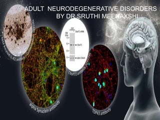
- 1. ADULT NEURODEGENERATIVE DISORDERS BY DR.SRUTHI MEENAXSHI
- 2. NEURODEGENERATIVE disorders, which are chronic and progressive, are characterized by selective and symmetric loss of neurons in motor, sensory, or cognitive systems.
- 3. Martin JB. N Engl J Med . Classification of Inherited Neurodegenerative Disorders.
- 4. Alzheimer’s disease usually begins in the seventh to ninth decades of life, but an early-onset familial form is now well recognized. The neuropathological hallmarks of neurofibrillary tangles and senile plaques were described by a German psychiatrist,Alois Alzheimer, in 1907. Neurofibrillary tangles are aggregations of the microtubular protein tau, which is hyperphosphorylated.
- 5. Senile (neuritic) plaques result from the accumulation of several proteins and an inflammatory reaction around deposits of b- amyloid. Degenerating nerve terminals in the plaques also contain tau. As these cellular changes progress, neurons are lost in the hippocampus, entorhinal cortex, and association areas of the neocortex.
- 9. Several illnesses characterized by dementia and often called Pick’s disease are associated with selective loss and atrophy of neurons in the frontal and temporal lobes The typical initial clinical findings are abnormal behavior without memory loss, followed by progressive dementia and, in some patients, parkinsonian symptoms of bradykinesia and rigidity. Brain imaging reveals severe frontal and temporal atrophy caused by neuronal loss in the cerebral cortex and amygdala, with less marked changes in the hippocampus. Tau-positive inclusions are found in neurons and glial cells, but senile plaques are not present.
- 10. Although usually sporadic in occurrence, a rare autosomal dominant inherited form of frontotemporal dementia associated with Parkinson’s disease was linked to the same region of chromosome 17q21– 22 as the gene for tau. Analysis of the tau gene in one family with frontotemporal dementia revealed nine polymorphisms, of which eight could be discounted because of their occurrence in normal subjects. One variation, however, Val279Met, was linked to the disease. Another mutation involves an alternative splice site downstream from exon10, six different mutations in this region have been identified in 13 families with frontotemporal dementia and parkinsonism. These findings suggest that errors in the splicing of exon 10 may result in the production of tau molecules that bind abnormally to microtubules. This error may cause tau to polymerize into neurofibrillary tangles.
- 13. Eight neurologic disorders are caused by an increase in the number of CAG repeats that encode expanded sequences of glutamine residues . These disorders are characterized by autosomal dominant or X-linked inheritance, onset in midlife, a progressive course, anticipation (a tendency toward earlier onset in successive generations), preponderance of unstable repeats from the paternal chromosome, and correlation of the number of CAG repeats with the severity of disease and the age at onset
- 15. Huntington’s disease is an autosomal dominant disorder with high penetrance. The characteristic findings of progressive chorea and dementia are caused by severe neuronal loss, initially in the neostriatum and later in the cerebral cortex. Huntington’s disease was linked initially to chromosome 4p16.3,8 in which one gene, initially labeled IT15 and now called HD, was found to contain an unstable CAG repeat in the open reading frame of its first exon. Normal subjects have a median of 19 CAG repeats (range, 11 to 34), whereas nearly all patients with Huntington’s disease have more than 40. Unstable or dynamic mutations have been identified in a few families, in which one parent has 34 to 38 CAG repeats and the progeny have more than 40. The HD gene encodes a protein named huntingtin
- 18. The Spinocerebellar ataxias (SCA) are a subset of hereditary cerebellar ataxias that are autosomal dominantly transmitted. They are progressive neurodegenerative diseases that share the clinical features of ataxia, which arise from the progressive degeneration of the cerebellum but can also affect other connected regions,including the brain stem
- 19. They are ahighly heterogenous group of disorders with a complex genotype– phenotype spectrum;many SCAs are caused by CAG nucleotide repeat expansions that encode polyglutamine,and therefore,involve the toxic polyglutamine protein (polyQ)
- 20. Spinocerebellar ataxia – An update ,Sullivian et al, Journal of Neurology https://doi.org/10.1007/s00415-018-9076-4
- 21. Spinocerebellar ataxia – An update ,Sullivian et al, Journal of Neurology https://doi.org/10.1007/s00415-018-9076-4
- 22. Spinocerebellar ataxia – An update ,Sullivian et al, Journal of Neurology https://doi.org/10.1007/s00415-018-9076-4
- 27. Prion diseases have a broad spectrum of clinical manifestations, including dementia, ataxia, insomnia, paraplegia, paresthesias, and deviant behavior. Neuropathological findings range from an absence of atrophy to widespread atrophy, from minimal to widespread neuronal loss, from sparse to widespread vacuolation or spongiform changes, from mild to severe reactive astrocytic gliosis, and from an absence ofPrPamyloid plaques to an abundance of plaques. None of these findings except the presence of PrP amyloid plaques is unequivocally diagnostic of a prion disease.The sporadic form of Creutzfeldt–Jakob disease, which is typically manifested as dementia and myoclonus, accounts forapproximately 85 percent of all cases of prion disease inhumans,whereas infectious and inherited prion diseases account for the rest.
- 28. Familial Creutzfeldt–Jakob disease, Gerstmann– Sträussler–Scheinker disease,and fatal familial insomnia are all dominantly inherited prion diseases caused by mutations in the prion protein gene PRNP Experiments that showed transmission of these diseases by filtrates of brain from familial cases were wrongly attributed to a virus. There is no Creutzfeldt– Jakob disease virus, and familial prion diseases are caused by mutations in PRNP
- 30. Friedreich's ataxia is an autosomal recessive disorder caused by an increase in the number of trinucleotide GAA repeats within the first intron of the FRDA gene on chromosome 9 that encodes the protein frataxin The disease is characterized clinically by the onset in the first two decades of life of limb ataxia, cerebellar dysarthria, a lack of deep- tendon reflexes, pyramidal signs, and sensory loss. Most patients have skeletal deformities and hypertrophic cardiomyopathy. They also have an increased incidence of blindness, deafness, and diabetes mellitus, suggesting that the disorder is systemic and is not limited to the nervous system.
- 31. Friedreich's ataxia is the most common of the hereditary ataxias The locus was mapped to chromosome 9 in 1988, and the FRDA gene was cloned in 1996. More than 95 percent of patients with classic Friedreich's ataxia are homozygous for the increase in GAA repeats, but a few have a combination of an increase in GAA repeats in one allele and a point mutation in the other allele, confirming that Friedreich's ataxia is a loss-of- function disorder. The disorder may be caused by interference by the intronic GAA repeat with transcription of the FRDA gene
- 32. The neural pathways affected in Friedreich's ataxia are those associated with large neuronal cell bodies and extensive axon elongations — the long tracts of the dorsal columns, pyramidal system, and peripheral nerves. Many of the clinical manifestations of the disease are mimicked by disorders of vitamin E metabolism. In affected patients, the concentrations of a partially degraded, 180-kd form of frataxin are abnormally low in muscle, cerebellar cortex, and cerebral cortex, and the frataxin is closely associated with mitochondria.
- 33. Frataxin contains 210 amino acids, and its N- terminal region contains an α-helix that might target the protein to mitochondria. The current hypothesis is that frataxin is a mitochondrial protein important for normal production of cellular energy. A defect in its action may result in abnormal accumulation of iron in mitochondria,followed by the death of the neurons that are most vulnerable to this abnormality.
- 35. Parkinson's disease is the second most common neurodegenerative disorder after Alzheimer's disease, with a prevalence of 2 percent among people over the age of 65 years. The characteristic symptoms of rigidity, bradykinesia, and tremor are associated with loss of cells in the substantia nigra and depletion of dopamine in the striatum. Large intracytoplasmic inclusions called Lewy bodies are the pathological hallmark of the disease, occurring predominantly in the melanin-containing neurons of the substantia nigra. Linkage data in a subgroup of families with autosomal dominant Parkinson's disease identified a disease locus on chromosome 4q21–23 and mutations in the gene for a synaptic protein, α- synuclein.
- 38. In patients with both inherited and sporadic Parkinson's disease, the Lewy bodies contain α-synuclein, ubiquitin, and proteasomal subunits. Mutations in another protein, ubiquitin carboxy-terminal hydrolase, have also been reported to cause familial Parkinson's disease. These mutations are hypothesized to lead to aberrations in the proteolytic pathway and to aggregation of proteins as Lewy bodies. In another common condition, diffuse Lewy body disease (also called Lewy body dementia), Lewy bodies are widely distributed in cortical neurons. The Lewy bodies in this condition are identical biochemically to those in sporadic or inherited Parkinson's disease.
- 39. Motor neurons extend from the brain stem throughout the spinal cord, forming the final common pathway for motor control. Lower motor neurons innervate muscles, and upper motor neurons carry impulses to lower motor neurons for voluntary muscle movements. Motor-neuron axons are among the longest in the nervous system. In normal persons, they are capable of surviving more than 100 years. Many inherited and sporadic misadventures may befall them, giving rise to muscle atrophy and weakness, and with the death of upper motor neurons, increased muscle tone (spasticity), hyperreflexia, and extensor plantar responses.
- 40. The most common motor-neuron disorder is amyotrophic lateral sclerosis, which usually begins in the fifth and sixth decades of life. In a typical patient, muscles innervated by both brain stem and spinal cord atrophy as lower motor neurons die, although those that control eye movements and bowel and bladder function are spared. The illness is usually sporadic, but in 1 to 10 percent of patients it is familial, being inherited as an autosomal dominant trait. The inherited and sporadic forms are indistinguishable clinically. In both forms the prognosis is grave, with death occurring in three to five years in 95 percent of patients. Neuropathological studies show loss of motor neurons throughout the neuraxis. The death of neurons is preceded by perikaryal shrinkage, the formation of webs of ubiquitin-positive threads, and axonal swellings that stain for ubiquitin and for α-synuclein.
- 41. The cause of motor-neuron loss in amyotrophic lateral sclerosis remains unknown, but a subgroup of patients with the familial form (less than 20 percent) have mutations in the superoxide dismutase type 1 (SOD1) gene on chromosome 21, which encodes a protein involved in the regulation of intracellular free radicals.
- 43. A consideration of motor-neuron disorders should not be limited to those with an onset in adult life. There are several inherited autosomal recessive disorders of childhood, grouped together as spinal muscular atrophy, in which lower motor neurons are predominantly affected. Two genes on chromosome 5q13 are associated with the disorder. The most important of these genes is one that encodes a novel protein, survival motor neuron, which appears to act by influencing the metabolism of messenger RNA. The survival motor neuron (SMN) gene is duplicated on chromosome 5q, with one copy centromeric and the other telomeric in position. The two copies differ only by single base pairs in exons 7 and 8.
- 45. Approximately 95 percent of patients with spinal muscular atrophy have deletions in the telomeric SMN gene Others have short deletions or missense mutations. There may be a balance between the numbers of copies expressed by the centromeric and telomeric SMN genes. In some mildly affected patients, there is a complete absence of expression of the telomeric SMN gene but overexpression of the centromeric SMN gene. The role of another gene in the 5q region that encodes a protein called neuronal apoptosis inhibitory protein (based on its homology with certain viral antiapoptotic proteins) remains controversial. It may be neuroprotective in the disorder, resulting in less severe symptoms when fully expressed, since it seems to be so in some patients with telomeric SMN mutations.
- 49. Spinal and bulbar muscular atrophy is an X- linked disorder with an onset in midlife in which increased numbers of CAG repeats are found in the androgen receptor gene Affected patients have symptoms of motor- neuron loss throughout the neuraxis, caused presumably by selective effects of the CAG repeats in neurons rich in androgen receptors.
- 50. Familial spastic paraparesis is inherited as either an autosomal dominant or X-linked condition that selectively affects upper motor neurons. Linkage studies point to genetic heterogeneity with loci on chromosomes 2p, 14q, and 15q. One form is reported to be associated with an increased number of CAG repeats in a gene on chromosome 2p21– 24, but this association has not been confirmed and the gene product has not been identified. The gene for an autosomal recessive variant of familial spastic paraparesis encodes a protein, paraplegin.
- 54. PROGRESSIVE NEUROLOGICALAND MENTAL DETERORIATION
- 56. Leukodystrophies currently are defined as genetically determined disorders that primarily affect the white matter of the central nervous system, regardless of the structural white matter component, molecular process, patient age group, and disease course involved. Genetic testing is of paramount importance
- 57. Leukodystrophies usually affect children, but in the last several decades, many instances of adult leukodystrophies have been reported in the medical literature. Because the clinical manifestation of these diseases can be nonspecific, MRI can help with establishing a diagnosis.
- 61. Metachromatic leukodystrophy is an autosomal recessive lysosomal condition due to arylsulfatase A (ARSA) gene mutations, resulting in deficiency of the enzyme arylsulfatase A (ASA) that leads to accumulation of 3-O-sulfogalactosylceramide (sulfatide) in oligodendrocytes, Schwann cells, and some neurons
- 62. Adults account for approximately 20% of patients with metachromatic leukodystrophy. The initial symptoms are often behavioral and psychiatric changes, followed by a slow decline in memory and intellectual abilities. Later, the onset of motor symptoms including spastic paraparesis and cerebellar ataxia and peripheral neuropathy occur.
- 63. MRI findings consist of confluent, symmetricT2 hyperintensity in the frontal or periventricular white matter (Fig 6). The subcortical U fibers are spared, and frequently some frontal predominance is present in patients with adult-onset metachromatic leukodystrophy. Loss of white matter volume results in brain atrophy in the late stages of the disease
- 65. Niemann-Pick type C disease (NPC) is a rare, neurovisceral, autosomal recessive disease, with an extremely heterogeneous clinical presentation. It is characterized by a wide range of symptoms that are not specific, such as neurological, systemic or psychiatric symptoms. The adult form of the disease concerns a small proportion (5 %) of the people affected and is usually expressed as a neurological form. A variety of progressive and disabling symptoms are encountered, mainly cerebellar signs (cerebellar ataxia, impaired gait, dysarthria), but also movement disorders, cataplexy, seizures and dysphagia. Patients face constant cognitive deterioration, which can result in severe dementia. Abnormal saccadic eye movement is often the first manifestation of the disease. Supranuclear gaze palsy is considered to be a specific sign and should be systematically searched for.
- 66. In terms of systemic signs, the usual infantile hepatosplenomegaly is very fickle in the adult form; if present, it is usually asymptomatic. Non-specific psychiatric symptoms are often associated with NPC disease. For one third of cases, it can also express as an isolated psychiatric- disorder form, such as schizophrenia-like psychosis (paranoid delusions, auditory hallucinations, interpretative thoughts, and disorganization), depression, bipolar disorder, obsessive-compulsive behaviour and behavioural problems (sleep disorders, hyperactivity, agitation, aggressiveness or self-mutilations). This psychiatric overview is mostly atypical and is accompanied by visual hallucinations, confusion, symptom fluctuations, treatment resistance or aggravation with neuroleptic drugs, catatonia, progressive cognitive decline, but also seizures.
- 67. NPC diagnosis is based on a filipin test on a fibroblast culture from a skin biopsy and also on a sequencing of the NPC1 and NPC2 genes. Routine laboratory biochemistry profiles are generally normal. The early diagnosis is fundamental to deploy the best follow-up care. The patient should therefore be in contact with a reference centre. Until recently, NPC treatment consisted in supportive therapies and symptomatic drugs, useful, however, with variable efficacy. The recent discovery of a medicine called Miglustat (N- butyldeoxynojirimycin; NB-DJN; Zavesca(®),Actelion Pharmaceuticals Ltd.) which improves the disease evolution, should encourage psychiatrists to look for it in every atypical psychosis.
- 68. 3-Hydroxy-3-methylglutaric aciduria (3-HMG, OMIN 246450) is a rare autosomal recessive metabolic disorder caused by a deficiency of 3-hydroxy-3-methylglutaryl-CoA lyase, a key enzyme in leucine metabolism and ketone body synthesis. Acute episodes of 3-HMG may be triggered by fasting or infection, and symptoms include vomiting, diarrhea, lethargy and hypotonia. If left untreated, prolonged hypoglycemia and metabolic acidosis may cause breathing problems, seizures, and coma. In addition, 3-HMG is associated with damage to the central nervous system, and there are several reports of white matter abnormality or cerebral atrophy
- 69. Wilson disease is an autosomal recessive condition, although it was not until 1985 that it was linked to chromosome 13. In 1993, 2 separate groups described mutations in the ATP7B gene in patients with Wilson disease. More than 500 ATP7B mutations have now been identified (http://www.wilsondisease.med.ualberta.ca/database.asp). Individual gene mutations have not been reliably associated with different presentations of Wilson disease although there is some evidence that truncating mutations may be associated with earlier onset than missense mutation and patients with frameshift mutations may be more likely to develop neurologic symptoms. Interestingly, there are case reports of monozygotic twins who are phenotypically discordant for Wilson disease. This finding strongly suggests there must be at least some contribution of environmental and epigenetic factors contributing toWilson disease
- 70. The copper metabolism disorder Wilson's disease was first defined in 1912. Wilson's disease can present with hepatic and neurological deficits, including dystonia and parkinsonism. Early-onset presentations in infancy and late-onset manifestations in adults older than 70 years of age are now well recognised. Direct genetic testing for ATP7B mutations are increasingly available to confirm the clinical diagnosis of Wilson's disease
- 73. Homocystinuria caused by cystathionine β- synthase (CBS) deficiency is characterized by involvement of the eye (ectopia lentis and/or severe myopia), skeletal system (excessive height, long limbs, scolioisis, and pectus excavatum), vascular system (thromboembolism), and CNS (developmental delay/intellectual disability). All four ‒ or only one ‒ of the systems can be involved; expressivity is variable for all of the clinical signs.
- 74. Two phenotypic variants are recognized, B6-responsive homocystinuria and B6-non-responsive homocystinuria. B6-responsive homocystinuria is usually milder than the non-responsive variant. Thromboembolism is the major cause of early death and morbidity. IQ in individuals with untreated homocystinuria ranges widely, from 10 to 138. In B6-responsive individuals the mean IQ is 79 versus 57 for those who are B6-non-responsive. Other features that may occur include: seizures, psychiatric problems, extrapyramidal signs (e.g., dystonia), hypopigmentation of the skin and hair, malar flush, livedo reticularis, and pancreatitis.
- 75. The cardinal biochemical features of homocystinuria include markedly increased concentrations of plasma total homocysteine and methionine. The diagnosis can be substantiated by detection of biallelic pathogenic variants in CBS, the gene encoding cystathionine β- synthase.
- 76. Prevention of primary manifestations: Individuals are treated to maintain normal or near-normal plasma total homocysteine concentrations using vitamin B6 (pyridoxine) therapy (if shown to be B6 responsive), a methionine-restricted diet, and folate and vitamin B12 supplementation. Betaine therapy is usually added to the therapeutic regimen; in adolescents and adults, betaine may be the major form of treatment, but it is preferable to remain on life-long metabolic diet.
- 78. The neuronal ceroid lipofuscinoses (NCLs) are at least 13 distinct progressive neurodegenerative disorders unified by the accumulation of lysosomal auto- fluorescent material called lipofuscin The only form that occurs via autosomal-dominant inheritance exhibits adult onset and is sometimes referred to as Parry type NCL. The manifestations may include behavioral symptoms followed by seizures, ataxia, dementia, and early death Mutations in the gene DNAJC5 that codes for the presynaptic co-chaperone cysteine string protein-α (CSPα) were recently reported in sporadic adult-onset cases and in families with dominant inheritance. The mutant CSPα protein may lead to disease progression by both loss and gain of function mechanisms. Iron chelation therapy may be considered as a possible pharmaceutical intervention
- 79. Clinically, most of these patients first presented with seizures. In our experience, other milder features such as behavioral changes, progressive tremor, myoclonus, memory loss, and frequent falls may precede the onset of seizures. The age of onset of clinical seizures ranges from mid-20s to mid-40s. All reported patients had abnormal EEG readings at the time of their initial clinical manifestations. The EEG abnormalities at earlier stages of the disease showed predominance of diffuse theta and delta slow waves during wakefulness, intermittent delta activity, and generalized spike-wave and poly spike-wave complexes indicative of epileptogenic potential. These changes become more prominent during the disease progression. After the onset of the seizures, there is slow, progressive cognitive decline, ataxia, memory loss, and speech impairment. Some patients show signs of depression. Age at death typically ranges from late 30s to 60 years and appears to be dependent upon the age of onset of the seizures.The usual cause of death is severe neurological compromise and resulting multi-organ failure
- 80. Alexander Disease.—Alexander disease is the result of an autosomal dominant mutation inthe glial fibrillary acidic protein (GFAP) gene. These mutations usually are de novo but may be hereditary in patients with adult-onset disease. This condition is characterized pathologically by diffuse Rosenthal fiber accumulation in astrocyte cytoplasms. Molecular testing is diagnostic, and a brain biopsy is no longer required to confirm the diagnosis (37,39).
- 81. The clinical presentation includes slowly progressive bulbar dysfunction (dysphagia, dysarthria, and dysphonia), pyramidal signs, and ataxia, with normal cognitive and intellectual functions. When present, palatal myoclonus is suggestive of this diagnosis (8,40,41). MRI findings in patients who develop Alexander disease in adulthood differ substantially from those of the early-onset form of the disease (Fig 9). White matter signal intensity abnormalities (increased signal intensity on T2-weighted and FLAIR MR images) and mild to severe atrophy of the medulla oblongata extending caudally to the upper cervical spinal cord are the hallmarks of this condition. Middle cerebellar peduncle signal intensity abnormalities also may be observed, and patchy enhancement may be present in affected regions.
- 83. Cerebrotendinous xanthomatosis (CTX) is a treatable autosomal recessive disorder and is characterized as a lipid storage disease caused by a mutation of the cytochrome P450 family 27 subfamily A member 1 (CYP27A1) gene, which leads to a deficiency of the mitochondrial enzyme 27-hydroxylase. This enzyme catalyzes one of the first steps in the metabolism of sterols. CTX results in the development of a tendinous xanthoma, early cataracts, diarrhea, and leukoencephalopathy. If left untreated, patients might develop progressive dementia and psychiatric symptoms The earliest and most relevant MRI features of CTX are involvement of the cerebellar white matter (Fig 11).
- 85. High signal intensity in the deep periventricular white matter and low or high signal intensity in the dentate nucleus onT2-weighted MR images is characteristic. T2- hypointense areas may be observed and probably represent hemosiderin deposition MR spectroscopy in patients with CTX shows increases in lactate and lipid peaks and a decreased N- acetyl aspartate peak, mainly in the cerebellum.The presence of lipid peaks is reasonable because CTX is a lipid storage disease.
- 86. Krabbe Disease.—Less commonly, Krabbe disease can manifest as diffuse periventricular white matter involvement
- 88. L-2-hydroxyglutaric aciduria is a rare neurometabolic disorder with autosomal recessive inheritance (L-2-hydroxyglutarate dehydrogenase [L2HGDH] gene) that is characterized by leukoencephalopathy that predominantly affects subcortical white matter (Fig 8) (14) . Patients initially may appear asymptomatic or may have a static encephalopathy .The neurologic symptoms are progressive and include cerebellar ataxia and intellectual decline . Hearing loss is another possible symptom of the disease. In certain patients, these features become apparent only in adulthood. Increased L2-hydroxyglutaric acid in urine is diagnostic (37)
- 89. . MRI in patients with L-2-hydroxyglutaric aciduria shows predominant subcortical white matter involvement, initially focal and evolving to become confluent. Periventricular white matter is spared. Increased signal intensity onT2-weighted or FLAIR MR images may be observed in the globus pallidus, and less importantly, in the caudate nucleus and putamen, with symmetric distribution. These same signal intensity abnormalities also may be observed in the dentate nucleus (38)
- 91. afora disease (LD) is an autosomal recessive progressive myoclonus epilepsy due to mutations in the EPM2A (laforin) and EPM2B (malin) genes, with no substantial genotype-phenotype differences between the two. Founder effects and recurrent mutations are common, and mostly isolated to specific ethnic groups and/or geographical locations. Pathologically, LD is characterized by distinctive polyglucosans, which are formations of abnormal glycogen. Polyglucosans, or Lafora bodies (LB) are typically found in the brain, periportal hepatocytes of the liver, skeletal and cardiac myocytes, and in the eccrine duct and apocrine myoepithelial cells of sweat glands.
- 92. With the exception of a few missense mutations LD is clinically homogeneous, with onset in adolescence. Symptoms begin with seizures, and neurological decline follows soon after. The disease course is progressive and fatal, with death occurring within 10 years of onset. Antiepileptic drugs are mostly non-effective, with none having a major influence on the progression of cognitive and behavioral symptoms. Diagnosis and genetic counseling are important aspects of LD, and social support is essential in disease management. Future therapeutics for LD will revolve around the pathogenesics of the disease. Currently, efforts at identifying compounds or approaches to reduce brain glycogen synthesis appear to be highly promising.
- 94. GM1 gangliosidosis is a rare disease that could manifest either during infancy (infantile form-type 1), as a juvenile (type 2), or in adulthood (type 3). There is a deficiency of the enzyme acid -galactosidase in both nervous and skeletal systems, but when it involves the skeletal system alone, it is called Morquio’s disease. The adult form (type 3) typically presents during childhood or adolescence as a slowly progressive dementia with prominent parkinsonian features and extrapyramidal disease, particularly dystonia. Marked phenotypic variability may occur.Age at death may vary greatly. Nervous system involvement in infantile and juvenile forms is generalized, but is localized in the adult form.
- 95. Similarly, skeletal involvement is generalized in the infantile form but localized in late infantile and adult forms of the disease. Pathology in adult (type 3) GM1 gangliosidosis is localized to basal ganglia with prominent involvement of caudate, putamen, and, to a lesser degree, amygdala and globus pallidum. Dystonia thus is a major neurological manifestation in adults. Adult (type 3) form of GM1 gangliosidosis shows higher residual activities of enzyme -galactosidase than infantile or juvenile forms2 and has the I51T mutation
- 97. Neurodegeneration with brain iron accumulation (NBIA) comprises a clinically and genetically heterogeneous group of disorders affecting children and adults. These rare disorders are often first suspected when increased basal ganglia iron is observed on brain magnetic resonance imaging. For the majority of NBIA disorders the genetic basis has been delineated, and clinical testing is available.
- 98. The four most common NBIA disorders include 1. pantothenate kinase-associated neurodegeneration (PKAN) due to mutations in PANK2, 2. phospholipase A2-associated neurodegeneration caused by mutation in PLA2G6, 3. mitochondrial membrane protein-associated neurodegeneration from mutations in C19orf12, 4. and beta-propeller protein-associated neurodegeneration due to mutations inWDR45.
- 99. The ultrarare NBIA disorders are caused by mutations in CoASY, ATP13A2, and FA2H (causing CoA synthase protein-associated neurodegeneration, Kufor-Rakeb disease, and fatty acid hydroxylase-associated neurodegeneration, respectively). Together, these genes account for disease in approximately 85% of patients diagnosed with an NBIA disorder
Notes de l'éditeur
- Table 1. Classification of Inherited Neurodegenerative Disorders.