Liver abscess
•Télécharger en tant que PPT, PDF•
20 j'aime•2,343 vues

a brief description on liver abscess, clinical features and management

Recommandé
Contenu connexe
Tendances
Tendances (20)
Similaire à Liver abscess
Similaire à Liver abscess (20)
Plus de Suman Baral
Dernier
Dernier (20)
Liver abscess
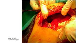
- 1. LIVER ABSCESS Suman Raj Baral MS, General Surgery
- 2. ANATOMY ?? ANATOMY - BASICS
- 4. Pyogenic Liver Abscess • Liver is the most common site of abdominal visceral abscess • PLA accounts for majority of hepatic abscess • NO significant gender, ethnic or geographic differences in disease frequency • Associated comorbid conditions are Cirrhosis, CRF and history of malignancy
- 5. Aetiology •E.coli : MC in western countries •Klebsiella pneumonia: MC in Asian countries •Staphylococcus : MC in children suffering from chronic granulomatous disease • Multiple Abscesses occur in those with biliary origin • Solitary Abscesses tend to be multiple and polymicrobial
- 6. Routes of Transmission •Biliary Tract (MC) • CBD stones leading to cholangitis (Asia) • Hilar Cholangiocarcinoma in western countries • CBD Strictures Portal Vein : 2nd MC Hepatic Artery: Hematogenous Spread, usually monomicrobial, staphylococcus or streptococcus Direct Extension: From Subdiaphragmatic Abscess, Suppurative Cholecystitis, empyema in chest, Perinephric abscess Penetrating or Blunt Trauma Cryptogenic: undiagnosed abdominal pathology, resolved infectious processes at the time of presentation or host factors such as diabetes or malignancy rendering liver for more susceptible to transient hepatic artery or portal vein bacteremia
- 7. Clinical Features • MC presentation is Fever. • MC LFT abnormality is raised ALP. • Classic Presentation: Fever, Jaundice(25%)and right upper quadrant pain and tenderness • MC presenting symptoms: Fever, Chills and Abdominal Pain • Usually single, involves right lobe • Malignancy, jaundice, deranged LFT and sepsis are associated with poor prognosis
- 8. Diagnosis • USG and CT are the main diagnostic modalities • USG- round, oval area that is less echogenic than the surrounding liver • CT- Similar findings of USG with and lesions are of lower attenuation than surrounding parenchyma • Confirmed by aspiration and culture. • Chest X-ray: Elevated Hemi-diaphragm, right sided pleural effusion or • atelectasis
- 9. Treatment • Percutaneous Catheter Drainage + IV Antibiotics : Treatment of Choice • After 2 weeks of parenteral antibiotics, oral agents to be continued for next 4 weeks LAPARATOMY ??
- 10. Amoebic Liver Abscess • Caused by Entamoeba histolytica, cysts acquired through faeco-oral route. • Usually Solitary and more common in right lobe • Trophozoites reach the liver through portal venous system • Majority of patients are young age groups
- 11. Pathogenesis • MC form of invasive disease is colitis, frequently affecting cecum and ascending colon • In colon- flask shaped ulcers seen. • Synchronous hepatic abscess seen in 1/3rd of patients with active amoebic colitis
- 12. Clinical Features • MC Symptom is abdominal pain. • Typical clinical picture- patient of 20-40 yrs age group, with history of travel to endemic area, presents with fever, chills, anorexia, right upper quadrant pain • Active colitis and abscess rarely occur simultaneously, as a rule colonic lesions are silent. • Raised PT(prothrombin time) is MC LFT abnormality.
- 13. Diagnosis • USG and CT are the main diagnostic modalities • CT- peripheral rim enhancement in CT distinguishes from • Pyogenic Liver Abscess. • Confirmed by serological tests(ELISA) for anti-amoebic antibodies • Cultures are usually sterile or negative • CXR- Elevated Hemi-diaphragm, right sided pleural effusion or atelectasis • - REDDISH –BROWN ANCHOVY SAUCE paste more reliable characteristic feature.
- 14. Treatment • Metronidazole (750mg TDS for 10-14 days)is the mainstay of treatment and curative in over 90% cases • Clinical improvement seen within 3 days • Luminal agents include iodoquinol, paromomycin and diloxanide furoate • Average time for radiological resolution of abscess is 3-9 months.
- 15. Indications for Aspiration • Diagnostic Uncertainty • Failure to respond to therapy in 3-5 days • Pyogenic Superinfection • High risk of rupture (size->5cm, left lobe abscess) • Pregnancy
- 16. Complications • Most frequent complication- Rupture into peritoneal cavity, pleural cavity or pericardium • Size appears to be the most important risk factor for rupture • Rupture into peritoneal cavity- Laparatomy • Rupture into pleural cavity- Thoracentesis
- 17. LET’S DIFFERENTIATE Clinical Features Amoebic Liver Abscess Pyogenic Liver Abscess Age(yrs) 20-40 >50 M:F >10:1 1.5:1 Solitary vs Multiple Solitary 80% Solitary 50% Location Usually Right Liver Usually right liver Travel to endemic area Yes No Diabetes Uncommon(2%) More Common Alcohol Use Common Common Jaundice Uncommon Common Elevated Bilirubin Uncommon Common Elevated ALP Common Common Positive Blood Culture No Common Positive Amoebic Serology Yes No
- 18. Thanks !!