Marchiafava–Bignami disease (MBD)
•Télécharger en tant que PPTX, PDF•
2 j'aime•2,325 vues


Recommandé
Contenu connexe
Tendances
Tendances (20)
Similaire à Marchiafava–Bignami disease (MBD)
Similaire à Marchiafava–Bignami disease (MBD) (20)
Plus de Dr Surendra Khosya
Plus de Dr Surendra Khosya (14)
Dernier
Dernier (20)
Marchiafava–Bignami disease (MBD)
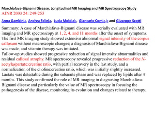
- 1. Marchiafava-Bignami Disease: Longitudinal MR Imaging and MR Spectroscopy Study AJNR 2003 24: 249-253 Anna Gambinia, Andrea Falinia, Lucia Moiolab, Giancarlo Comia,b and Giuseppe Scotti Summary: A case of Marchiafava-Bignami disease was serially evaluated with MR imaging and MR spectroscopy at 1, 2, 4, and 11 months after the onset of symptoms. The first MR imaging study showed extensive abnormal signal intensity of the corpus callosum without macroscopic changes; a diagnosis of Marchiafava-Bignami disease was made, and vitamin therapy was initiated. Follow-up studies showed progressive reduction of signal intensity abnormalities and residual callosal atrophy. MR spectroscopy revealed progressive reduction of the Nacetylaspartate:creatine ratio, with partial recovery in the last study, and a normalization of the choline:creatine ratio, which was initially slightly increased. Lactate was detectable during the subacute phase and was replaced by lipids after 4 months. This study confirmed the role of MR imaging in diagnosing MarchiafavaBignami disease and particularly the value of MR spectroscopy in focusing the pathogenesis of the disease, monitoring its evolution and changes related to therapy.
- 2. MR Imaging Findings in 56 Patients with Wernicke Encephalopathy: Nonalcoholics May Differ from Alcoholics AJNR January 2009 30: 171-176 G. Zuccolia, D. Santa Cruzd, M. Bertolinib, A. Rovirae, M. Galluccif, C. Carollogand N. Pipitonec BACKGROUND AND PURPOSE: Wernicke encephalopathy (WE) is a severe neurologic disorder resulting from dietary vitamin B1 deficiency. This study was undertaken to analyze and compare MR imaging findings and neurologic manifestations at clinical presentations of patients with WE with and without a history of alcohol abuse. MATERIALS AND METHODS: WE patients were identified using diagnostic neurologic data bases. 56 patients (29 females, 27 males) diagnosed between 1999 and 2008 with WE who improved within 1 month from the onset of thiamine administration were included in the analysis. Patients’ records were reviewed for clinical manifestations and imaging studies’ findings. MR imaging was performed in the acute phase of the disease at a field strength of 1T (16 patients) and 1.5T (40 patients). All MR images were of acceptable to good quality and were retrospectively reviewed. We compared imaging findings and clinical presentation in the alcoholic (AL) group versus the non-alcoholic (NA) group using the 2-tailed Fisher exact test and the Phi coefficient as appropriate. RESULTS: Forty-three percent of the patients were in the AL group, whereas 57% were in the NA group. Eighty-nine percent showed changes in consciousness, 75% had ocular manifestations, and 54% had ataxia. On MR imaging, 80% of the patients had evidence of symmetric lesions in the medial thalami and in the periventricular region of the third ventricle; 59%, in the periaqueductal area; 45%, in the mamillary bodies; 36%, in the tectal plate; and 7%, in the periventricular gray matter located anteriorly to the fourth ventricle. Signal-intensity alterations in areas considered atypical for the disease were noted only in the NA group and always in association with the typical findings. Contrast enhancement of the thalamus and mamillary bodies was significantly associated with alcohol abuse. CONCLUSIONS: Contrast enhancement in the mamillary bodies and thalamus is a typical finding of the disease in AL patients. Atypical MR imaging findings characterize NA patients.
- 3. Topographic distribution of the lesions in AL and NA patients with WE† Patient Group Thal (%) Periaq (%) Mam Bodies (%) Tectal Plate (%) CNN (%) Fvgm (%) Cer (%) DN (%) Vermis (%) AL 63 46 33 13 0 4 0 0 0 NA 94* 68 52 52* 32* 9 9 3 6 Neurologic symptoms at clinical onset in the AL and NA groups Group CC (%) OA (%) AT (%) T (%) AL 83 92 71 54 NA 94 69 41 34 * * CC indicates changes in consciousness; OA, ocular abnormalities; AT, ataxia; T, classic triad of the disease.
- 4. American Journal of Roentgenology 2009 192:2, 501-508 Review Neuroimaging Findings in Acute Wernicke's Encephalopathy: Review of the Literature Giulio Zuccoli1 and Nicolò Pipitone2 ABSTRACT : OBJECTIVE. Wernicke's encephalopathy is an acute neurologicalsyndrome resulting from thiamine (vitamin B1) deficiency. Early recognition is important because timely thiamine supplementation can reverse the clinical features of the disease. The aim of this article is to provide an update on the typical and atypical neuroimaging findings of the acute phase of the disease. CONCLUSION. Wernicke's encephalopathy is characterized by a quite distinct pattern of MR alterations, which include symmetrical alterations in the thalami, mamillary bodies, tectal plate, and periaqueductal area, but atypical alterations may also been seen. A thorough knowledge of the neuroimaging findings of Wernicke's encephalopathy will assist in arriving at an early diagnosis, thus reducing the morbidity and mortality associated with this disease.
- 5. 61-year-old alcoholic man with Wernicke encephalopathy during acute phase of disease. A, Axial T2-weighted image shows asymmetric edema of mamillary bodies (arrows). B, Multiplanar gradient-recalled image shows blooming consistent with hemorrhage (arrow) in left mamillary body. C, Symmetric involvement of medial thalami (arrows) is seen on T2-weighted image. D, Contrast enhancement of mamillary bodies (arrows) is seen on T1-weighted image. www.ajronline.org/doi/full/10.2214/AJR.09.4130
- 6. 53-year-old alcoholic man affected by Marchiafava-Bignami disease. A, Multiple cavitations and atrophy of corpus callosum are noted (arrows) on sagittal T1weighted images. B, Axial FLAIR image shows cavitations of splenium of corpus callosum www.ajronline.org/doi/full/10.2214/AJR.09.4130
- 7. 53-year-old alcoholic woman with Marchiafava-Bignami disease during subacute phase. A, Sagittal FLAIR image shows signal intensity alteration involving inferior aspect of corpus callosum (arrows). B, Axial FLAIR image depicts two curvilinear hyperintensities (arrows) in splenium of corpus callosum. http://www.ajronline.org/doi/full/10.2214/AJR.09.4130
- 8. Acute demyelination in Marchiafava bignami syndrome: Axial DW images (a and b) show symmetric hyperintensities in the splenium of corpus callosum, which show reduced diffusivity
- 9. Marchiafava bignami syndrome: Axial DW image (a) shows symmetric hyperintensities in the splen T2-w axial (b), coronal (c) and sagittal (f), and, FLAIR axial (d) and sagittal (e) images show
- 10. (A), diffusion-weighted image (DWI) (B), and the apparent diffusion coefficient (ADC) map (C), showing hyperintense lesions in the precentral gyrus bilaterally and the splenium of the corpus callosum.
- 11. (A), DWI(B), and ADC map (C) 17 days after admission, showing disappearance of the hyperintense lesions.
- 12. T2 hyperintensity in the splenium of the corpus callosum. Bhat A et al. Pract Neurol doi:10.1136/practneurol-2013000657 ©2013 by BMJ Publishing Group Ltd
- 13. Corresponding fibre disruption on diffusion-tensor imaging in the splenium of the corpus callosum. Bhat A et al. Pract Neurol doi:10.1136/practneurol-2013000657 ©2013 by BMJ Publishing Group Ltd
- 14. T2 hyperintensity in the genu of the corpus callosum. Bhat A et al. Pract Neurol doi:10.1136/practneurol-2013000657 ©2013 by BMJ Publishing Group Ltd
- 15. Corresponding fibre disruption on diffusion-tensor imaging in the genu of the corpus callosum. Bhat A et al. Pract Neurol doi:10.1136/practneurol-2013000657 ©2013 by BMJ Publishing Group Ltd
- 16. (Left) Sagittal T2WI MR shows a swollen corpus callosum splenium with high signal in the middle white matter layers (arrow) and peripheral sparing, classic for acute MarchiafavaBignami disease. (Right) Axial FLAIR MR in the same case shows high signal in the corpus callosum splenium (arrows) without other identifiable abnormalities. Male patient with a history of alcohol abuse and seizures.
- 17. (Left) Axial T2WI MR shows symmetric hyperintensities in the medial thalami (arrows). Patient also had hyperintensities in the periaqueductal gray matter (not shown), classic for WE. (Right) Coronal T1 C+ MR in the same case shows enhancement in the tectal plate bilaterally (arrows). Nonalcoholic WE seen in a patient who had had a bone marrow transplant and hyperalimentation.
- 18. This MRI shows typical high signal intensities (SIs) in the medial thalamus (A), periaqueductal gray (B), mamillary bodies (C), cerebellar vermis (B, C, D), and paravermian superior cerebellum (D). All the lesions represent high SIs on the DWI (E–H). The ADC images of the cerebellar vermis (K, L) and paravermian superior cerebellum (L) show low SIs (arrowheads), whereas other described areas (I J) show iso-SIs (arrows). Neurology. Apr 8 2008;70(15):e48.
- 19. Marchiafawa bignami disease possibly related to consumption of a locally brewed alcoholic beverage: Report of two cases Jagdeo P. Rawat, Charles Pinto1, Kapil S. Kulkarni, M. Ananthi K. Muthusamy, Malay D. Dave www.indianjpsychiatry.org DOI:10.4103/0019-5545.124720 Marchiafava Bignami disease is a rare toxic disease seen mostly in chronic alcoholics, resulting in progressive demyelination and necrosis of the corpus callosum. Initially it was thought to be specific to individuals in central Italy, consuming large amounts of Chianti red wine; however, alcoholic beverages worldwide are presently implicated. In our case series of two cases, locally made “illicit” liquor (Mahuwa Alcohol) could be the causative factor. In radiological point of view typically the corpus callosum is affected, with involvement of the body, genu, and splenium in order of occurrence. Occasionally the entire callosum may be also involved. Clinical presentation varies from case to case.
Notes de l'éditeur
- Acute demyelination in Marchiafavabignami syndrome: Axial DW images (a and b) show symmetric hyperintensities in the splenium of corpus callosum, which show reduced diffusivity on the ADC maps