Contenu connexe
Similaire à Duchenne y distrofina_patogenesis_y_oport_de_tto (20)
Plus de Tamara Jorquiera (17)
Duchenne y distrofina_patogenesis_y_oport_de_tto
- 1. review
review
Duchenne muscular dystrophy and dystrophin:
pathogenesis and opportunities for treatment
Third in Molecular Medicine Review Series
Kristen J. Nowak† & Kay E. Davies+
MRC Functional Genetics Unit, University of Oxford, UK
Duchenne muscular dystrophy (DMD) is caused by mutations in protein. Becker muscular dystrophy (BMD; OMIM 300376)—a
the gene that encodes the 427-kDa cytoskeletal protein dys- much milder form of the disease—is caused by a reduction in the
trophin. Increased knowledge of the function of dystrophin and amount, or alteration in the size, of the dystrophin protein. The
its role in muscle has led to a greater understanding of the high incidence of sporadic cases of DMD (1 in 10,000 sperm or
pathogenesis of DMD. This, together with advances in the eggs) means that genetic screening will never eliminate this dis-
genetic toolkit of the molecular biologist, are leading to many ease, so an effective therapy is highly desirable. This review sum-
different approaches to treatment. Gene therapy can be marizes our understanding of the disease and the strategies that are
achieved using plasmids or viruses, mutations can be corrected being developed for an effective treatment (Fig 1).
using chimaeraplasts and short DNA fragments, exon skipping
of mutations can be induced using oligonucleotides and Pathogenesis
readthrough of nonsense mutations can be achieved using Dystrophin has a major structural role in muscle as it links the
aminoglycoside antibiotics. Blocking the proteasome degrada- internal cytoskeleton to the extracellular matrix. The amino-terminus
tion pathway can stabilize any truncated dystrophin protein, of dystrophin binds to F-actin and the carboxyl terminus to the
and upregulation of other proteins can also prevent the dys- dystrophin-associated protein complex (DAPC) at the sarcolemma
trophic process. Muscle can be repopulated with myoblasts or (Fig 2; Blake et al, 2002). The DAPC includes the dystroglycans,
stem cells. All, or a combination, of these approaches hold great sarcoglycans, integrins and caveolin, and mutations in any of
promise for the treatment of this devastating disease. these components cause autosomally inherited muscular dystro-
Keywords: Duchenne muscular dystrophy; DMD; gene therapy; phies (Dalkilic & Kunkel, 2003). The DAPC is destabilized when
muscle dystrophin is absent, which results in diminished levels of the
EMBO reports (2004) 5, 872–876. doi:10.1038/sj.embor.7400221 member proteins (Straub et al, 1997). This in turn leads to progres-
sive fibre damage and membrane leakage. The DAPC has a sig-
Introduction nalling role, the loss of which also contributes to pathogenesis
Duchenne muscular dystrophy (DMD; OMIM 310200) is an X-linked (Blake et al, 2002). DMD patients are usually wheelchair-bound
recessive disorder that affects 1 in 3,500 males and is caused by by 12 years of age and die of respiratory failure in their late teens
mutations in the dystrophin gene (Blake et al, 2002). The gene is or early twenties. Many boys have an abnormal electrocardio-
the largest in the human genome, encompassing 2.6 million base gram by the age of 18, indicating that any therapeutic agent must
pairs of DNA and containing 79 exons. Approximately 60% of dys- also target the diaphragm and cardiac muscle.
trophin mutations are large insertions or deletions that lead to
frameshift errors downstream, whereas approximately 40% are Animal models
point mutations or small frameshift rearrangements (Hoffman, Animal models are valuable resources for studying the patho-
2001). The vast majority of DMD patients lack the dystrophin genesis of disease, and provide a test-bed for pre-clinical trials.
Two of the most widely used animal models for DMD research
MRC Functional Genetics Unit, Department of Human Anatomy and Genetics, are the mdx mouse and the golden retriever muscular dystrophy
University of Oxford, South Parks Road, Oxford OX1 3QX, UK
†
(GRMD) dog, both of which are dystrophin negative (Collins &
Present address: Centre for Neuromuscular and Neurological Disorders and Centre for
Medical Research, University of Western Australia, Queen Elizabeth II Medical Centre, Morgan, 2003). However, the mdx mouse only has a mild pheno-
Western Australia 6009, Australia type (most likely due to the high regenerative capacity of mouse
+
Corresponding author. Tel: +44 1865 272179; Fax: +44 1865 272420; muscle), and thus mice that are null for both dystrophin and
E-mail: kay.davies@anat.ox.ac.uk
another muscle protein, utrophin, more closely resemble the
Submitted 7 June 2004; accepted 14 July 2004 human phenotype (Collins & Morgan, 2003).
8 7 2 EMBO reports VOL 5 | NO 9 | 2004 ©2004 EUROPEAN MOLECULAR BIOLOGY ORGANIZATION
- 2. DMD: pathogenesis and treatment
K.J. Nowak & K.E. Davies review
Gene replacement therapy humans because of revertant fibres, which are muscle fibres that
One problem that might be encountered during the expression of a express a smaller, but functional, dystrophin protein due to exon
previously missing gene product is the onset of an immunological skipping. To be effective, it is necessary to have long-term delivery
reaction. However, recent investigations by Ferrer and colleagues of the missing gene, or persistent gene correction, in the vast major-
(Ferrer et al, 2004) suggest that this should not be a problem in ity of muscle fibres of a DMD patient (approximately 20% of the
total body mass). On average, an injection into muscle will trans-
duce cells only within a couple of centimetres of the injection site
Intramuscular injection (Hartigan-O’Connor & Chamberlain, 2000; O’Hara et al, 2001).
or systemic delivery
This suggests that multiple injections would be required for the
Virus treatment of a whole muscle (for example, hundreds of injections
Plasmid for the heart or diaphragm), which is not a realistic prospect.
The approach of using either viruses or plasmids to deliver
dystrophin sequences has made significant progress (Scott et al,
Patient Donor
2002; Gregorevic & Chamberlain, 2003). Advances in viral
delivery, such as functional dystrophin mini- and microgenes and
Genetic gutted vectors with large insert capacity and lowered immuno-
modification genicity, have led to good results in mdx mice. One study reported
52% of fibres expressing dystrophin after one year (Dudley et al,
Stem cells
2004), and another showed that around 40% of muscle function
Myoblasts improvement can occur if 25–30% of fibres express dystrophin
(DelloRusso et al, 2002). Recent exciting results show that the
adeno-associated virus type 6 (AAV-6) serotype provides very
efficient delivery to many muscles by administration through the
tail vein (Gregorevic et al, 2004). Systemic delivery of nonviral
Proteasome vectors has led to dystrophin expression in 40% of fibres (Liu
inhibitor Antibody
blockade et al, 2001), with widespread transgene expression in approxi-
• myostatin mately 10% of fibres in all leg muscles after injection into the
tail artery or vein (Liang et al, 2004). Phase I human trials using
plasmid DNA delivery of dystrophin have been initiated (Romero
Nucleus
et al, 2002).
Upregulation
therapy
Myoblast transplantation
• utrophin
Muscle cell • IGF-1 The regenerative capacity of muscle means that during the early
• α7 Integrin phases of DMD pathology, the fusion of resident myoblasts (or
• GalNAc transferase satellite cells) leads to the formation of new muscle fibres, but
• ADAM12
• nNOS
eventually the skeletal muscle is replaced by connective tissue as
• Calpastatin the proliferative potential of satellite cells is exhausted. Therefore,
• Anti-TNFα the delivery of normal or genetically modified myogenic and
satellite cells (both devoted to the myogenic lineage) or stem cells
Chimaeraplast have been explored as potential therapies. Despite initial promis-
ing results for myoblast transfer into mdx muscle (Partridge et al,
1989), most human clinical trials have given disappointing results
due to a lack of cell survival, immune rejection and limited dis-
Aminoglycosides
persal. Skuk and colleagues (Skuk et al, 2004) used donor
myoblasts to perform 25 injections into the tibialis anterior mus-
cle of three immunosuppressed DMD patients in a 1-cm3 region.
Transcription After four weeks, biopsies at the injected site showed that
Antisense between 6.8% and 11% of donor cells expressed dystrophin.
oligonucleotide
Short
fragment Stem-cell therapy
homologous Self-renewing, immune-privileged stem cells have been shown
repair
to proliferate longer than myoblast cells, to migrate from the cir-
culatory system after intra-arterial injection and to be more
DNA RNA
(dystrophin) (dystrophin) effective than myoblast cells in muscle regeneration and dys-
trophin expression after implantation (Peng & Huard, 2004).
Intra-arterial injection of muscle-derived stem cells into the
Fig 1 | Summary of the wide range of approaches being used to treat hindlimbs of mdx mice resulted in their migration from the circu-
Duchenne muscular dystrophy. IGF1, insulin-like growth factor 1; NOS, latory system with subsequent co-expression of LacZ and dys-
nitric oxide synthase; TNF-α, tumour necrosis factor-α. trophin in all muscles (Torrente et al, 2001). Recently identified,
©2004 EUROPEAN MOLECULAR BIOLOGY ORGANIZATION EMBO reports VOL 5 | NO 9 | 2004 8 7 3
- 3. review DMD: pathogenesis and treatment
K.J. Nowak & K.E. Davies
Laminin-2
Collagen Extracellular
Extracellular matrix
Biglycan Sarcospan
Dystroglycans α
Caveolin-3
Sarcolemma β α β γ δ
nNOS Intracellular
α1 β1 Syntrophins
C’ terminus
Dystrophin
Sarcoglycans Dystrobrevin
Dysbindin
Syncoilin
us
rmin
N’ te
Desmin
Actin cytoskeleton
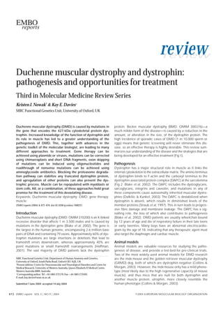
Fig 2 | The dystrophin-associated protein complex in muscle linking the internal cytoskeleton to the extracellular matrix. NOS, nitric oxide synthase.
vesselassociated fetal stem cells known as ‘mesoangioblasts’ Antisense oligonucleotides
have been shown to provide widespread rescue of dystrophy in Antisense oligonucleotides can sterically inhibit gene expression
α-sarcoglycan-negative mice after femoral artery delivery; more- by hybridizing to target mRNA sequences at sites such as
over, lentiviral transduction of mesoangioblasts isolated from exon–intron boundaries, translation inhibition codons and
dystrophic mice before injection gave similarly optimistic results sequences downstream of the initiation codon. The identification
(Sampaolesi et al, 2003). of revertant fibres in dystrophic muscle that express the dystrophin
protein by exon skipping has guided the use of antisense oligo-
Aminoglycoside antibiotics nucleotides for the genetic therapy of DMD (van Deutekom & van
Between 5% and 15% of DMD cases are caused by premature Ommen, 2003). Researchers have tried to redirect dystrophin
stop codons, and so the use of aminoglycoside antibiotics (for splicing to exclude an exon that contains a premature stop codon
example, gentamycin and negamycin), which promote trans- (for example, exon 23 in the mdx mouse model), in an effort to
lational readthrough of stop codons, has been investigated. restore the reading frame and to produce a slightly shorter, but
Despite hopeful results in mdx mice (6% dystrophin-positive hopefully partially functional protein. Successful skipping has
fibres, Arakawa et al, 2003; 10–20% of normal dystrophin levels, been demonstrated in cultured mdx myotubes (Wilton et al, 1999),
Barton-Davis et al, 1999), no dystrophin expression has been the mdx mouse (Lu et al, 2003), and cultured muscle cells derived
achieved in human studies of DMD and BMD patients and a repli- from DMD patients (Aartsma-Rus et al, 2003). Hyaluronidase-
cation of the mdx results have not been forthcoming (Dunant et al, enhanced electrotransfer delivery of antisense olignucleotides has
2003). Recent cell-culture experiments using eight different been shown to result in dystrophin expression in 20–30% of fibres
patient mutations indicate that some sequences are better suppressed in the tibialis anterior muscle of the mdx mouse after one injection
by aminoglycosides than others (Howard et al, 2004). (Wells et al, 2003). Recent investigations into double-exon and
multi-exon skipping (skipping of numerous successive exons) have
Precise correction of the mutation enhanced the technique to treat a greater number of dystrophin
The precise correction of a dystrophin mutation can occur through mutations with the same antisense oligonucleotides (Aartsma-Rus
the use of short fragments or chimaeraplasts (double-stranded et al, 2004).
RNA–DNA chimaeric oligonucleotides), which are designed to
contain the correct nucleotide. Unfortunately, intramuscular injec- Proteasome inhibitors
tions of chimaeraplasts have produced limited dystrophin protein in Bonuccelli and coworkers (Bonucelli et al, 2003) explored the use
the GRMD dog and mdx mouse, with dystrophin-positive cells of proteasome inhibitors as a therapy for DMD on the premise that,
restricted to the area surrounding the injection site. Advances in the absence of dystrophin, members of the DAPC are degraded
include high conversion efficiencies in mdx muscle precursor cells through an unknown pathway that leads to their reduction in dys-
in vitro (2–15%, Bertoni et al, 2002) and induced exon skipping, trophic muscle. Continuous systemic treatment of the proteasome
which led to a range of functional transcripts, protein expression inhibitor MG-132—using a subcutaneously implanted osmotic
and localization (Bertoni et al, 2003). A 603-bp PCR product cor- pump over eight days—resulted in decreased damage of the muscle
rected the mutant base in 15–20% of mdx myoblasts, but despite membrane and improved muscle integrity. The dystrophin protein
the persistence of the corrected nucleotide for 3–4 weeks, the present at the plasma membrane after treatment lacked the C-termi-
transfected cells lost viability and did not express any full-length nal domain due to the presence of the nonsense mutation in exon
dystrophin transcript (Kapsa et al, 2001). 23, and hence was a truncated form. These findings corroborate the
8 7 4 EMBO reports VOL 5 | NO 9 | 2004 ©2004 EUROPEAN MOLECULAR BIOLOGY ORGANIZATION
- 4. DMD: pathogenesis and treatment
K.J. Nowak & K.E. Davies review
proposal that protein degradation in dystrophin-deficient muscle is Conclusions
mediated by the proteasomal pathway and open up a new avenue A successful treatment for DMD and BMD is essential for sufferers
for therapeutic emphasis. of these diseases, but the main stumbling block for many thera-
peutic approaches is the delivery of the treatment to a sufficient
Upregulation therapy proportion of muscle mass to provide benefit. The systemic deliv-
Upregulation therapy is based on increasing the expression of ery of stem cells that leads to their migration into muscle tissue
alternative genes to replace a defective gene and is particularly and particularly into areas of damage is a cause for optimism and
beneficial when an immune response is mounted against a previ- has been shown to be safe. The precise correction of dystrophin
ously absent protein. Upregulation of utrophin, an autosomal mutations or the splicing out of the exon containing such a muta-
paralogue of dystrophin, has been proposed as a potential thera- tion holds promise, although these methods require optimization
py for DMD (Perkins & Davies, 2002; Khurana & Davies, 2003). for almost every different mutation in the gene. In addition, the
When utrophin is overexpressed in transgenic mdx mice it local- cost of agents such as chimaeraplasts and antisense oligo-
izes to the sarcolemma of muscle cells and restores the compo- nucleotides is high and not feasible for widespread use.
nents of the DAPC, which prevents dystrophic development and Aminoglycosides are only applicable to nonsense mutations, and
in turn leads to functional improvement of skeletal muscle recent evidence suggests that only a subset of these might respond
(Rybakova et al, 2002). Adenoviral delivery of utrophin in the to such treatment. Moreover, the side effects of these drugs need
dog has been shown to prevent pathology (Cerletti et al, 2003). to be further explored and managed. Blocking of the proteasome
Commencement of increased utrophin expression shortly after pathway has yielded exciting results and has shown that restora-
birth in the mouse model can be effective, and no toxicity is tion of the DAPC can occur by the correct localization of
observed when utrophin is ubiquitously expressed, which is dystrophin, albeit a truncated form. Upregulation of a range of
promising for the translation of this therapy to humans. proteins has also produced optimistic results with recovery of nor-
Upregulation of endogenous utrophin to sufficient levels to mal muscle function despite an absence of dystrophin protein.
decrease pathology might be achievable by the delivery of small, Increased expression of these genes might be achievable by sys-
diffusible compounds. Detailed analyses of the two utrophin pro- temic delivery of small molecules. It is foreseen that a fusion of
moters have given some insight into the mechanisms of utrophin these varied therapeutic methods might be successfully used in
expression, and in turn have provided the prospect of designing the future, such as viral transduction of stem cells sourced from a
specific small chemicals to interfere with or enhance these DMD patient and subsequent intravenous reintroduction
mechanisms (Khurana & Davies, 2003). (Bachrach et al, 2004), or perhaps upregulation of utrophin
Experiments to increase the expression levels of other genes expression partnered with inducing increased levels of IGF1 and
have also been successful in improving the pathology of mdx- the blocking of myostatin.
cultured myotubes and/or mdx mice, namely nitric oxide syn-
thetase (NOS; Wehling et al, 2001), L-arginine, which is a NOS ACKNOWLEDGEMENTS
We apologize to our fellow researchers whose work we could not reference
substrate (Chaubourt et al, 2002), α7β1-integrin (Burkin et al,
due to space restrictions. We are grateful to the Muscular Dystrophy
2001), synaptic cytotoxic T-cell GalNAc transferase (Galgt2; Campaign (UK), the Muscular Dystrophy Association (USA) and the
Nguyen et al, 2002), insulin-like growth factor 1 (IGF1; Barton et al, Association Française contre les Myopathies for their support. K.J.N. is an
2002), calpastatin (Spencer et al, 2002) and a disintegrin and Australian NHMRC CJ Martin Fellow (212086.
metalloprotease ADAM12 (Moghadaszadeh et al, 2003).
Interestingly, following the overexpression of many of these REFERENCES
Aartsma-Rus A, Janson AA, Kaman WE, Bremmer-Bout M, den Dunnen JT,
genes, there was an increase not only in the levels of many
Baas F, Van Ommen GJ, den Dunnen JT, van Deutekom JC (2003)
dystrophin-associated proteins, but also in the amount of Therapeutic antisense-induced exon skipping in cultured muscle cells
utrophin. The use of antibodies to specifically block the action of from six different DMD patients. Hum Mol Genet 12: 907–914
either myostatin (a member of the transforming growth factor-β Aartsma-Rus A, Janson AA, Kaman WE, Bremmer-Bout M, van Ommen G,
(TGF-β) superfamily; Bogdanovich et al, 2002), or tumour necro- den Dunnen JT, van Deutekom JCT (2004) Antisense-induced
multiexon skipping for Duchenne muscular dystrophy makes more
sis factor-α (TNF-α; Grounds & Torrisi, 2004), has led to the func- sense. Am J Hum Genet 74: 83–92
tional improvement of dystrophic muscle in the mouse. The exact Arakawa M et al (2003) Negamycin restores dystrophin expression in
molecular mechanisms for the improvement of dystrophic fea- skeletal muscle of mdx mice. J Biochem 134: 751–758
tures for the above experiments are unknown, but it is thought Bachrach E, Li S, Perez AL, Schienda J, Liadaki K, Volinski J, Flint A,
Chamberlain J, Kunkel LM (2004) Systemic delivery of human
that the mode of action of these ‘rescue’ molecules is to sustain
microdystrophin to regenerating mouse dystrophic muscle by muscle
regeneration and reduce fibrosis (myostatin blockade, ADAM12, progenitor cells. Proc Natl Acad Sci USA 101: 3581–3586
IGF1), promoting cell adhesion and muscle stability (Galgt2, α7- Barresi R et al (2004) LARGE can functionally bypass α-dystroglycan
integrin, ADAM12) and preventing necrosis (calpastatin, TNF-α; glycosylation defects in distinct congenital muscular dystrophies. Nat
Engvall & Wewer, 2003). These experiments illustrate the range Med 10: 696–703
Barton ER, Morris L, Musaro A, Rosenthal N, Sweeney HL (2002) Muscle-
of possible pathways that might be targeted for the alleviation of specific expression of insulin-like growth factor I counters muscle
dystrophic pathology that is caused by defects in the dystrophin decline in mdx mice. J Cell Biol 157: 137–148
gene, without actually correcting the gene or expressing dys- Barton-Davis ER, Cordier L, Shoturma DI, Leland SE, Sweeney HL (1999)
trophin from another source. Interestingly, overexpression of the Aminoglycoside antibiotics restore dystrophin function to skeletal
muscles of mdx mice. J Clin Invest 104: 375–381
glycosyltransferase LARGE can functionally bypass α-dystroglycan
Bertoni C, Rando TA (2002) Dystrophin gene repair in mdx muscle
glycosylation defects in distinct congenital muscular dystrophy precursor cells in vitro and in vivo mediated by RNA–DNA chimeric
(Barresi et al, 2004). oligonucleotides. Hum Gene Ther 13: 707–718
©2004 EUROPEAN MOLECULAR BIOLOGY ORGANIZATION EMBO reports VOL 5 | NO 9 | 2004 8 7 5
- 5. review DMD: pathogenesis and treatment
K.J. Nowak & K.E. Davies
Bertoni C, Lau C, Rando TA (2003) Restoration of dystrophin expression in mdx Lu QL, Mann CJ, Lou F, Bou-Gharios G, Morris GE, Xue SA, Fletcher S, Partridge
muscle cells by chimeraplast-mediated exon skipping. Hum Mol Genet 12: TA, Wilton SD (2003) Functional amounts of dystrophin produced by
1087–1099 skipping the mutated exon in the mdx dystrophic mouse. Nat Med 9:
Blake DJ, Weir A, Newey SE, Davies KE (2002) Function and genetics of 1009–1014
dystrophin and dystrophin-related proteins in muscle. Physiol Rev 82: Moghadaszadeh B et al (2003) Compensation for dystrophin-deficiency:
291–329 ADAM12 overexpression in skeletal muscle results in increased α7 integrin,
Bogdanovich S, Krag TO, Barton ER, Morris LD, Whittemore LA, Ahima RS, utrophin and associated glycoproteins. Hum Mol Genet 12: 2467–2479
Khurana T (2002) Functional improvement of dystrophic muscle by Nguyen HH, Jayasinha V, Xia B, Hoyte K, Martin PT (2002) Overexpression of the
myostatin blockade. Nature 420: 418–421 cytotoxic T cell Ga/NAc transferase in skeletal muscle inhibits muscular
Bonuccelli G, Sotgia F, Schubert W, Park DS, Frank PG, Woodman SE, Insabato L, dystrophy in mdx mice. Proc Natl Acad Sci USA 99: 5616–5621
Cammer M, Minetti C, Lisanti MP (2003) Proteasome inhibitor (MG-132) O’Hara AJ, Howell JM, Taplin RH, Fletcher S, Lloyd F, Kakulas B, Lochmuller H,
treatment of mdx mice rescues the expression and membrane localization of Karpati G (2001) The spread of transgene expression at the site of gene
dystrophin and dystrophin-associated proteins. Am J Pathol 163: 1663–1675 construct injection. Muscle Nerve 24: 488–495
Burkin DJ, Wallace GQ, Nicol KJ, Kaufman DJ, Kaufman SJ (2001) Enhanced Partridge TA, Morgan JE, Coulton GR, Hoffman EP, Kunkel LM (1989) Conversion
expression of the α7β1 intergrin reduces muscular dystrophy and restores of mdx myofibers from dystrophin negative to positive by injection of normal
viability in dystrophic mice. J Cell Biol 152: 1207–1218 myoblasts. Nature 337: 176–179
Cerletti M et al (2003) Dystrophic phenotype of canine X-linked muscular Peng H, Huard J (2004) Muscle-derived stem cells for musculoskeletal tissue
dystrophy is mitigated by adenovirus-mediated utrophin gene transfer. Gene regeneration and repair. Transpl Immunol 12: 311–319
Ther 10: 750–757 Perkins KJ, Davies KE (2002) The role of utrophin in the potential therapy of
Chaubourt E, Voisin V, Fossier P, Baux G, Israel M, De La Porte S (2002) Muscular Duchenne muscular dystrophy. Neuromuscul Disord S1: S78–S89
nitric oxide synthase (muNOS) and utrophin. J Physiol Paris 96: 43–52 Romero N, Benveniste O, Payan C, Braun S, Squiban P, Herson S, Fardeau M
Collins CA, Morgan JE (2003) Duchenne’s muscular dystrophy: animal models (2002) Current protocol of a research phase I clinical trial of full-length
used to investigate pathogenesis and develop therapeutic strategies. Int J Exp dystrophin plasmid DNA in Duchenne/Becker muscular dystrophies: part II,
Pathol 84: 165–172 clinical protocol. Neuromuscul Disord 12: S45–S48
Dalkilic I, Kunkel LM (2003) Muscular dystrophies: genes to pathogenesis. Curr Rybakova IN, Patel JR, Davies KE, Yurhcenco PD, Ervasti JM (2002) Utrophin
Opin Genet Dev 13: 231–238 binds laterally along actin filaments and can couple costameric actin with
DelloRusso C, Scott J, Hartigan-O’Connor D, Salvatori G, Barjot C, Robinson AS, the sarcolemma when overexpressed in dystrophin-deficient muscles of
Crawford RW, Brooks SV, Chamberlain JS (2002) Functional correction of mice. Mol Biol Cell 13: 1512–1521
adult mdx mouse muscle using gutted adenoviral vectors expressing full- Sampaolesi M et al (2003) Cell therapy of α-sarcoglycan null dystrophic mice
length dystrophin. Proc Natl Acad Sci USA 99: 12979–12984 through intra-arterial delivery of mesoangioblasts. Science 301: 487–492
Dudley RWR, Lu Y, Gilbert R, Matecki S, Nalbantoglu J, Petrof BJ, Karpati G Scott JM, Li S, Harper SQ, Welikson R, Bourque D, DelloRusso C, Hauschka SD,
(2004) Sustained improvement of muscle function on year after full-length Chamberlain JS (2002) Viral vectors for gene transfer of micro-, mini-, or
dystrophin gene transfer into mdx mice by a gutted helper-dependent full-length dystrophin. Neuromuscul Disord 12: S23–S29
adenoviral vector. Hum Gene Ther 15: 145–156 Skuk D et al (2004) Dystrophin expression in myofibers of Duchenne muscular
Dunant P, Walter MC, Karpati G, Lochmüller H (2003) Gentamicin fails to dystrophy patients following intramuscular injections of normal myogenic
increase dystrophin expression in dystrophin-deficient muscle. Muscle cells. Mol Ther 9: 475–482
Nerve 27: 624–627 Spencer MJ, Meligren RL (2002) Overexpression of a calpastatin transgene in
Engvall E, Wewer UM (2003) The new frontier in muscular dystrophy research: mdx muscle reduces dystrophic pathology. Hum Mol Genet 11: 2645–2655
booster genes. FASEB J 17: 1579–1584 Straub V, Campbell KP (1997) Muscular dystrophies and the
Ferrer A, Foster H, Wells KE, Dickson G, Wells DJ (2004) Long-term expression of dystrophin–glycoprotein complex. Curr Opin Neurol 10: 168–175
full-length human dystrophin in transgenic mdx mice expressing internally Torrente Y et al (2001) Intraarterial injection of muscle-derived CD34+ Sca-1+
deleted human dystrophins. Gene Ther 11: 884–893 stem cells restores dystrophin in mdx mice. J Cell Biol 152: 335–348
Gregorevic P, Chamberlain JS (2003) Gene therapy for muscular dystrophy—a van Deutekom JC, van Ommen GJ (2003) Advances in Duchenne muscular
review of promising progress. Expert Opin Biol Ther 3: 803–814 dystrophy gene therapy. Nat Rev Genet 4: 774–783
Gregorevic P, Blankenship MJ, Allen JM, Meuse L, Oakley S, Miller D, Russell D, Wehling M, Spencer MJ, Tidball JG (2001) A nitric oxide synthase transgene
Chamberlain JS (2004) Systemic gene transfer to striated muscles using ameliorates muscular dystrophy in mdx mice. J Cell Biol 155: 123–131
adeno-associated viral vectors. Nat Med (in press) Wells KE, Fletcher S, Mann CJ, Wilton SD, Wells DJ (2003) Enhanced in vivo
Grounds MD, Torrisi J (2004) Anti-TNFα (Remicade) therapy protects dystrophic delivery of antisense oligonucleotides to restore dystrophin expression in
skeletal muscle from necrosis. FASEB J 18: 676–682 adult mdx mouse muscle. FEBS Lett 552: 145–149
Hartigan-O’Connor D, Chamberlain JS (2000) Developments in gene therapy for Wilton SD, Lloyd F, Carville K, Fletcher S, Honeyman K, Agrawal S, Kole R (1999)
muscular dystrophy. Microsc Res Tech 48: 223–238 Specific removal of the nonsense mutation from the mdx dystrophin mRNA
Hoffman EP, Dressman D (2001) Molecular pathophysiology and targeted using antisense oligonucleotides. Neuromuscul Disord 9: 330–338
therapeutics for muscular dystrophy. Trends Pharmacol Sci 22: 465–470
Howard MT, Anderson CB, Fass U, Khatris S, Gesteland RF, Atkins JF, Flanigan
KM (2004) Readthrough of dystrophin stop codon mutations induced by
aminoglycosides. Ann Neurol 55: 422–426
Kapsa R, Quigley A, Lynch GS, Steeper K, Kornberg AJ, Gregorevic P, Austin L,
Byrne E (2001) In vivo and in vitro correction of the mdx dystrophin gene
nonsense mutation by short fragment homologous replacement. Hum Gene
Ther 12: 629–642
Khurana TS, Davies KE (2003) Pharmacological strategies for muscular dystrophy.
Nat Rev Drug Discov 2: 379–390
Liang KW, Nishikawa M, Liu F, Sun B, Ye Q, Huang L (2004) Restoration of
dystrophin expression in mdx mice by intravascular injection of naked DNA
containing full-length dystrophin cDNA. Gene Ther 11: 901–908
Liu F, Nishikawa M, Clemens PR, Huang L (2001) Transfer of full-length Dmd to
the diaphragm muscular of Dmd (mdx/mdx) mice through systemic
administration of plasmid DNA. Mol Ther 4: 45–51 Kristen J. Nowak Kay E. Davies
8 7 6 EMBO reports VOL 5 | NO 9 | 2004 ©2004 EUROPEAN MOLECULAR BIOLOGY ORGANIZATION