2. 3958 JOURNAL OF CELL SCIENCE 114 (22)
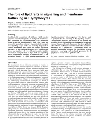
Fig. 1. Model of lipid-raft structure and function in
biological membranes. (A) Rafts are membrane A
microdomains formed by high concentrations of
sphingolipids (dark-brown-headed structures) and
cholesterol (red bean-shaped structures) immersed
in a phospholipid-rich (light-brown-headed
structures) environment. Glycolipids and
sphingomyelin are restricted to the outer leaflet of
the bilayer, whereas cholesterol and phospholipids
are in both leaflets. Note that lipids in the rafts
usually have long and saturated fatty acyl chains
(red two-legged shapes), whereas those in lipids
excluded from these microdomains are shorter and B
unsaturated (green two-legged shapes).
(B) Principles of selective recruitment of proteins
in rafts. Recruitment of membrane proteins in
phospholipid-rich membrane regions takes place
through protein-protein interactions. However, in Out
rafts this process takes place through interactions
between the lipids within the rafts and the
transmembrane domain of integral membrane In
proteins (lipid-protein interaction) or the lipid Lipid raft
moiety of proteins attached to the membrane by a
lipid modification (lipid-lipid interaction). The Lipid-lipid interaction
recruitment of cytosolic proteins by protein- Protein-lipid interaction
Protein-protein interaction
protein interactions through modular domains
(SH2 domains, SH3 domains, etc.) can take place in both raft and non-raft membranes. Proteins excluded from rafts are in yellow; proteins
included in rafts are in blue (integral membrane proteins), light brown (GPI-anchored proteins) or pink (acylated, cytosolically-oriented,
proteins such as Src family kinases, Ras and heterotrimeric G proteins).
Rafts as platforms for assembly of the T cell Fig. 2. Lipid raft reorganisation after TCR engagement. At steady
signalling machinery state, CD4, Lck, LAT and CD3ζ are associated with small rafts (red)
Pioneering work described the isolation of detergent-resistant in T cells. Upon triggering, lipid rafts concentrate in the
membrane complexes enriched in GPI-anchored proteins from immunological synapse, gathering together specific membrane
T lymphocytes (Hoessli and Rungger-Brandle, 1983). These proteins. Lck becomes activated and phosphorylates immunoreceptor
membranes were then characterised as large non-covalent tyrosine-based activation motifs (ITAMs) within the CD3 subunits.
complexes containing GPI-anchored proteins and Src family These phosphorylated motifs become docking sites for the tandem
SH2 domains of the tyrosine kinase ZAP70, which is subsequently
tyrosine kinases (Stefanova et al., 1991; Cinek and Horejsi,
activated by tyrosine phosphorylation, probably by Lck. Activated
1992). The observation that GPI-anchored proteins can ZAP70 phosphorylates the tyrosine residues present in the
transduce activation signals to internal Src family kinases transmembrane adapter LAT, which recruits Gads, phospholipase
(Bamezai et al., 1989; Gunter et al., 1987; Korty et al., 1991) Cγ1 (PLCγ1) and Grb2. As a consequence, different processes are
despite the molecules residing in the opposite leaflets of the triggered: (1) LAT-associated Gads bring the adapter protein SLP-76
lipid bilayer was regarded for a long time as a puzzling and to the rafts, and this adapter becomes a substrate for ZAP70.
intriguing phenomenon occurring in rafts (Brown, 1993). It is Phosphorylated SLP-76 recruits the Tec family protein tyrosine
well known that T cell antigen receptor (TCR) engagement kinase Itk, the guanine-nucleotide-exchange factor Vav and the
triggers the assembly of a large macromolecular complex adapter molecule Nck. Subsequently, Nck recruits the PAK and
containing a variety of signalling molecules and adapters, but WASP proteins through its SH3 domains. PAK and WASP are
regulated by Vav and in turn regulate the reorganisation of the
only recently have investigators postulated that rafts are
cytoskeleton. (2) PLCγ1 recruited to LAT is activated through
platforms for this signalling complex (Montixi et al., 1998; tyrosine phosphorylation by ZAP70 and Itk. Activated PLCγ1
Xavier et al., 1998; Zhang et al., 1998). In resting T cells, rafts converts phosphatidylinositol (4,5)-bisphosphate (PtdIns(4,5)P2) into
are highly enriched in the Src kinases Lck and Fyn (Montixi diacylglycerol (DAG) and inositol (3,4,5)-trisphosphate
et al., 1998; Xavier et al., 1998) and the linker for activation (Ins(1,4,5)P3). Subsequently, DAG activates protein kinase C and
of T cells (LAT) transmembrane adapter (Zhang et al., 1998). Ras guanyl-nucleotide-releasing protein (RasGRP), and Ins(1,4,5)P3
The co-receptor CD4 is detected in raft fractions to a minor activates the transcription factor NF-AT by promoting Ca2+
extent, and CD3ζ is also partially associated with rafts mobilization and calcineurin activation. (3) Grb2 associated with
(Montixi et al., 1998; Xavier et al., 1998) (Fig. 2, top panel). LAT recruits Sos to the rafts, and this attracts Ras and subsequently
Extensive crosslinking of the TCR with antibodies promotes other machinery, which results in activation of MAP kinases.
Although represented as excluded in resting cells and included in
the rapid activation of Src kinases and subsequent
activated cells, the presence in rafts of components of the TCR-CD3
accumulation in rafts of a series of newly tyrosine- (other than CD3ζ) before and after triggering is controversial (see
phosphorylated substrates (Kane et al., 2000; Langlet et al., text). Curved arrows in dark blue indicate relevant tyrosine
2000; Leo and Schraven, 2001), including virtually all the phosphorylation events occurring upon activation. Horizontal brown
hyperphosphorylated p23 CD3ζ molecules (Montixi et al., arrows indicate tyrosine dephosphorylation events carried out by
1998; Xavier et al., 1998; Kosugi et al., 1999), the activated CD45 molecules present close to the raft edge.
3. Lipid rafts in T cells 3959
forms of the ZAP70 tyrosine kinase and phospholipase C γ1 polyoxyethylene ether (Brij) detergent series (Montixi et al.,
(PLCγ1), phosphoinositide 3-kinase (PI3-K), the Vav 1998; Galbiati et al., 2001) or in situ by immunofluorescence
Rac/CDC42 exchange factor (Montixi et al., 1998; Xavier et analysis in the absence of detergent (Janes et al., 1999). As a
al., 1998) and LAT (Brdicka et al., 1998; Zhang et al., 1998) consequence of both raft redistribution and cytoskeletal
(Fig. 2, bottom panel). The presence of the TCR-CD3 reorganisation, a supramolecular activation complex (the
complex in the rafts before and after engagement is immunological synapse) containing the assembled signalling
controversial (Montixi et al., 1998; Janes et al., 1999; Kosugi machinery is formed at the interface of the T lymphocyte and
et al., 1999). It appears that the association is weak and the antigen-presenting cell (APC) (Monks et al., 1998;
sensitive to most non-ionic detergents but is readily Grakoui et al., 1999; Dustin and Chan, 2000). Upon assembly
detectable by biochemical means using certain of the signalling machinery, the cytoskeleton is reorganised,
RESTING
TCR-CD3
CD45
CD45
CD4
α β
γ δ
δ ε ζζ
Lck
LAT
ACTIVATED
TCR-CD3
CD45
CD45
CD4
α β
γ ε δ ε ζζ
Ras
Lck
LAT
Sos Raf
Grb2
-Y P
ZAP70
-Y P (3)
-Y P
PLCγ1
MAPK
Gads
P Y- -Y P
activation
SLP-76
(2) ITK
DAG -Y P Vav
Ins(1,4,5)P3 P Y- (1)
2+ PKC -Y P Nck PAK
Ca
activation WASP
Raft RasGRP
NF-AT activation Actin
activation reorganisation
4. 3960 JOURNAL OF CELL SCIENCE 114 (22)
and the Ras/MAPK and PLCγ1 cascades are activated within interactions are necessary for T cell activation under these
the rafts, which produce signals that stimulate T cell conditions. CD28 functions as the major T cell costimulatory
proliferation (Lin and Weiss, 2000). receptor, but other T cell surface molecules can induce T cell
The tyrosine kinase activity of Src kinases is essential for T costimulation as well. The nature of these signals has been
cell activation. This activity is regulated negatively by Csk, a recently explained in terms of raft reorganisation. Thus,
tyrosine kinase that phosphorylates Src kinases at an inhibitory whereas the crosslinking of the TCR alone by suboptimal
C-terminal tyrosine residue. Csk is recruited into rafts through amounts of anti-CD3 antibodies does not result in clustering
an interaction between its SH2 domain and the cytoplasmic of the rafts, coengagement of CD28 redistributes lipid rafts to
domain of PAG/Cbp, a transmembrane adapter protein the contact site (Viola et al., 1999). Reorganisation of the actin
constitutively present in rafts, upon phosphorylation of a specific cytoskeleton to the contact site is required for the sustained
tyrosine residue present in the cytoplasmic domain (Awabuchi signalling that leads to T cell activation (Valitutti et al., 1995).
et al., 2000; Brdicka et al., 2000). This phosphorylation is In addition to promoting translocation of the rafts,
probably mediated by a Src kinase, thus providing an costimulation through CD28 induces movement of receptors
autoregulatory loop for Src kinases. Proline-enriched protein linked to the actin cytoskeleton to the T-cell–APC interface
tyrosine phosphatase (PEP), a tyrosine phosphatase associated (Wülfing and Davis, 1999). Similarly, costimulation through
with Csk that dephosphorylates the activating phosphorylation the raft-associated GPI-anchored molecule CD48 enhances the
sites of Lck and Fyn (Cloutier and Veillette, 1999), might be recruitment of hyperphosphorylated CD3ζ to the rafts and
involved in PAG/Cbp dephosphorylation (Torgersen et al., targets TCR-CD3 elements to the cytoskeleton (Moran and
2001). The CD45 protein tyrosine phosphatase, an integral Miceli, 1998). Thus, costimulation through accessory
protein excluded from rafts, can both positively and negatively molecules appears to involve a dynamic reorganisation of rafts
regulate Lck molecules present at the edge of the rafts by to surround the TCR molecules, a process requiring
dephosphorylating the C-terminal and autophosphoylation sites, simultaneous engagement of the TCR (Yashiro-Ohtani et al.,
respectively (Rodgers and Rose, 1996). Other possible substrates 2000). Although the mechanism of CD28-costimulation-
of CD45 are LAT, CD3ζ and other subunits of the CD3 complex. dependent migration of rafts to the contact site has not yet been
SH2-domain-containing protein tyrosine phosphatases SHP-1 elucidated, this process is known to be disrupted by the
and SHP-2 might also participate in the dephosphorylation of expression of kinase-active/SH3-impaired Lck mutants (Patel
substrates through recruitment into rafts by interactions with et al., 2001) and negatively regulated by the Cbl-b adapter
transmembrane adapter proteins (Kosugi et al., 2001; Wei-Chih (Krawczyk et al., 2000). The observations that, unlike the TCR
et al., 2001). present in mature T cells, the pre-TCR of CD4– CD8–
In addition to the TCR, other multichain immune thymocytes is constitutively present in rafts (Saint-Ruf et al.,
recognition receptors, such as the B cell antigen receptor 2000) and that the coalescence of rafts triggered by TCR and
(BCR) and the high-affinity IgE receptor (FcεRI) of mast cells, CD28 costimulation takes place in mature T cells but not in
appear to use lipid rafts for signalling (Langlet et al., 2000; inmature CD4+ CD8+ thymocytes (Ebert et al., 2000) indicate
Cherukuri et al., 2001). Using high-resolution transmission that the use of rafts is regulated during T cell differentiation.
electron microscopic analysis, Wilson and co-workers recently
showed that in resting cells FcεRI colocalises loosely with the
Src family kinase Lyn in small clusters, whereas LAT occurs The role of rafts in T cell activation
in clusters distinct from those containing the receptor (Wilson It is increasingly clear that lipid-raft aggregation accompanies
et al., 2000; Wilson et al., 2001). Upon FcεRI crosslinking, two signalling following TCR engagement (Janes et al., 1999), that
different processes take place: (1) FcεRI redistributes into the activation process requires machinery able to access rafts
specialised domains that exclude Lyn and accumulate the (Kabouridis et al., 1997; Lin et al., 1999) and that raft integrity
tyrosine kinase Syk, PLCγ2 and a portion of the p85 subunit is necessary for efficient binding of TCR to MHC class I
of PI3-K and other signalling molecules; and (2) LAT clusters molecules (Drake and Braciale, 2001). The exact role of the
rapidly enlarge without mixing extensively with the FcεRI rafts in the activation process, however, is still under debate.
clusters, and LAT associates with PLCγ1 and p85. Biochemical Treatment of T cells with polyunsaturated fatty acids impairs
analysis indicated that both FcεRI and LAT are present in rafts activation signals (Stülnig et al., 1998), but whether this
in mast cells (Wilson et al., 2001). Therefore, mast cells might treatment is specific to the lipid rafts and, if so, how rafts are
propagate activation signals from two distinct types of raft perturbed is not known. Conflicting effects of methyl-β-
subdomain: primary subdomains organised around FcεRI; and cyclodextrin (a cholesterol-sequestering agent) on T cell
secondary subdomains, including clusters organised around activation have been reported. Seed and co-workers found that
LAT. Whether a similar topographical segregation of signalling treatment with methyl-β-cyclodextrin impairs activation
subdomains applies also to T and B lymphocytes remains to processes, suggesting that raft integrity is required for T cell
be established. activation (Xavier et al., 1998). However, Kabouridis et al.
have reported that treatment with methyl-β-cyclodextrin itself
induces different activation pathways in T cells, suggesting that
Mobilisation of lipid rafts upon T cell costimulation rafts are required only to keep apart the activation machinery,
Unlike triggering by extensive TCR crosslinking with an which would otherwise form an ensemble without TCR
excess of anti-CD3 antibodies, triggering by APCs involves engagement (Kabouridis et al., 2000). Moreover, the
displaying limited amounts of processed antigen peptides, coalescence with the TCR-CD3 complex of rafts containing
which cannot produce massive TCR engagement directly. Lck, GM1 and cholesterol, but not LAT, has recently been
Costimulatory signals provided by other receptor-ligand questioned in studies using immunoisolation of plasma
5. Lipid rafts in T cells 3961
Fig. 3. Transport pathways in
polarised epithelial MDCK cells
and T lymphocytes. (A) In
A MDCK CELL
C CAVEOLAE FORMATION
MDCK epithelial cells, newly
synthesised proteins are
Apical
segregated after passage through
the Golgi in different vesicular Tight
carriers destined for the apical junction
(red) and basolateral (green) Caveola
subdomains, which have different
protein compositions and
functions. Partitioning of proteins
Nucleus
into rafts appears to mediate the
sorting of at least some apical Raft
membrane proteins, such as HA,
whereas basolateral sorting
(green arrow) is dependent on the Caveolin-1 MAL
existence of a specific signal in
the cytoplasmic tail of membrane
proteins. Caveolae are raft- B T LYMPHOCYTE
D TRANSPORT VESICLE
containing invaginated structures
FORMATION
exclusively located in the
Uropod
basolateral surface. MAL and
caveolin-1 are machinery
involved in raft-dependent apical
transport (straight arrow in red)
and caveolae formation (curved
arrow in red), respectively.
(B) Polarised migrating T
Nucleus
lymphocytes display two poles:
the leading edge at the front and Raft
a membrane protrusion (the
uropod) at the trailing edge, each
of which has a specific protein Leading edge MAL
composition and function. HA
appears to employ rafts for
biosynthetic transport (red arrow) to the uropod, which contains rafts. T cells lack caveolin-1 but do express MAL. (C) Caveolin-1 is necessary
for caveolae formation and organises lipid rafts to build the caveolar architecture. (D) MAL is necessary for apical transport and appears to
organise lipid rafts for the formation of the transport vesicles.
membrane subdomains containing TCR-CD3 complexes certain functions, such as migration or cell-cell interactions
prepared in the absence of detergent (Harder and Kuhn, 2000). (Sánchez-Madrid and del Pozo, 1999). The poles of a
migrating T cell display specific features related to the
specialised function of these cells in the immune response.
Rafts in membrane trafficking in epithelial MDCK Thus, in addition to its general role in adhesion to the substrate
cells and T lymphocytes during migration, the leading edge in T lymphocytes
The existence of rafts was originally postulated to explain the constitutes a zone of high sensitivity to antigen and
specific sorting of glycolipids and proteins to the cell surface chemotactic cytokines (Negulescu et al., 1996; Nieto et al.,
of polarised MDCK epithelial cells (Simons and Wandinger- 1997). The trailing end forms a characteristic membrane
Ness, 1990) (Fig. 3A). Their model was mainly based on the protrusion, the uropod, that selectively concentrates molecules
following findings: (1) the preferential targeting of both involved in intercellular adhesion, such as ICAM-1, ICAM-2
influenza virus hemagglutinin (HA) and glycolipids to the and ICAM-3, CD43 and CD44 (Sánchez-Madrid and del Pozo,
apical surface in polarised epithelial MDCK cells; (2) the 1999) (Fig. 3B). In common with targeting of HA to the apical
insolubility of glycolipid-enriched membranes in non-ionic surface, HA becomes integrated into rafts soon after
detergents at low temperatures; and (3) the fact that newly biosynthesis (Millán et al., 2002) and is selectively sorted to
synthesised HA becomes insoluble during biosynthetic the uropod protrusion (Fig. 4). Thus, T lymphocytes appear to
transport to the cell surface. The last observation was have a transport route reminiscent of that of the apical pathway
interpreted as meaning that HA associates in the Golgi with in MDCK cells, which could target specific proteins to the
glycolipid and cholesterol-containing vesicular carriers uropod. Note that all surface HA is detected in raft lipids
destined for the apical surface. This model drew experimental (Millán et al., 2002), indicating that the uropod tip is rich in
support from the observation that transport of HA to the apical rafts. The selective targeting of HA suggests that the uropod
surface is impaired by the disruption of raft integrity by rafts have a lipid composition different from those containing
cholesterol sequestration (Keller and Simons, 1998). In T cells, TCR-signalling-sensitive molecules at the leading edge. The
a polarised morphology is evident when the cells carry out raft-mediated pathway of transport to the uropod might be
6. 3962 JOURNAL OF CELL SCIENCE 114 (22)
Fig. 4. Vectorial transport of HA in T lymphocytes. Polarised T lymphoblasts infected with the influenza virus were fixed and subjected to
double-label immunofluorescence analysis with antibodies specific to HA and to ICAM-3, a uropod protein marker, in the absence of a
permeabilization step. The bright field image is depicted to show the cell morphology. The uropod and the direction of migration are indicated
by an arrowhead and an arrow, respectively. Controls to assess the specificity of the labelling included incubations with control primary
antibodies or omission of the primary antibodies. Bar, 5 µm.
involved in the generation of a raft reservoir at the T cell with caveolin-1 include Src family kinases, Ras, eNOS, PKCα,
surface, which could subsequently be used for the formation PKA, MEK/ERK and heterotrimeric G proteins (Smart et al.,
of the immunological synapse and in the specific delivery of 1999). Upon ligand binding, a number of membrane receptors
intracellular raft proteins and lipids to this site after TCR migrate to the caveolae to exploit the pre-assembled signalling
engagement. In support of this view, TCR triggering induces machinery stored in these structures. The existence of a family
transport of the GM1 ganglioside and Lck from an intracellular of raft-associated proteins similar to caveolin-1, the caveolin
store to the plasma membrane (Tuosto et al., 2001) and family, suggests that caveolins are elements of the machinery
translocation of Lck-associated protein kinase C-θ to rafts (Bi involved in raft organisation (Razani et al., 2000).
et al., 2001), which localise to the T cell synapse. These MAL is an integral membrane proteolipid protein that
findings imply that there is a link between the exocytic/ selectively resides in lipid rafts in polarised epithelial cells
endocytic trafficking of lipids and proteins and T cell (Zacchetti et al., 1995; Martín-Belmonte et al., 1998). An
signalling. essential role for MAL in apical sorting has recently been
In addition to the role of the rafts in exocytic transport, an demonstrated: depletion of endogenous MAL severely reduces
endocytic pathway involving rafts mediates the internalisation transport of HA and GPI-anchored proteins to the apical
of GPI-anchored proteins and interleukin 2 receptors by a surface in epithelial MDCK cells (Cheong et al., 1999;
clathrin-independent mechanism (Bamezai et al., 1992; Puertollano et al., 1999; Martín-Belmonte et al., 2000). MAL
Deckert et al., 1996; Lamaze et al., 2001). Therefore, it is continuously cycles from the Golgi to the plasma membrane
conceivable that, in addition to laterally diffusing along the and endosomes (Puertollano and Alonso, 1999). Consensus
plasma membrane, raft proteins and lipids are internalised and sorting motifs in the MAL C-terminus appear to regulate the
transported to the contact site to build the immunological shuttling of the vesicles and, hence, cargo transport
synapse. (Puertollano et al., 2001). These findings, together with the
observation that overexpression of MAL is able to direct the
de novo formation of vesicles (Puertollano et al., 1997), are
Lessons from other cell types: specific protein interpreted as signifying that MAL organises internal rafts for
machinery for raft-mediated processes formation of the apical transport carriers (Puertollano et al.,
In epithelial cells and fibroblasts, raft reorganisation for 2001) (Fig. 3D). The central role of MAL in apical transport,
transport or signalling processes involves specialised protein the existence of a family of proteins that have significant
machinery that recruits and structures the appropriate rafts. overall sequence identity with MAL (Pérez et al., 1997) and
Raft-containing vesicular invaginations of the plasma the observation that a new member of this family, BENE, is
membrane known as caveolae are involved in signalling and present in lipid rafts in endothelial-like ECV304 cells (de
clathrin-independent endocytosis in epithelial cells and Marco et al., 2001) are all consistent with the idea that the
fibroblasts (Anderson, 1998). In polarised epithelial cells, MAL family of proteins constitutes machinery for raft
caveolae are restricted to the basolateral surface (Scheiffele organisation.
et al., 1998) (Fig. 3A). Caveolin-1 is a multifunctional raft-
associated protein primarily identified as a component of the
caveolar architecture (Smart et al., 1999; Razani et al., 2000). Specific machinery for raft organisation in T cells?
Caveolin-1 and another member of the caveolin family, Extensive aggregation of GPI-anchored proteins is not able to
caveolin-2, are involved in the biogenesis of caveolae, which induce full activation signals in T cells, which indicates that
probably involves a raft-mediated pathway from the trans- clustering of surface rafts alone might not be sufficient to elicit
Golgi network to the basolateral membrane (Scheiffele et al., the activation process (Moran et al., 1998; Millán et al., 2001).
1998). Caveolin-1 directs the organisation of rafts into Therefore, it appears that rafts need to be reorganised in
caveolae-like vesicles (Fra et al., 1995) (Fig. 3C) and forms a specific ways for activation. The LAT adapter (Wilson et al.,
scaffold onto which many classes of signalling molecule are 2001) and members of the flotillin/reggie protein family
recruited to generate pre-assembled signalling complexes (Stuermer et al., 2001; Volonté et al., 1999) are candidates for
within caveolae. Signal transducing proteins known to interact elements of the machinery involved in raft remodelling in T
7. Lipid rafts in T cells 3963
lymphocytes (Galbiati et al., 2001). Although T cells are able M. J. and Altman, A. (2001). Antigen-induced translocation of PKC-θ to
to assemble signalling- and transport-competent rafts, membrane rafts is required for T cell activation. Nat. Immunol. 2, 556-563.
Brdicka, T., Cerny, J. and Horejsi, V. (1998). T cell receptor signalling
structures that have a morphology characteristic of caveolae are
results in rapid tyrosine phosphorylation of the linker protein LAT present
conspicuously absent in these cells (Fra et al., 1994). in detergent-resistant membrane microdomains. Biochem. Biophys. Res.
Moreover, they do not express caveolin-1, and no expression Commun. 248, 356-360.
of members of the caveolin family has yet been described in T Brdicka, T., Pavlistova, D., Leo, A., Bruyns, E., Korinek, V., Angelisova.
cells. Thus, in contrast to other cell types, T cells do not use P., Scherer, J., Shevchenko, A., Hilgert, I., Cerny, J. et al. (2000).
Phosphoprotein associated with glycosphingolipid-enriched microdomains
caveolins to organise rafts. (PAG), a novel ubiquitously expressed transmembrane adapter protein,
Despite its restricted range of tissue expression, the MAL binds the protein tyrosine kinase Csk and is involved in regulation of T cell
gene is expressed in T lymphocytes and polarised epithelial activation. J. Exp. Med. 191, 1591-1604.
cells (Martín-Belmonte et al., 1998). In both cell types, MAL Brown, D. (1993). The tyrosine kinase connection: how GPI-anchored
proteins activate T cells. Curr. Opin. Immunol. 5, 349-354.
resides in lipid rafts located in the perinuclear region and on Brown, D. A. and London, E. (2000). Structure and function of sphingolipid-
the plasma membrane (Zacchetti et al., 1995; Martín-Belmonte and cholesterol-rich membrane rafts. J. Biol. Chem. 275, 17221-17224.
et al., 1998; Millán et al., 1997). Given the demonstrated role Brown, D. A. and Rose, J. K. (1992). Sorting of GPI-anchored proteins to
of MAL in raft-mediated transport in MDCK cells, one obvious glycolipid-enriched membrane subdomains during transport to the apical
hypothesis is that MAL is involved in raft-mediated trafficking cell surface. Cell 68, 533-544.
Brown, R. E. (1998). Sphingolipid organization in biomembranes: what
in T lymphocytes. Indeed, we have identified MAL, together physical studies of model membranes reveal. J. Cell Sci. 111, 1-9.
with Lck, in rafts isolated from an endosomal fraction in the Cerny, J., Stockinger, H. and Horejsi, V. (1996). Noncovalent associations
Jurkat T cell line (Millán and Alonso, 1998). MAL might of T lymphocyte surface proteins. Eur. J. Immunol. 26, 2335-2343.
thus be involved in translocating Lck and raft lipids to the Cheong, K. H., Zacchetti, D., Schneeberger, E. E. and Simons, K. (1999).
VIP17/MAL, a lipid raft-associated protein, is involved in apical transport
immunological synapse upon TCR engagement. in MDCK cells. Proc. Natl. Acad. Sci. USA 96, 6241-6262.
Cherukuri, A., Dykstra, M. and Pierce, S. K. (2001). Floating the raft
hypothesis: lipid rafts play a role in immune cell activation. Immunity 14,
Conclusion and perspectives 657-660.
Recent research has yielded a plethora of information on the Cinek, T. and Horejsi, V. (1992). The nature of large noncovalent
complexes containing glycosyl-phosphatidylinositol-anchored membrane
role of lipid rafts in T cell activation. Most of the work done glycoproteins and protein tyrosine kinases. J. Immunol. 149, 2262-2270.
on T cells has developed in parallel to and independently of Cloutier, J. F. and Veillette, A. (1999). Cooperative inhibition of T-cell
that in other cell systems. The identification of the protein antigen receptor signaling complex between a kinase and a phosphatase. J.
machinery involved in the organisation of rafts in epithelial Exp. Med. 189, 111-121.
de Marco, M. C., Kremer, L., Albar, J. P., Martínez-Menárguez, J. A.,
cells and fibroblasts has been an important contribution to the Ballesta, J., García-López, M. A., Marazuela, M., Puertollano, R. and
field. However, the role in T cells of similar or novel machinery Alonso, M. A. (2001). BENE, a novel raft-associated protein of the MAL
that organises lipid rafts has scarcely been investigated. Now proteolipid family, interacts with caveolin-1 in human endothelial-like
might be the time to examine in T cells which type of rafts ECV304 cells. J. Biol. Chem. 276, 23009-23017.
intervene in the activation process, how rafts are structured and Deckert, M., Ticchioni, M. and Bernard, A. (1996). Endocytosis of GPI-
anchored proteins in human T lymphocytes: role of glycolipid-based
stabilised, what role is played by the exocytic and endocytic domains, actin cytoskeleton, and protein kinases. J. Cell Biol. 133, 791-799.
pathways in surface raft reorganisation, what machinery is Drake III, D. R. and Braciale, T. J. (2001). Lipid raft integrity affects the
involved, and whether or not there is a structural link between efficiency of MHC class I tetramer binding and cell surface TCR
the machinery involved in the organisation of lipid rafts in T arrangement on CD8+ T cells. J. Immunol. 166, 7009-7013.
Dupree, P., Parton, R. G., Raposo, G., Kurzchalia, T. V. and Simons, K.
lymphocytes and that used in other cell types. (1993). Caveolae and sorting in the trans-Golgi network of epithelial cells.
EMBO J. 12, 1597-1605.
We thank P. Aganzo for his help with the artwork, and B. Alarcón Dustin, M. L. and Chan, A. C. (2000). Signaling takes shape in the immune
for critically reading the manuscript. J.M. is the recipient of a system. Cell 103, 283-294.
postdoctoral fellowship from the Comunidad de Madrid. This work Ebert, P. J. R., Baker, J. F. and Punt, J. A. (2000). Immature CD4+CD8+
was supported by grants from the Ministerio de Ciencia y Tecnología thymocytes do not polarize lipid rafts in response to TCR-mediated signals.
(PM99-0092), the Comunidad de Madrid (08.3/0025/2000) and the J. Immunol. 165, 5435-5442.
Fondo de Investigación Sanitaria (01/0085-01). Fra, A. M., Williamson, E., Simons, K. and Parton, R. G. (1994). Detergent-
insoluble glycolipid microdomains in lymphocytes in the absence of
caveolae. J. Biol. Chem. 269, 30745-30748.
Fra, A. M., Williamson, E., Simons, K. and Parton, R. G. (1995). De novo
References formation of caveolae in lymphocytes by expression of VIP21-caveolin.
Anderson, R. G. W. (1998). The caveolae membrane system. Annu. Rev. Proc. Natl. Acad. Sci. USA 92, 8655-8659.
Biochem. 67, 199-225. Friedrichson, T. and Kurzchalia, T. V. (1998). Microdomains of GPI-
Awabuchi, M., Satomi, Y., Takao, T., Shimonishi, Y., Nada, S., Nagai, K., anchored proteins in living cells revealed by crosslinking. Nature 394, 802-
Tarakhovsky, A. and Okada, M. (2000). Transmembrane phosphoprotein 805.
Cbp regulates the activities of Src-family tyrosine kinases. Nature 27, 945- Gagescu, R., Demaurex, N., Parton, R. G., Hunziker, W., Huber, L. A. and
947. Gruenberg, J. (2000). The recycling endosome of Madin-Darby canine
Bamezai, A., Goldmacher, V., Reiser, H. and Rock, K. L. (1989). kidney cells is a mildly acidic compartment rich in raft components. Mol.
Internalization of phosphatidylinositol-anchored lymphocyte proteins. I. Biol. Cell 11, 2775-2791.
Documentation and potential significance for T cell stimulation. J. Immunol. Galbiati, F., Razani, B. and Lisanti, M. P. (2001). Emerging themes in lipid
143, 3107-3116. rafts and caveolae. Cell 106, 403-411.
Bamezai, A., Goldmacher, V. S. and Rock, K. L. (1992). Internalization of Grakoui, A., Bromley, S. K., Sumen, C., Davis, M. M., Shaw, A. S., Allen,
glycosyl-phosphatidylinositol (GPI)-anchored lymphocyte proteins. II. GPI- P. M. and Dustin, M. L. (1999). The immunological synapse: a molecular
anchored and transmembrane molecules internalize through distinct machine controlling T cell activation. Science 285, 221-227.
pathways. Eur. J. Immunol. 22, 15-21. Gunter, K. C., Germain, R. N., Kroczek, R. A., Saito, T., Yokoyama, W.
Bi, K., Tanaka, Y., Coudronniere, N., Sugie, K., Hong, S., van Stipdonk, M., Chan, C., Weiss, A. and Shevach, E. M. (1987). Thy-1-mediated T-
8. 3964 JOURNAL OF CELL SCIENCE 114 (22)
cell activation requires co-expression of CD3/Ti complex. Nature 326, 505- uropod subdomain and are necessary for uropod integrity and function. Blood
507. (in press).
Harder, T. and Kuhn, M. (2000). Selective accumulation of raft-associated Millán, J., Qaidi, M. and Alonso, M. A. (2001). Segregation of costimulatory
membrane protein LAT in T cell receptor signaling assemblies. J. Cell Biol. components into specific T-cell surface lipid rafts. Eur. J. Immunol. 31, 467-
151, 199-207. 473.
Harder, T. and Simons, K. (1997). Caveolae, DIGs, and the dynamics Monks, C. R. F., Freiberg, B. A., Kupfer, H., Sciaky, N. and Kupfer, A.
of sphingolipid-cholesterol microdomains. Curr. Opin. Cell Biol. 9, 534- (1998). Three-dimensional segregation of supramolecular activation clusters
542. in T cells. Nature 395, 82-86.
Hoessli, D. C. and Rungger-Brandle, E. (1983). Isolation of plasma Montixi, C., Langlet, C., Bernard, A.-M., Thimonier, J., Dubois, C.,
membrane domains from murine T lymphocytes. Proc. Natl. Acad. Sci. USA Wurbel, M. A., Chauvin, J. P., Pierres, M. and He, H. T. (1998).
80, 439-443. Engagement of T cell receptor triggers its recruitment to low density
Jacobson, K. and Dietrich, C. (1999). Looking at lipid rafts? Trends Cell detergent-insoluble membrane domains. EMBO J. 17, 5334-5348
Biol. 9, 87-91. Moran, M. and Miceli, C. (1998). Engagement of GPI-linked CD48
Janes, P. W., Ley, S. C. and Magee, A. I. (1999). Aggregation of lipid rafts contributes to TCR signals and cytoskeletal reorganization: a role for lipid
accompanies signaling via the T cell antigen receptor. J. Cell Biol. 147, 447- rafts in T cell activation. Immunity 9, 787-796.
461. Negulescu, P. A., Krasieva, T. B., Khan, A., Kerschbaum, H. H. and
Kabouridis, P. D., Magee, A. L. and Ley, S. C. (1997). S-acylation of LCK Cahalan, M. D. (1996). Polarity of T cell shape, motility and sensitivity to
protein tyrosine kinase is essential for signaling function in T lymphocytes. antigen. Immunity 4, 421-430.
EMBO J. 16, 4983-4998. Nichols, B. J., Kenworthy, A. K., Polishchuk, R. S., Lodge, R., Roberts, T.
Kabouridis, P. D., Janzen, J., Magee, A. L. and Ley, S. C. (2000). Cholesterol H., Hirschberg, K., Phair, R. D. and Lippincott-Schwartz, J. (2001).
depletion disrupts lipid rafts and modulates the activity of multiple signaling Rapid cycling of lipid raft markers between the cell surface and Golgi
pathways in T lymphocytes. Eur. J. Immunol. 30, 954-963. complex. J. Cell Biol. 153, 529-541.
Kane, L. P., Lin, J. and Weiss, A. (2000). Signal transduction by the TCR Nieto, M., Frade, J. M. R., Sancho, D., Mellado, M., Martinez-A., C. and
for antigen. Curr. Opin. Immunol. 12, 242-249. Sánchez-Madrid, F. (1997). Polarization of chemokine receptors to the
Keller, P. and Simons, K. (1998). Cholesterol is required for surface transport leading edge during lymphocyte chemotaxis. J. Exp. Med. 186, 153-158.
of influenza virus hemagglutinin. J. Cell Biol. 140, 1357-1367. Patel, V. P., Moran, M., Low, T. A. and Miceli, M. C. (2001). A molecular
Korty, P. E., Brando, C. and Shevach, E. M. (1991). CD59 functions as a framework for two-step T cell signalling: Lck Src homology 3 mutations
signal-transducing molecule for human T cell activation. J. Immunol. 146, discriminate distinctly regulated lipid raft reorganizations events. J.
4092-4098. Immunol. 166, 754-764.
Kosugi, A., Saitoh, S., Noda, S., Yasuda, K., Hayashi, F., Ogata, M. and Pérez, P., Puertollano, R. and Alonso, M. A. (1997). Structural and
Hamoka, T. (1999). Translocation of tyrosine-phosphorylated TCRζ chain biochemical similarities reveal a family of proteins related to the MAL
to glycolipid-enriched membrane domains upon T cell activation. Int. proteolipid, a component of detergent-insoluble membrane microdomains.
Immunol. 11, 1395-1401. Biochem. Biophys. Res. Commun. 232, 618-621
Kosugi, A., Sakakura, J., Yasuda, K., Ogata, M. and Hamaoka, T. (2001). Pralle, A., Keller, P., Florin, E.-L., Simons, K. and Hörber, J. K. H. (2000).
Involvement of SHP-1 tyrosine phosphatase in TCR-mediated signaling Sphingolipid-cholesterol rafts diffuse as small entities in the plasma
pathways in lipid rafts. Immunity 14, 669-680. membrane of mammalian cells. J. Cell Biol. 148, 997-1007.
Krawczyk, C., Bachmaier, K., Sasaki, T., Jones, R. G., Snapper, S. B., Puertollano, R. and Alonso, M. A. (1999). MAL, an integral element of the
Bouchard, D., Kozieradzki, I., Ohashi, P. S., Alt, F. W. and Penninger, apical sorting machinery, is an itinerant protein that cycles between the trans-
J. M. (2000). Cbl-b is a negative regulator of receptor clustering and raft Golgi network and the plasma membrane. Mol. Biol. Cell 10, 3435-3477.
aggregation in T cell. Immunity 13, 463-473. Puertollano, R., Li, S., Lisanti, M. P. and Alonso, M. A. (1997).
Lamaze, C., Dujeancourt, A., Baba, T., Lo, C. G., Benmerah, A. and Recombinant expression of the MAL proteolipid, a component of
Dautry-Varsat, A. (2001). Interleukin 2 receptors and detergent-resistant glycolipid-enriched membrane microdomains, induces the formation of
membrane domains define a clathrin-independent endocytic pathway. Mol. vesicular structures in insect cells. J. Biol. Chem. 272, 18311-18315.
Cell 7, 661-671. Puertollano, R., Martín-Belmonte, F., Millán, J., de Marco, M. C., Albar,
Langlet, C., Bernard, A. M., Drevot, P. and He, H. T. (2000). Membrane J. P., Kremer, L. and Alonso, M. A. (1999). The MAL proteolipid is
rafts and signaling by the multichain immune recognition receptors. Curr. necessary for normal apical transport and accurate sorting of the influenza
Opin. Immunol. 12, 250-255. virus hemagglutinin in Madin-Darby canine kidney cells. J. Cell Biol. 145,
Leo, A. and Schraven, B. (2001). Adapters in lymphocyte signalling. Curr. 141-145.
Opin. Immunol. 13, 307-316. Puertollano, R., Martínez-Menárguez, J. A., Batista, A., Ballesta, J. and
Lin, J. and Weiss, A. (2000). T cell receptor signalling. J. Cell Sci. 114, 243- Alonso, M. A. (2001). An intact dilysine-like motif in the carboxyl terminus
244. of MAL is required for normal apical transport of the influenza virus
Lin, J., Weiss, A. and Finco, T. S. (1999). Localization of LAT in glycolipid- hemagglutinin cargo protein in epithelial Madin-Darby canine kidney cells.
enriched microdomains is required for T cell activation. J. Biol. Chem. 274, Mol. Biol. Cell 12, 1869-1883.
28861-28864. Razani, B., Schlegel, A. and Lisanti, M. P. (2000). Caveolin proteins in
Martín-Belmonte, F., Kremer, L., Albar, P. J., Marazuela, M. and Alonso, signaling, oncogenic transformation and muscular dystrophy. J. Cell Sci.
M. A. (1998). Expression of the MAL gene in the thyroid: the MAL 113, 2103-2109.
proteolipid, a component of glycolipid-enriched membranes, is apically Rodgers, W. and Rose, J. K. (1996). Exclusion of CD45 inhibits activity of
distributed in thyroid follicles. Endocrinology 139, 2077-2084. p56lck associated with glycolipid-enriched membrane domains. J. Cell Biol.
Martín-Belmonte, F., Puertollano, R., Millán, J. and Alonso, M. A. (2000). 135, 1515-1523.
The MAL proteolipid is necessary for the overall apical delivery of Röper, K., Corbeil, D. and Hutter, W. B. (2000). Retention of prominin in
membrane proteins in the polarized epithelial Madin-Darby canine kidney microvilli reveals distinct cholesterol-based lipid microdomains in the apical
and Fischer Rat thyroid cell lines. Mol. Biol. Cell 11, 2033-2045. plasma membrane. Nature Cell Biol. 2, 582-592.
Millán J., Puertollano, R., Fan, L., Rancaño, C. and Alonso, M. A. (1997). Saint-Ruf, C., Panigada, M., Azogui, O., Debey, P., von Boehmer, H.
The MAL proteolipid is a component of the detergent-insoluble membrane Grassi, F. (2000). Different initiation of pre-TCR and γδTCR signalling.
subdomains of human T lymphocytes. Biochem. J. 321, 247-252. Nature 406, 524-527.
Millán, J. and Alonso, M. A. (1998). MAL, a novel integral membrane protein Sánchez-Madrid, F. and del Pozo, M. A. (1999). Leukocyte polarization in
of human T lymphocytes, associates with glycosylphosphatidylinositol- cell migration and immune interactions. EMBO J. 18, 501-511.
anchored proteins and Src-like tyrosine kinases. Eur. J. Immunol. 28, 3675- Scheiffele, P., Verkade, P., Fra, A. M., Virta, H., Simons, K. and Ikonen,
3684. E. (1998). Caveolin-1 and -2 in the exocytic pathway of MDCK cells. J.
Millán, J., Cerny, J., Horejsi, V. and Alonso, M. A. (1999). CD4 segregates Cell Biol. 140, 795-806.
into specific detergent-resistant T-cell membrane microdomains. Tissue Simons, K. and Ikonen, E. (1997). Functional rafts in cell membranes. Nature
Antigens 53, 33-40. 387, 569-572.
Millán, J., Montoya, M. C., Sancho, D., Sánchez-Madrid, F. and Alonso, M. Simons, K. and Wandinger-Ness, A. (1990). Polarized sorting in epithelia.
A. (2002). Lipid rafts mediate biosynthetic transport to the T lymphocyte Cell 62, 207-210.
9. Lipid rafts in T cells 3965
Smart, E. J., Graf, G. A., McNiven, M. A., Sessa, W. C., Engelman, J. A., Volonté, D., Galbiati, F., Li, S., Nishiyama, K., Okamoto, T. and Lisanti,
Scherer, P. E., Okamoto, T. and Lisanti, M. P. (1999). Caveolins, liquid- M. P. (1999). Flotillins/cavatellins are differentially expressed in cells and
ordered domains, and signal transduction. Mol. Cell. Biol. 19, 7289-7304. tissues and form a hetero-oligomeric complex with caveolins in vivo. J. Biol.
Stefanova, I., Horejsi, V., Ansotegui, I., Knapp, W. and Stockinger, H. Chem. 274, 12702-12709.
(1991). GPI-anchored cell-surface molecules complexed to protein tyrosine Wei-Chih, M., Yu, C. L., Burakoff, S. J. and Jin, Y. J. (2001). Targeting Src
kinases. Science 254, 1016-1019. homology 2 domain-containing tyrosine phosphatase (SHP-1) into lipid
Stuermer, C. A. O., Lang, D. M., Kirsch, F., Wiechers, M., Deininger, S- rafts inhibits CD3-induced T cell activation. J. Immunol. 166, 3975-3982.
O. and Plattner, H. (2001). GPI-anchored proteins and fyn kinase assemble Wilson, B. S., Pfeiffer, J. R. and Oliver, J. M. (2000). Observing FcεRI
in non-caveolar plasma membrane microdomains defined by reggie-1 and signaling from the inside of the mast cell membrane. J. Cell Biol. 149, 1131-
-2. Mol. Biol. Cell 12 (in press). 1142.
Stülnig, T. M., Berger, M., Sigmund, T., Raederstorff, D., Stockinger, H. Wilson, B. S., Pfeiffer, J. R., Surviladze, Z., Gaudet, E. A. and Oliver, J.
and Waldhäusl, W. (1998). Polyunsaturated fatty acids inhibit T cell signal M. (2001). High resolution mapping of mast cell membranes reveals
transduction by modification of detergent-insoluble membrane domains. J. primary and secondary domains of FcεRI and LAT. J. Cell Biol. 154, 645-
Cell Biol. 143, 637-644. 658.
Torgersen, K. M., Vang, T., Abrahamsen, H., Yaqub, S., Horejsi, V., Wülfing, C. and Davis, M. M. (1999). A receptor/cytoskeletal movement
Schraven, B., Rolstad, B., Mustelin, T. and Taskén, K. (2001). Release triggered by costimulation during T cell activation. Science 282, 2266-2269.
fromtonic inhibition of T cell activation through transisent displacement of C- Xavier, R., Brennan, T., Li, Q., McCormack, C. and Seed, B. (1998).
terminal Src kinase (Csk) from lipid rafts. J. Biol. Chem. 276, 29313-29318. Membrane compartmentation is required for efficient T cell activation.
Tuosto, L., Parolini, I., Schroder, S., Sargiacomo, M., Lanzavecchia, A. Immunity 8, 723-732.
and Viola, A. (2001). Organization of plasma membrane functional rafts Yashiro-Ohtani, Y., Zhou, X. Y., Toyo-oka, K., Tai, X. G., Park, C. S.,
upon T cell activation. Eur. J. Immunol. 31, 345-349. Hamaoka, T., Abe, R., Miyake, K. and Fujiwara, H. (2000). Non-CD28
Valitutti, S., Dessing, M., Aktories, K., Gallati, H. and Lanzavecchia, A. costimulatory molecules present in T cell rafts induce T cell costimulation
(1995). Sustained signaling leading to T cell activation results from by enhancing the association of TCR with rafts. J. Immunol. 164, 1251-
prolonged T cell receptor occupancy. Role of T cell actin cytoskeleton. J. 1259.
Exp. Med. 181, 577-584. Zacchetti, D., Peranen, J., Murata, M., Fiedler, K. and Simons, K. (1995).
Varma, R. and Mayor, S. (1998). GPI-anchored proteins are organized in VIP17/MAL, a proteolipid in apical transport vesicles. FEBS Lett. 377, 465-
submicron domains at the cell surface. Nature 394, 798-801. 469.
Viola, A., Schroeder, S., Sakakibara, Y. and Lanzavecchia, A. (1999). T Zhang, W., Trible, R. P. and Samelson, L. E. (1998). LAT palmitoylation:
lymphocyte costimulation mediated by reorganization of membrane its essential role in membrane microdomain targeting and tyrosine
microdomains. Science 283, 680-682. phosphorylation during T cell activation. Immunity 9, 239-246.